. 1998 Jul 7;95(14):8211-5.
doi: 10.1073/pnas.95.14.8211.

Efficient adenoviral infection with IkappaB alpha reveals that macrophage tumor necrosis factor alpha production in rheumatoid arthritis is NF-kappaB dependent

Affiliations

Efficient adenoviral infection with IkappaB alpha reveals that macrophage tumor necrosis factor alpha production in rheumatoid arthritis is NF-kappaB dependent

B Foxwell et al. Proc Natl Acad Sci U S A. .

Abstract

Tumor necrosis factor (TNF) alpha has been shown to be a major therapeutic target in rheumatoid arthritis with the success of anti-TNFalpha antibody clinical trials. Although signaling pathways leading to TNFalpha expression have been studied in some detail, there is evidence for considerable differences between individual cell types. This prompted us to investigate the intracellular signaling pathways that result in increased TNFalpha synthesis from macrophages in the diseased synovial joint tissue. Using an adenoviral system in vitro we report the successful delivery of genes to more than 95% of normal human macrophages. This permitted us to show, by using adenoviral transfer of IkappaB alpha, the natural inhibitor of NF-kappaB, that induction of TNFalpha in normal human macrophages by lipopolysaccharide, but not by some other stimuli, was inhibited by 80%. Furthermore the spontaneous production of TNFalpha from human rheumatoid joint cell cultures was inhibited by 75%, indicating that the NF-kappaB pathway is an essential step for TNFalpha synthesis in synovial macrophages and demonstrating that NF-kappaB should be an effective therapeutic target in this disease.


Figures
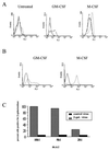
Figure 1
Figure 1
The effect of cytokine treatment on integrin expression and adenoviral infection of primary human monocytes. Monocytes were untreated or treated with GM-CSF and M-CSF for 48 hr followed by assay for (A) expression of integrins αVβ3 (broken line), αVβ5 (dotted line), or the negative control OX14 (solid line) using indirect immunofluoresence and FACS (FL1); or β-gal activity subsequent to infection with Advβgal (dotted line) at a moi of 100 (B) or various moi (C) assayed by FACS.
Figure 2
Figure 2
Expression of IκBα in AdvIκBα-infected macrophages and RAW 264.7 cells. RAW 264.7 cells (A) or M-CSF-treated human monocytes (B) were untreated or infected with control adenovirus (Adv0) or infected with the IκBα adenovirus (AdvIκBα) at the given moi. After 48 hr, cytosolic (A and B) and nuclear (C) extracts were prepared and assayed for IκBα expression by Western blotting. Blots were stripped and reprobed for p42/44MAPK as a control.
Figure 3
Figure 3
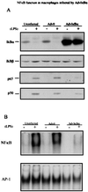
Inhibition of NF-κB function in AdvIκBα-infected macrophages. M-CSF-treated monocytes were infected with AdvO or AdvIκBα at a moi of 50. A third group was untreated. After an additional 3 days in M-CSF the cells were washed, and half of each group was treated (+) or otherwise (−) with 10 ng/ml LPS for 30 min. (A) The cells then were lysed, and cytosolic and nuclear extracts were prepared and analyzed for IκBα/β expression in the cytosol, or p65/p50 expression in the nucleus, by immunowestern blotting. Protein (150 μg and 50 μg) was analyzed for each cytosol or nuclear sample, respectively. (B) Nuclear extracts were prepared and 20 μg protein analyzed for NF-κB (Upper) or AP-1 (Lower) activity by electrophoretic mobility-shift assay.
Figure 4
Figure 4
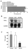
AdvIκBα infection inhibits TNFα induction in primary cells and rheumatoid joint cell cultures. Human monocytes cultured in the presence of M-CSF for 48 hr were untreated, infected with control adenovirus (Adv0), or infected with AdvIκBα at various moi. Two days after infection, cells were replated at 5 × 10⁵ cells per well on a 96-well plate (A) or at 5–10 × 106 cells per 100 mm Petri dish (B) and stimulated with 10 ng/ml LPS for 2 hr (A) or 16 hr (B). (A) Supernatants were removed and analyzed for TNFα by ELISA. (B) Cells were harvested and mRNA extracted and subjected to Northern analysis with the indicated probes. A represents results from seven different blood donors. Data in A and B are expressed as a percentage of the TNFα production from LPS-activated uninfected cells. Using a paired comparisons Student’s t test, there was significant (P < 0.001) inhibition at 20:1, 40:1, and 80:1 of AdvIκB. (C) Cells from rheumatoid synovium were cultured in 48-well plates at 1 × 10⁶ cells and infected with either AdvIκBα or AdvO at a moi of 40 to 1. The supernatants were taken for ELISA assay at 48 hr; pooled data from the five patients is shown (±SD). The null hypothesis that there was no change in TNF production whether cells were infected with AdvIκB or Adv0 could be rejected (P < 0.05) using a Wilcoxon’s signed rank test on paired differences compared with untreated cells on each patient.
Figure 5
Figure 5
Lack of apoptosis with TNFα in IκBα-infected macrophages. Human monocytes treated with M-CSF for 48 hr (A) or Hela cells (B) were plated on 60-mm Petri dishes at a density of 2–3 × 10⁶ cells per dish. They were left untreated or infected with control adenovirus or AdvIκBα at a moi of 40. Two days after infection, TNFα (20 ng/ml) and cycloheximide (2 μg/ml) were added as indicated. After 16 hr cells were stained for 30 min in 1.5 ml of a hypotonic fluorochrome solution (50 μg/ml of propidium iodide in 0.1% sodium citrate plus 0.1% Triton X-100), and the resulting propidium iodide-stained nuclei were analyzed by flow cytometry. The percentage of cells in each sector is indicated.

Similar articles

Cited by

References

    1. Buchan G, Barrett K, Turner M, Chantry D, Maini R N, Feldmann M. Clin Exp Immunol. 1988;73:449–455. - PMC - PubMed
    1. Brennan F M, Chantry D, Jackson A, Maini R, Feldmann M. Lancet. 1989;2:244–247. - PubMed
    1. Feldmann M, Brennan F M, Maini R N. Annu Rev Immunol. 1996;14:397–440. - PubMed
    1. Thorbecke G J, Shah R, Leu C H, Kuruvilla A P, Hardison A M, Palladino M A. Proc Natl Acad Sci USA. 1992;89:7375–7379. - PMC - PubMed
    1. Williams R O, Feldmann M, Maini R N. Proc Natl Acad Sci USA. 1992;89:9784–9788. - PMC - PubMed

Publication types

MeSH terms