. 1998 Jul 7;95(14):8227-32.
doi: 10.1073/pnas.95.14.8227.

Human parvovirus B19 as a causative agent for rheumatoid arthritis

Affiliations

Human parvovirus B19 as a causative agent for rheumatoid arthritis

Y Takahashi et al. Proc Natl Acad Sci U S A. .

Abstract

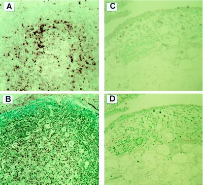
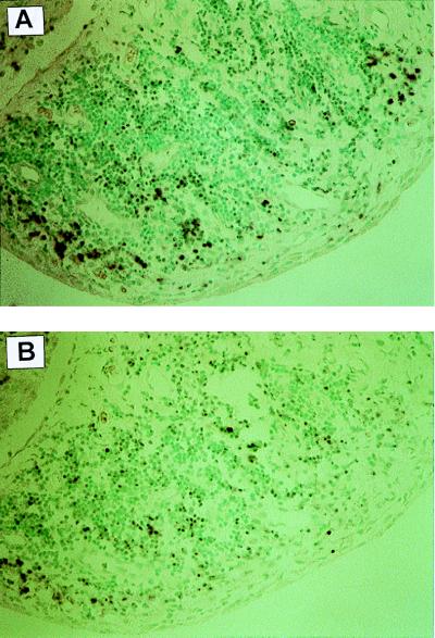
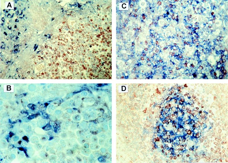
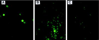
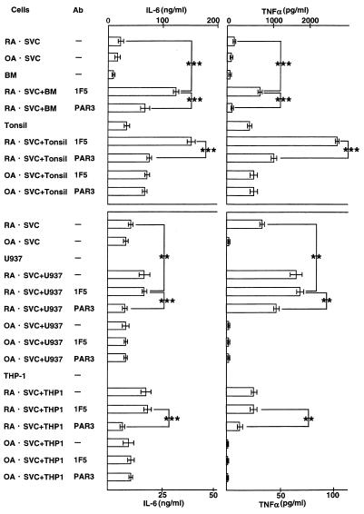
Human parvovirus B19 (B19) DNA was detected in the synovial tissues in 30 of 39 patients with rheumatoid arthritis (RA), and infrequently in those with osteoarthritis and traumatic joints. On the other hand, the expression of the B19 antigen VP-1 was specific (27/27) in RA synovium with active synovial lesions, but not in osteoarthritis and controls. The target cells of B19 were macrophages, follicular dendritic cells, T cells, and B cells, but not synovial lining cells in the synovium. B19-negative bone marrow cells, tonsil cells, and macrophage cell line U-937 cells became positive for the expression of VP-1, and more productive for interleukin 6 and tumor necrosis factor alpha when cocultured with RA synovial cells. The expression of VP-1 and the production of interleukin 6 and tumor necrosis factor alpha was significantly inhibited by the addition of neutralizing antibody for B19, suggesting that B19 detected in RA synovial cells is infective. B19 is involved in the initiation and perpetuation of RA synovitis, leading to joint lesions.


Figures

Figure 1
Figure 1
B19 DNA and VP-1 in synovial specimens. (A) ISH of RA synovium shows nuclear stain for B19 DNA and RNA (purple) in the cells mainly in lymphoid follicle. (B) Stain for anti-VP-1 antibody. The composed cells in the germinal center of lymphoid follicle and the mononuclear cells infiltrated in the sublining layer are stained on the RA synovium (brown). (C) The composed cells of OA synovium are negative for B19 DNA and RNA, when determined by ISH. (D) Stain for anti-VP-1 antibody. The SVC are not stained for anti-VP-1 antibody on OA synovium. All original magnifications ×400.
Figure 2
Figure 2
ISH for the detection of B19 DNA and RNA in RA synovial specimens. RA synovial specimens were treated with (B) or without RNase (A), and subsequently hybridized with digoxigenin-labeled B19 probe. The numbers of B19 DNA and RNA-positive signals (purple) in mononuclear cells infiltrated in the sublining layer decreased after treatment with RNase (B). B19 DNA and/or RNA are negative in synovial lining cells even before the treatment with RNase. Original magnification ×400.
Figure 3
Figure 3
Immunohistochemical detection of VP-1 and cell markers in RA SVC by double staining. (A) Synovial lining cells are stained for KP-1 (blue), but not for anti-VP-1 (red). Original magnification ×400. (B) Mononuclear cells in the sublining layer are stained for both KP-1 (blue) and anti-VP-1 (red) antibody. Original magnification ×1,000. (C) Lymphocytes in lymphoid follicle are stained for both CD20 (blue) and anti-VP-1 (red) antibody. Original magnification ×400. (D) Mononuclear cells in the germinal center are stained for both Ki-M4P (a follicular dendritic cell marker, blue) and anti-VP-1 (red) antibody. Antigen-presenting cells (macrophages and follicular dendritic cells) and lymphocytes are positive for VP-1. Original magnification ×400.
Figure 4
Figure 4
An immunofluorescence analysis of the infectivity of B19. B19-negative tonsil cells or U937 cells were cocultured with SVC at double-chamber culture system using Cell Culture Insert (Falcon), washed three times at 3 days, and then tested for the reactivity with F(ab)′2 fragments of VP-1 (14). Tonsil cells (A) or U937 cell (B) cocultured with RA-derived SVC are positive for VP-1. But U937 cells (C) do not show immunoreactivity for VP-1 when cocultured with OA-derived SVC.
Figure 5
Figure 5
Inhibition of the production of IL-6 or TNFα by anti-VP-1 antibody PAR3. The experimental system is the same as that in Fig. 4. SVC from RA induce or enhance IL-6 and TNFα production by bone marrow cells (BM), tonsil cells (tonsil), a macrophage cell line U937, or THP-1 when tested at 3 days after the coculture, but those from OA do not. The addition of F(ab)′2 fragments of neutralizing antibody of B19 PAR3 (14), but not that of 1F5 with irrelevant specificity causes marked decrease of IL-6 or TNFα production in the culture supernatant. ∗∗∗, P < 0.01, ∗∗, P < 0.02.

References

    1. White D G, Woolf A D, Mortimer P P, Cohen B J, Blake D R, Bacon P A. Lancet. 1985;i:419–421. - PubMed
    1. Reid D M, Reid T M S, Brown T, Rennie J A N, Eastmond C J. Lancet. 1985;i:422–425. - PubMed
    1. Woolf A D, Campion G V, Chishic K A, Wise S, Cohen B J, Klouda P T, Caul O, Dieppe P A. Arch Intern Med. 1989;149:1153–1156. - PubMed
    1. Nishioka K, Maruyama I, Sato K, Kitajima I, Nakajima Y, Osame M. Lancet. 1989;1:441. - PubMed
    1. Naides S J. Curr Opin Rheumatol. 1994;6:423–428. - PubMed

Publication types

MeSH terms

Substances