. 1998 Jul 7;95(14):8257-61.
doi: 10.1073/pnas.95.14.8257.

Evidence for multiclonality in multicentric Kaposi's sarcoma

Affiliations

Evidence for multiclonality in multicentric Kaposi's sarcoma

P S Gill et al. Proc Natl Acad Sci U S A. .

Abstract

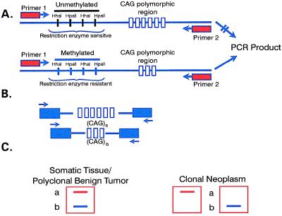
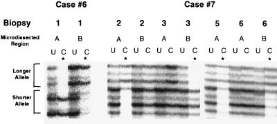
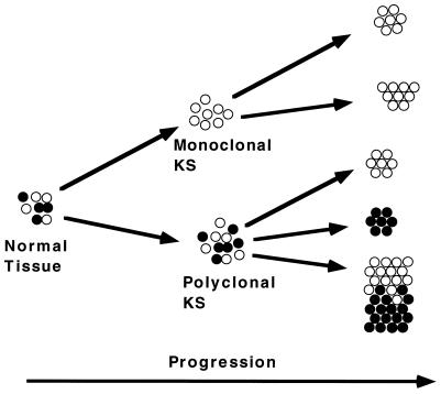
Kaposi's sarcoma (KS) develops in a variety of clinical states and is the most common tumor seen in patients with HIV-1 infection. KS develops as a multifocal mucocutaneous disease with subsequent spread to visceral organs, and it has been argued to be a benign proliferation caused by its multifocality at initial presentation, lack of aneuploidy, and spontaneous regression upon withdrawal of immunosuppressive agents in iatrogenically induced disease. We wished to determine whether KS lesions are clonal, indicative of a true neoplasm. Also, we tested whether multifocal KS lesions are clonally related, derived from a common progenitor cell or of independent cellular origin. We studied the X-chromosome inactivation pattern of the human androgen receptor gene in tumor biopsies of women with KS. This procedure tests for the clonality of a tissue specimen, a hallmark of neoplasia. Each specimen was microdissected to minimize normal cell contamination. Of 12 evaluable cases, 10 were HIV-seropositive and 2 were HIV-seronegative. Twenty-four biopsies from the 12 patients were examined. Five cases were consistent with individual KS lesions being clonal. In two cases, multiple KS specimens derived from the individual patients had different androgen receptor alleles inactivated, proving unequivocally that these KS lesions arose independently from distinct transformed cells. In seven cases, only a polyclonal pattern of inactivation was observed, whereas two others had tumor areas of both clonal and polyclonal inactivation patterns. These findings suggest that KS can be a clonal neoplasm, and in some of the cases multiple KS lesions in a given patient can arise from independent cellular origins and acquire clonal characteristics. The polyclonal inactivation pattern observed in other KS lesions may represent a premalignant stage or false negative results.


Figures

Figure 1
Figure 1
Schema of clonality analysis based on x-chromosome inactivation of the AR. (A) Top allele contains unmethylated restriction sites that will undergo cutting of DNA with the methylation-sensitive enzymes and thus fail to amplify during PCR. Bottom allele contains methylated restriction sites, and thus DNA will not be cut with methylation-sensitive enzymes, resulting in amplification during PCR. (B) Polymorphism of CAG repeats of the AR. (C) Amplification of AR allele from somatic cells with specific primers flanking the CAG repeat will result in two products of different size. In a clonally derived cell population, the same allele is inactivated by methylation in all the cells. Amplification of only the endonuclease insensitive allele can occur leading to the product size of one of the two alleles.
Figure 2
Figure 2
Clonal analysis of representative KS lesions. The biopsy number and microdissected region of the KS lesions are given above their respective autoradiograph. U, uncut control; C, digested (or cut) with methylation-sensitive restriction enzyme HhaI prior to PCR amplification of the AR gene. The presence of a single AR gene in the digested (C) lane of the KS lesion indicates clonality (e.g., biopsy 2, region A of case 7). The presence of both AR alleles in the digested (C) lane (e.g., biopsy 2, region B of case 7) indicates a polyclonal pattern. Different AR alleles inactivated (e.g., region A and B of case 6; biopsy 2, region A; biopsy 3, region B of case 7) indicate these KS lesions arose independently and are not clonally related either in the same biopsy (case 6) or different biopsies (case 7).
Figure 3
Figure 3
(A and B) Histologic examination of two KS regions from case 7 that are shown for their X-chromosome inactivation pattern in Fig. 2. The left column represents biopsy 2, and the right column represents biopsy 3. Histologic examinations at low power (×20 magnification, Top), intermediate power (×100 magnification, Middle), and high power (×400 magnification, Bottom) are shown. Discrete tumor regions with minimal cellularity surrounded by normal tissue are noted. The lesions display characteristic features of KS with proliferation of spindle cells and slit-like vascular structures with extravasated red blood cells.
Figure 4
Figure 4
Model for KS progression. Normal tissue is a mosaic of cells with different methylated AR alleles (represented by open and filled circles) caused by random X-chromosome inactivation. Neoplasms typically develop from clonal outgrowth of a single cell into a monoclonal tumor with subsequent metastasis of the same clone (monoclonal KS pathway). Alternatively, as evidence from our data and shown in the polyclonal KS pathway, individual lesions may have a polyclonal/hyperplastic stage, followed by the outgrowth of distinct clonal neoplasm(s) (seen as groups of either open or closed circles, e.g., case 7). This process may also occur at distinct sites within the same lesion (shown as defined areas of both open and closed circles in the same group, e.g., case 6).

References

    1. Geddes M, Francheschi S, Barchielli A, Falcini F, Carli S, Cocconi G, Conti E, Crosignani P, Gata L, Giarelli L. Br J Cancer. 1994;69:333–336. - PMC - PubMed
    1. Wahman A, Melnick S, Rhame F S, Potter J D. Epidemiol Rev. 1991;13:178–199. - PubMed
    1. Penn I. Transplantation. 1979;27:8–11. - PubMed
    1. Lifson A R, Darrow W W, Hessol N A, O’Malley P M, Barnhart L, Jaffe H J, Rutherford G W. Am J Epidemiol. 1990;131:221–231. - PubMed
    1. Gill P S, Hamilton A, Naidu Y. AIDS Updates. 1994;7:1–11.

Substances