. 1998 Jul 7;95(14):8274-9.
doi: 10.1073/pnas.95.14.8274.

Nanobacteria: an alternative mechanism for pathogenic intra- and extracellular calcification and stone formation

Affiliations

Nanobacteria: an alternative mechanism for pathogenic intra- and extracellular calcification and stone formation

E O Kajander et al. Proc Natl Acad Sci U S A. .

Abstract

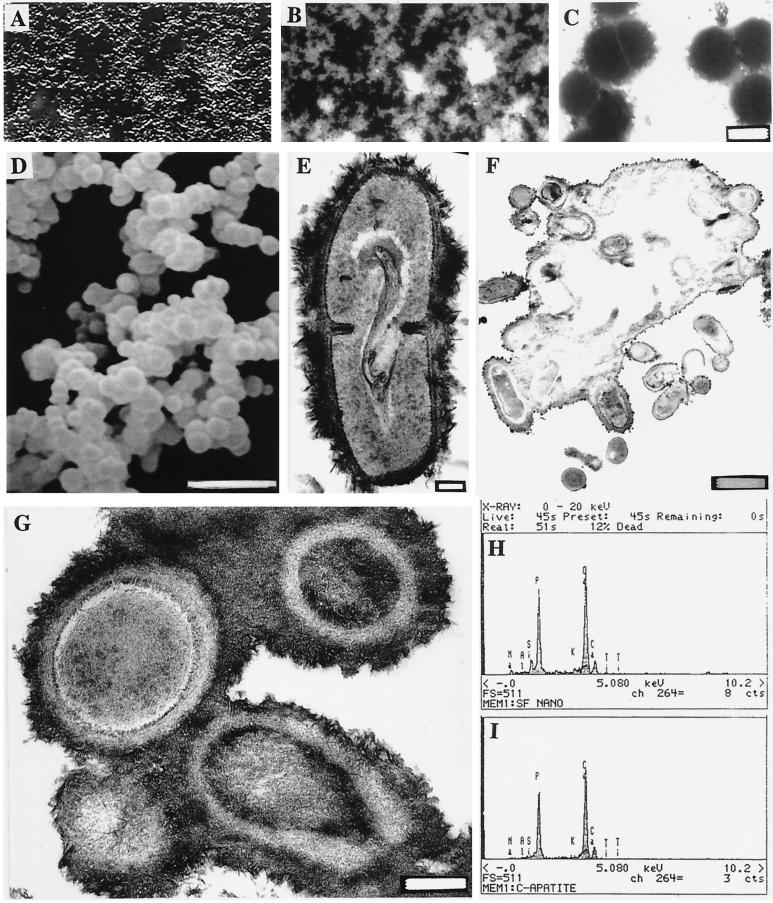
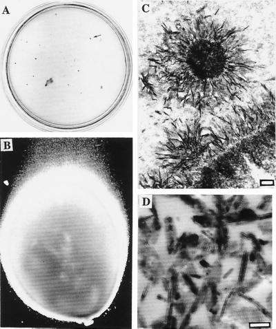
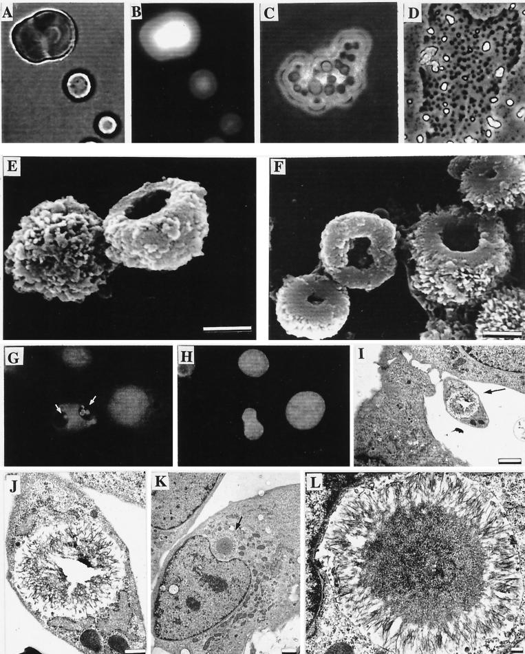
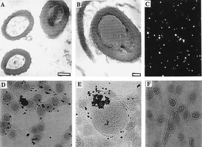
Calcium phosphate is deposited in many diseases, but formation mechanisms remain speculative. Nanobacteria are the smallest cell-walled bacteria, only recently discovered in human and cow blood and commercial cell culture serum. In this study, we identified with energy-dispersive x-ray microanalysis and chemical analysis that all growth phases of nanobacteria produce biogenic apatite on their cell envelope. Fourier transform IR spectroscopy revealed the mineral as carbonate apatite. The biomineralization in cell culture media resulted in biofilms and mineral aggregates closely resembling those found in tissue calcification and kidney stones. In nanobacteria-infected fibroblasts, electron microscopy revealed intra- and extracellular acicular crystal deposits, stainable with von Kossa staining and resembling calcospherules found in pathological calcification. Previous models for stone formation have led to an hypothesis that elevated pH due to urease and/or alkaline phosphatase activity is a lithogenic factor. Our results indicate that carbonate apatite can be formed without these factors at pH 7.4, at physiological phosphate and calcium concentrations. Nanobacteria can produce apatite in media mimicking tissue fluids and glomerular filtrate and provide a unique model for in vitro studies on calcification.


Figures

Figure 1
Figure 1
Light and electron microscopic images of nanobacteria and their analyses with EDX. (A) DIC image of bottom-attached nanobacteria after a 2-mo culture period. (B) DNA staining of the same area (×1600) with the modified Hoechst method. (C) Negative staining of nanobacteria isolated directly from FBS. (Bar = 200 nm.) (D) SEM micrograph showing their variable size. (Bar = 1 μm.) (E) A dividing nanobacterium covered with a “hairy” apatite layer. (Bar = 100 nm.) (F) TEM micrograph of nanobacteria buried in an apatite layer after a 3-mo culture period (bar = 1 μm) and G at higher magnification (bar = 200 nm). White central areas in F are artifacts due to loss of the mineral layer in sectioning. (H) EDX analysis in SEM of nanobacteria showing Ca and P peaks similar to those of hydroxyapatite (I).
Figure 2
Figure 2
Nanobacterial stony colonies and comparison to hydroxyapatite. (A) Colonies on modified Loeffler medium in a 10-cm plate. The colonies penetrated through the medium forming stony pillars. Arrow shows one typical grayish brown colony depicted in B. (×40.) (C) Needle-like crystal deposits in the pillar revealed by TEM. (Bar = 200 nm.) (D) TEM image of reference apatite crystals. (Bar = 100 nm.)
Figure 3
Figure 3
Nanobacteria cultured under SF conditions and their interaction with cells. (A) Light microscopic micrograph. (B) DNA staining of the same area with the modified Hoechst staining method. (C) DIC images of nanobacteria inside a common apatite shelter. (D) A partly demineralized nanobacterial group (A–D, ×860). (E and F) SEM micrographs of nanobacterial dwellings detached from the culture vessel. (Bars = 1 μm.) (G) IIFS of internalized mineralized nanobacteria (white arrows) in 3T6 cells. (H) DNA staining of the same area with standard Hoechst method. (×540.) (I–L) TEM micrographs of intracellular calcifications in 3T6 cells caused by SF nanobacteria. (Bars: I and K = 2 μm; J = 500 nm; L = 200 nm.)
Figure 4
Figure 4
Examples of extra- and intracellular calcification by nanobacteria. TEM micrograph of cultured nanobacteria (bar = 200 nm) from FBS (A) and a bacterium in a kidney stone after demineralization (B; bar = 50 nm). (C) IIFS of the same kidney stone with anti-nanobacteria mAb. (D and E) von Kossa staining results of 3T6 cells exposed to SF nanobacteria for 24 hr. (F) Negative control. (×270.)

References

    1. Robey P G, Boskey A L. In: Osteoporosis. Marcus R, Feldman D, Kelsey J, editors. San Diego: Academic; 1996. pp. 95–183.
    1. Folk R L. J Sediment Petrol. 1993;63:990–999.
    1. Sillitoe R H, Folk R L, Saric N. Science (Washington DC) 1996;272:1153–1155. - PubMed
    1. Mojzsis S J, Arrhenius G, McKeegan K D, Harrison T M, Nutman A P, Friend C R L. Nature (London) 1996;384:55–59. - PubMed
    1. Çiftçioglu N, Kajander E O. Pathophysiology. 1998;4:259–270.

LinkOut - more resources