. 1998 Jul 7;95(14):8322-7.
doi: 10.1073/pnas.95.14.8322.

Different patterns of truncated prion protein fragments correlate with distinct phenotypes in P102L Gerstmann-Sträussler-Scheinker disease

Affiliations

Different patterns of truncated prion protein fragments correlate with distinct phenotypes in P102L Gerstmann-Sträussler-Scheinker disease

P Parchi et al. Proc Natl Acad Sci U S A. .

Abstract

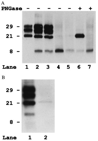
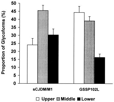
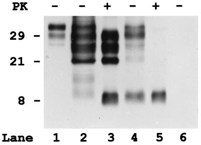
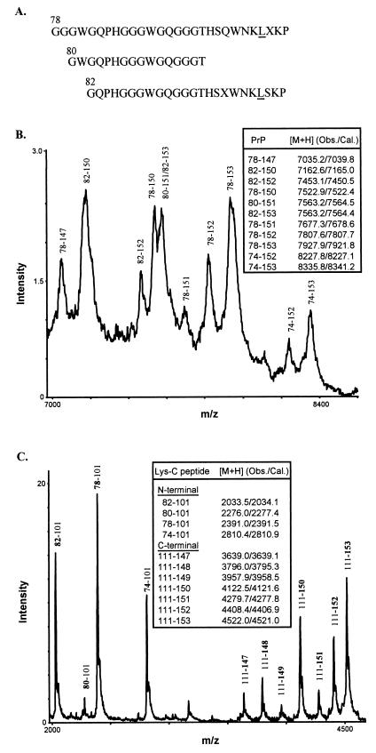
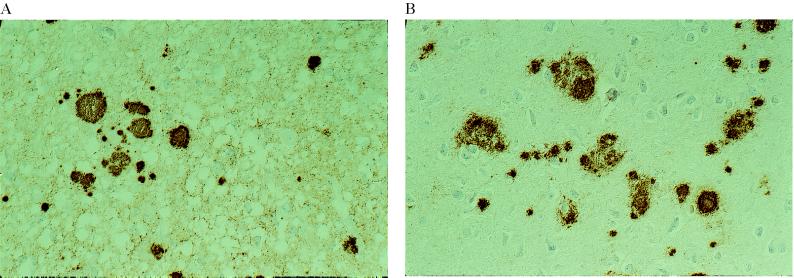
The clinicopathological phenotype of the Gerstmann-Sträussler-Scheinker disease (GSS) variant linked to the codon 102 mutation in the prion protein (PrP) gene (GSS P102L) shows a high heterogeneity. This variability also is observed in subjects with the same prion protein gene PRNP haplotype and is independent from the duration of the disease. Immunoblot analysis of brain homogenates from GSS P102L patients showed two major protease-resistant PrP fragments (PrP-res) with molecular masses of approximately 21 and 8 kDa, respectively. The 21-kDa fragment, similar to the PrP-res type 1 described in Creutzfeldt-Jakob disease, was found in five of the seven subjects and correlated with the presence of spongiform degeneration and "synaptic" pattern of PrP deposition whereas the 8-kDa fragment, similar to those described in other variants of GSS, was found in all subjects in brain regions showing PrP-positive multicentric amyloid deposits. These data further indicate that the neuropathology of prion diseases largely depends on the type of PrP-res fragment that forms in vivo. Because the formation of PrP-res fragments of 7-8 kDa with ragged N and C termini is not a feature of Creutzfeldt-Jakob disease or fatal familial insomnia but appears to be shared by most GSS subtypes, it may represent a molecular marker for this disorder.


Figures

Figure 1
Figure 1
(A) Immunoblot analysis with the 3F4 antibody of the PrP-res extracted from the cerebral cortex of four subjects with GSS P102L (lanes 2–7) and one subjects with sporadic CJD (lane 1). Lanes 6 and 7 include samples that also were deglycosylated with PNGase F (PNGase, lanes +). Lanes: 1, sporadic CJD, codon 129 M/M, and PrP-res type1; 2 and 6, subject 2; 3, subject 5; 4 and 7, subject 7; 5, subject 6. (B) Immunoblot analysis of PrP-res from subjects 2 and 7 probed with the anti-C antiserum.
Figure 2
Figure 2
Relative proportion of the three PrP-res glycoforms of the 21-kDa fragment (PrP-res type 1) in GSS P102L compared with that of sporadic CJD (sCJD). Mean ± SD. Upper, high-molecular mass glycoform; middle, low-molecular-mass glycoform; low, unglycosylated form.
Figure 3
Figure 3
Immunoblot analysis of purified detergent insoluble fractions (P3) of PrP from the cerebral cortex of one control subject (lane 6) and two subjects (2 and 7) with GSS P102L (lanes 2–5), before (lanes 1, 2, 4, and 6) and after PK digestion (lanes 3 and 5). Although in the control (lane 6), no PrP is detectable in the insoluble fraction not treated with PK, in both GSS samples, significant amount of truncated PrP fragments, in addition to some full-length PrP, are present. However, pattern and properties of the truncated PrP peptides are different in the two GSS cases. In subject 2 (lane 2), there are large amount of 29-, 27-, and 21-kDa fragments that comigrate with the PrP-res peptides visible after PK treatment. In addition, two bands migrating at 11- and 9-kDa are present. Patient 7 (lane 4) shows similar bands at 29, 27, and 21 kDa, but only before PK treatment, indicating that these peptides are insoluble but not protease resistant. In addition, a band at 8 kDa that has similar intensity and comigrates with the PrP-res fragment is visible. Full-length PrP from a crude homogenate of the control subject is represented in lane 1.
Figure 4
Figure 4
Characterization of 8-kDa PrP-res by protein sequencing and mass spectrometry. (A) N-terminal sequences of purified 8-kDa PrP-res. The experimentally determined amino acid signals at each Edman degradation cycle were aligned with the known human PrP sequence to derive three major N-terminal species, with the residue positions shown above the first amino acids and the mutant residue at position 102 underlined. Unassigned residues were indicated as X. (B) Spectrum of 8-kDa PrP-res obtained by matrix-assisted laser desorption/ionization mass spectrometry. Major PrP species having Leu at position 102 and inclusive of N and C termini were assigned according to the observed and calculated protonated mass signals ([M+H] values shown in Inset). (C) Matrix-assisted laser desorption/ionization mass spectrum of the Lys-C digest of 8-kDa PrP-res. The N- and C-terminal PrP peptides generated by Lys-C digestion were assigned as in B. The N-terminal peptides corresponded to fragments that begin (bold) at positions 74, 78, 80, and 82, respectively, and end at the cleavage site at position 101 (the presence of Leu at position 102 introduces a specific cleavage at position 101). No wild-type N-terminal peptides were observed because, if present, they would correspond to PrP peptides having Pro at position 102 and spanning residues 74–106, 78–106, 80–106, and 82–106 (calculated [M+H] values 3348.6, 2929.2, 2815.1, 2571.8, respectively). The C-terminal peptides corresponded to fragments that begin at the cleavage site at positions 111 and end (bold) at positions 147, 148, 149, 150, 151, 152, and 153, respectively.
Figure 5
Figure 5
PrP immunoreactivity in two GSS P102L subjects showing distinct patterns of PrP-res on immunoblot. (A) Cerebral cortex of patient 5. A diffuse, synaptic staining is seen in association with multiple PrP immunopositive plaques. Immunolabeling with 3F4, ×250. (B) Cerebral cortex of patient 6. There are multiple PrP immunopositive plaques. No synaptic staining is visible. Immunolabeling with 3F4, ×250.

References

    1. Parchi P, Piccardo P, Gambetti P, Ghetti B. In: Progress in Pathology 4. Kirkham N, Lemoine N R, editors. Edimburgh: Churchill Livingstone; 1998. pp. 39–77.
    1. Ghetti B, Piccardo P, Frangione B, Bugiani O, Giaccone G, Young K, Prelli F, Farlow M R, Dlouhy S R, Tagliavini F. Brain Pathol. 1996;6:127–145. - PubMed
    1. Monari L, Chen S G, Brown P, Parchi P, Petersen R B, Mikol J, Gray F, Cortelli P, Montagna P, Ghetti B, et al. Proc Natl Acad Sci USA. 1994;91:2839–2842. - PMC - PubMed
    1. Parchi P, Castellani R, Capellari S, Ghetti B, Young K, Chen S G, Farlow M, Dickson D W, Sima A A F, Trojanowski J Q, et al. Ann Neurol. 1996;39:767–778. - PubMed
    1. Parchi P, Capellari S, Chen S G, Petersen R B, Gambetti P, Kopp N, Brown P, Kitamoto T, Tateishi J, Giese A, et al. Nature (London) 1997;386:232–234. - PubMed

Publication types