. 1998 Jul 7;95(14):8357-62.
doi: 10.1073/pnas.95.14.8357.

Normal retina releases a diffusible factor stimulating cone survival in the retinal degeneration mouse

Affiliations

Normal retina releases a diffusible factor stimulating cone survival in the retinal degeneration mouse

S Mohand-Said et al. Proc Natl Acad Sci U S A. .

Abstract

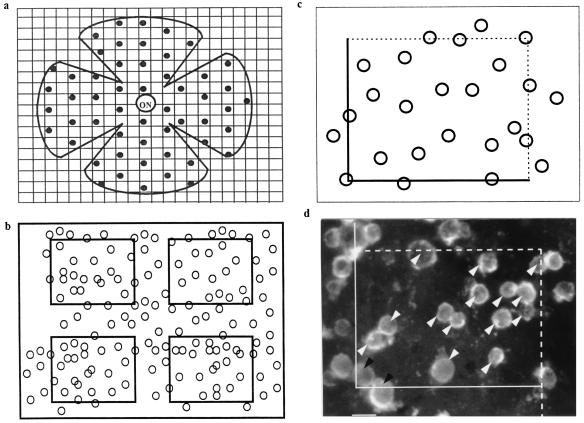
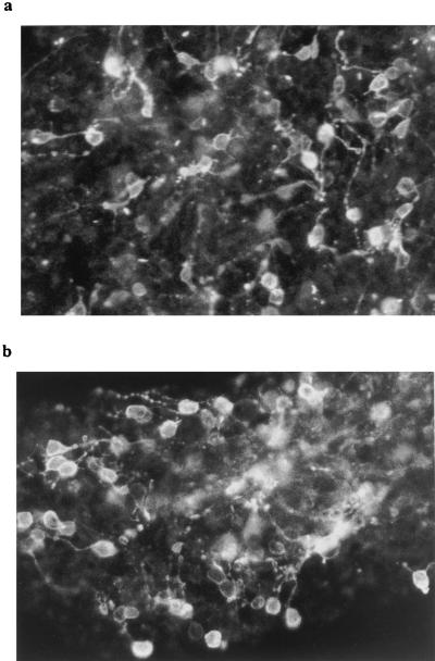
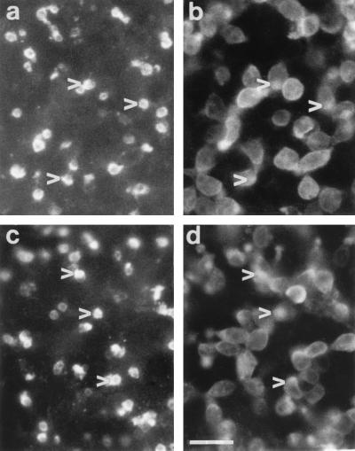
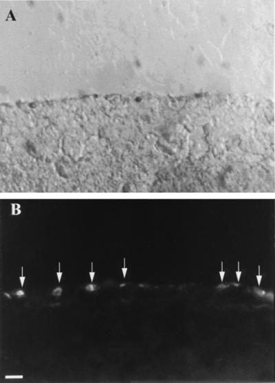
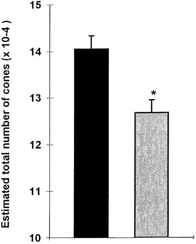
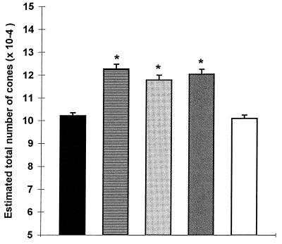
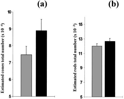
The role of cellular interactions in the mechanism of secondary cone photoreceptor degeneration in inherited retinal degenerations in which the mutation specifically affects rod photoreceptors was studied. We developed an organ culture model of whole retinas from 5-week-old mice carrying the retinal degeneration mutation, which at this age contain few remaining rods and numerous surviving cones cocultured with primary cultures of mixed cells from postnatal day 8 normal-sighted mice (C57BL/6) retinas or retinal explants from normal (C57BL/6) or dystrophic (C3H/He) 5-week-old mice. After 7 days, the numbers of residual cone photoreceptors were quantified after specific peanut lectin or anti-arrestin antibody labeling by using an unbiased stereological approach. Examination of organ cultured retinas revealed significantly greater numbers of surviving cones (15-20%) if cultured in the presence of retinas containing normal rods as compared with controls or cocultures with rod-deprived retinas. These data indicate the existence of a diffusible trophic factor released from retinas containing rod cells and acting on retinas in which only cones are present. Because cones are responsible for high acuity and color vision, such data could have important implications not only for eventual therapeutic approaches to human retinal degenerations but also to define interactions between retinal photoreceptor types.


Figures

Figure 1
Figure 1
(a) Systematic random sampling of the retinal surface for stereological counting. ON, optic nerve. (b) Each sample was subdivided into four counting fields after focusing and digitization. (c) Counting was performed by using a stereological dissector. (d) Example of a counted sample from a 5-week-old rd mouse retina cocultured for 8 days with dissociated cells from 8-day-old C57 rod-containing retina. White arrow head, counted cells; black arrow head, excluded cells. (Bar = 5 μm.)
Figure 2
Figure 2
Five-day in vitro cultures of normal (a) and rd (b) mice retinal cells from postnatal day 8 animals, stained with rho-4D2 anti-rod opsin antibody. In both cases, numerous immunoreactive cells are visible. (Bar = 10 μm.)
Figure 3
Figure 3
Explanted rd mice retinas cultured for 1 week in DMEM alone (a and b) or in the presence of dissociated young retinal cells (c and d), then double immunolabeled with PNA lectin (a and c) and arrestin antiserum (b and d). The same fields are shown in a and b and in c and d and reveal that PNA lectin labels rounded matrix-associated structures protruding from the retinal surface whereas arrestin is present throughout the photoreceptor cell bodies and outer segments. The differing focal plane of the two labels makes superimposition difficult, but the overlapping nature of the staining pattern can be observed (corresponding arrowheads in paired fields a and b and in paired fields c and d). (Bar = 10 μm.)
Figure 4
Figure 4
Cross section through a PNA-labeled flat-mounted retina, showing that labeling is restricted to cones. (a) Nomarski image of outer 50 μm of retina; (b) The same field viewed by fluorescence microscopy, showing lectin staining present only at the surface (arrows). (Bar = 10 μm.)
Figure 5
Figure 5
Estimated total number of cones (mean ± SD) in retinas from 5-week and 6-week rd mice in vivo. *P < 0.01.
Figure 6
Figure 6
Estimated total number of cones (mean ± SD) in 5-week-old rd (C3H/He/J) mice retinas cultured in DMEM alone and from 5-week-old rd mice retinas cocultured with cells from 8-day-old normal mice retinas with cells from 8-day-old rd mice retinas, with retinal explants from 5-week-old normal mice, and with retinal explants from 5-week-old rd mice. *P < 0.0001.
Figure 7
Figure 7
Estimated total number (mean ± SD) of cones (a) and rods (b) in retinas from 5-week-old rd mice (C3H/He/N) cultured in DMEM alone and in retinas from 5-week-old rd mice cocultured with 8-day-old normal C57BL/6 mice. *P < 0.0001. There was no significant difference between rod numbers.

Similar articles

Cited by

References

    1. Berson E L. Invest Ophthalmol Visual Sci. 1993;34:1659–1676. - PubMed
    1. Adler R. Arch Ophthalmol. 1996;114:79–83. - PubMed
    1. Chang G Q, Hao Y, Wong F. Neuron. 1993;11:595–605. - PubMed
    1. Gregory C Y, Bird A C. Br J Ophthalmol. 1995;79:186–190. - PMC - PubMed
    1. Al-Maghtheh M, Gregory C, Inglehearn C, Hardcastle A, Bhattacharya S. Hum Mutat. 1993;2:249–255. - PubMed

Publication types

Substances

LinkOut - more resources