

Inhibition in verbal working memory revealed by brain activation
- PMID: 9653200
- PMCID: PMC20989
- DOI: 10.1073/pnas.95.14.8410
Inhibition in verbal working memory revealed by brain activation
Abstract
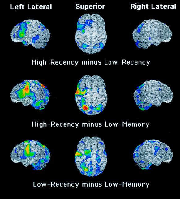
There are many occasions in which humans and other animals must inhibit the production of some behavior or inhibit the processing of some internal representation. Success in inhibitory processing under normal circumstances can be revealed by the fact that certain brain pathologies render inhibitory processing ineffective. These pathologies often have been associated with damage to frontal cortex, including lateral and inferior aspects. We provide behavioral evidence of a verbal working memory task that, by hypothesis, engaged inhibitory processing, and we show (by using positron emission tomograpny) that the inhibitory processing is associated with a lateral portion of the left prefrontal cortex. The task in which subjects engaged was item-recognition: Four target letters were presented for storage followed, after a brief interval, by a probe letter that could match a target letter or not. On some trials, when the probe did not match a target letter and therefore required a "no" response, the probe had matched a target letter of the previous trial, so on these trials a "yes" response was prepotent and had to be inhibited, by hypothesis. Compared with a condition in which no prepotent response was created, this condition yielded brain activation in left inferior frontal gyrus, in the region of Brodmann's area 45.
Figures
References
Publication types
MeSH terms
LinkOut - more resources
Full Text Sources
Medical
