. 1998 Jul;180(14):3522-8.
doi: 10.1128/JB.180.14.3522-3528.1998.

Identification of cpxR as a positive regulator essential for expression of the Shigella sonnei virF gene

Affiliations

Identification of cpxR as a positive regulator essential for expression of the Shigella sonnei virF gene

S i Nakayama et al. J Bacteriol. 1998 Jul.

Abstract

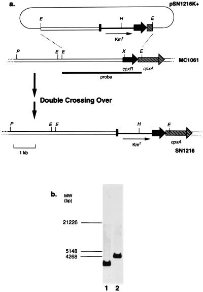
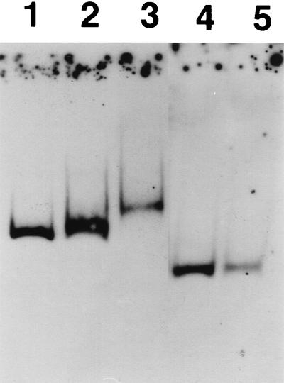
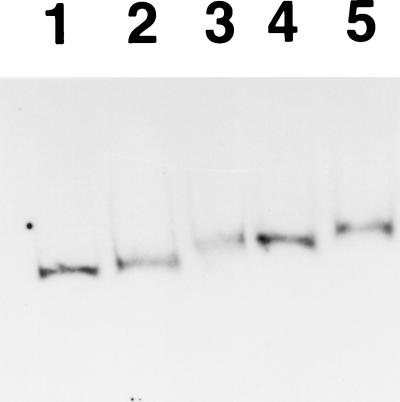
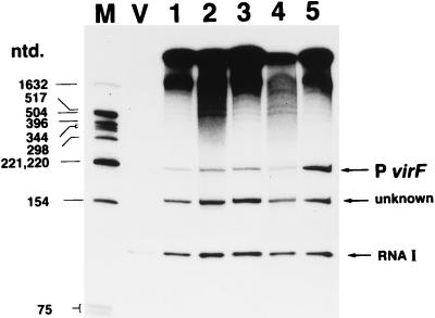
virF is the master regulator which activates the virulence determinant genes of Shigella spp. such as ipaBCD and virG. We previously reported that expression of virF itself is regulated in a pH-dependent manner and that cpxA, a sensor of a two-component regulatory system, is involved in this regulation (S. Nakayama and H. Watanabe, J. Bacteriol. 177:5062-5069, 1995). Disruption of cpxR, which has been thought to be the cognate response regulator of cpxA (J. Dong, S. Iuchi, H.-S. Kwan, Z. Lue, and E. C. C. Lin, Gene 136:227-230, 1993), abolished virF expression almost completely. Purified CpxR bound directly to the upstream region of virF. Binding capacity was enhanced when CpxR was phosphorylated by coincubation with acetyl phosphate in vitro. Furthermore, we observed that phosphorylated CpxR could activate virF transcription in vitro. These results clearly indicated that CpxR was an essential activator for virF expression and strongly suggested that the binding of phosphorylated CpxR to the target site upstream of the virF gene induced a direct activation of virF transcription.


Figures

FIG. 1
FIG. 1
(a) Schematic drawings of the constructs of plasmid pSN1216K+ and of chromosomes around the cpxR-cpxA regions in MC1061 and SN1216. Thick arrows indicate the locations and direction of cpxR and cpxA genes. Thin arrows in pSN1216K+ and SN1216 show the sites and the direction of the inserted Kmr cassette. The location of the 3-kb EcoRI probe used in the Southern hybridization analysis is shown by a thick line under the MC1061 drawing. Abbreviations: E, EcoRI; H, HindIII; P, PstI; X, XhoI. (b) Confirmation of chromosomal constructions of MC1061 and SN1216 by Southern hybridization with the probe described above. Ten micrograms of the chromosome DNA of MC1061 (lane 1) and SN1216 (lane 2) was completely digested by EcoRI and run on a 0.7% agarose gel containing 1× TAE buffer. After the DNA was blotted onto a nylon membrane, hybridization was performed with the horseradish peroxidase-labeled probe, followed by washing and signal detection (see Materials and Methods for the detailed protocol). The positions of DNA molecular size markers in base pairs are indicated on the left.
FIG. 2
FIG. 2
Gel shift assay. Eight femtomoles of probe C consisting of nt −103 to +110, with the virF transcription start site as nt +1 (lanes 1, 2, and 3), and probe D, nt −37 to +110 (lanes 4 and 5), was generated by PCR, and the 3′ termini were labeled with DIG–11-ddUTP. Probes were incubated at 25°C for 30 min in 20 mM HEPES (pH 7.6)–1 mM EDTA–10 mM (NH4)2SO4–1 mM DTT–0.2% (wt/vol) Tween 20–30 mM KCl–1 μg of poly(dI-dC) per 20 μl–0.1 μg of poly-l-lysine per 20 μl in the absence of CpxR (lanes 1 and 4) or the presence of 0.9 (lane 2) or 1.8 μM (lanes 3 and 5) (final concentrations) CpxR sample in a final reaction volume of 20 μl. After electrophoresis on a 6% polyacrylamide gel containing 0.25× TBE, DNA was blotted onto a nylon membrane, followed by incubation with anti-DIG Fab–alkaline phosphatase conjugate and signal detection (see Materials and Methods for the detailed protocol).
FIG. 3
FIG. 3
Gel shift assay with phosphorylated or nonphosphorylated CpxR. Eight femtomoles of DIG-labeled probe C (see legend to Fig. 2) (lanes 1 to 5) was incubated under the same condition as those for Fig. 2 in the absence of CpxR (lane 1) or the presence of 0.9 (lanes 2 and 4) or 1.8 μM (lanes 3 and 5) (final concentrations) CpxR samples preincubated in 50 mM acetyl phosphate at 37°C for 30 min (lanes 4 and 5) or in phosphorylation buffer only (lanes 2 and 3) (see Materials and Methods for the detailed conditions of the phosphorylation of CpxR). After the electrophoresis, the same protocol as that of Fig. 2 was performed.
FIG. 4
FIG. 4
In vitro transcription assay of virF transcript. pSN600-T (lanes 1 to 5) or pHSG397 (lane V) (0.5 pmol) was preincubated without CpxR (lanes V and 1) or with nonphosphorylated (lanes 2 and 3) or phosphorylated (lanes 4 and 5) CpxR samples in 40 mM Tris-HCl (pH 7.9)–10 mM MgCl2–0.1 mM EDTA–150 mM KCl–5% glycerol–2 mM DTT at 37°C for 5 min, followed by the addition of 1 U of E. coli RNA polymerase (for the phosphorylation of CpxR samples, see the legend to Fig. 3 and Materials and Methods). The final concentrations of CpxR were 0 (lanes V and 1), 0.14 (lanes 2 and 4), and 1.4 μM (lanes 3 and 5). After further incubation at 37°C for 5 min, transcription was started by the addition of nucleoside triphosphates and [α-32P]UTP. The final volume of the reaction mixture was adjusted to 50 μl. After the reaction, the samples were extracted with phenol-CHCl3, precipitated by EtOH, and denatured as described in Materials and Methods. RNA was analyzed by electrophoresis on a urea–denatured 8% polyacrylamide gel containing 1× TBE, followed by autoradiography. The positions of the virF transcript (P virF), internal control RNAI, and a cryptic RNA about 150 nt long of unknown origin are indicated by horizontal arrows on the right. The positions of denatured DNA molecular size markers in nucleotides (ntd.) (lane M) are indicated on the left.

References

    1. Adler B, Sasakawa C, Tobe T, Makino S, Komatsu K, Yoshikawa M. A dual transcriptional activation system for the 230kb plasmid genes coding for virulence-associated antigens of Shigella flexneri. Mol Microbiol. 1989;3:627–635. - PubMed
    1. Albright L M, Huala E, Ausubel F M. Prokaryotic signal transduction mediated by sensor and regulator protein pairs. Annu Rev Genet. 1989;23:311–336. - PubMed
    1. Bernardini M L, Fontaine A, Sansonetti P J. The two-component regulatory system OmpR-EnvZ controls the virulence of Shigella flexneri. J Bacteriol. 1990;172:6274–6281. - PMC - PubMed
    1. Brosius J, Dull T J, Sleeter D D, Noller H F. Gene organization and primary structure of a ribosomal RNA operon from Escherichia coli. J Mol Biol. 1981;148:107–127. - PubMed
    1. Chang A C Y, Cohen S N. Construction and characterization of amplifiable multicopy DNA cloning vehicles derived from the p15A cryptic miniplasmid. J Bacteriol. 1978;134:1141–1156. - PMC - PubMed

Publication types

MeSH terms

LinkOut - more resources