. 1998 Aug;72(8):6291-7.
doi: 10.1128/JVI.72.8.6291-6297.1998.

The herpesvirus transactivator VP16 mimics a human basic domain leucine zipper protein, luman, in its interaction with HCF

Affiliations

The herpesvirus transactivator VP16 mimics a human basic domain leucine zipper protein, luman, in its interaction with HCF

R Lu et al. J Virol. 1998 Aug.

Abstract

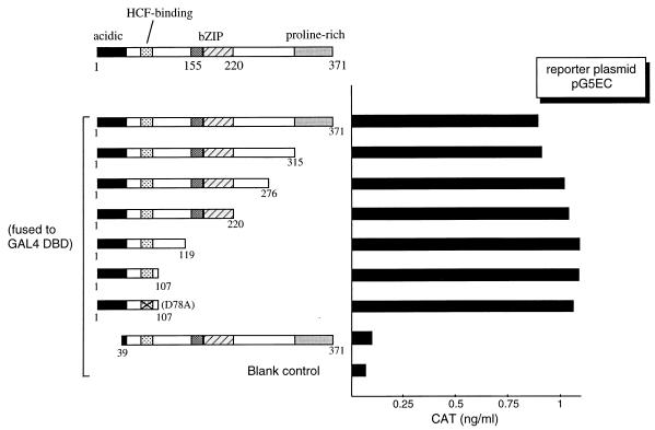
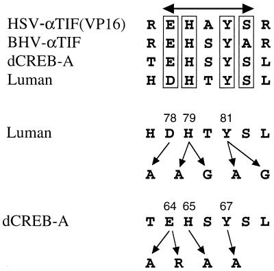
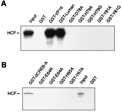
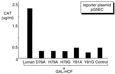
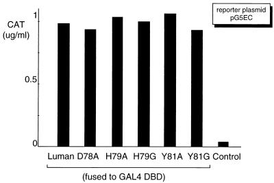
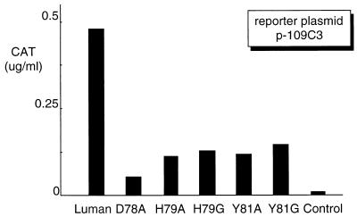
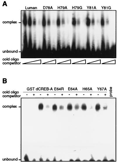
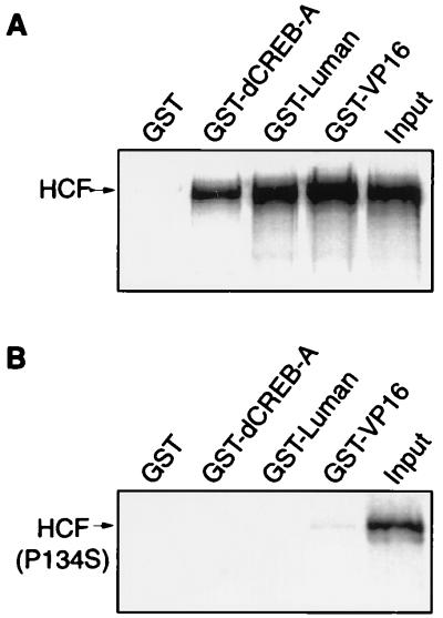
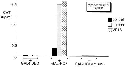
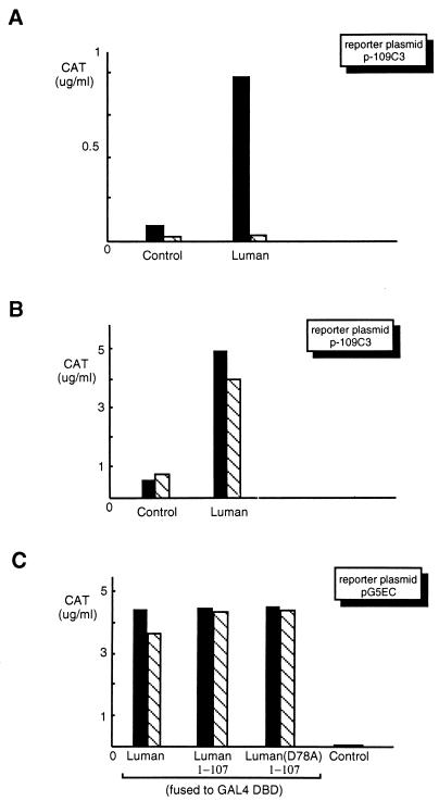
In human cells infected with herpes simplex virus (HSV), viral gene expression is initiated by the virion protein VP16. VP16 does not bind DNA directly but forms a multiprotein complex on the viral immediate-early gene promoters with two cellular proteins: the POU domain protein Oct-1 and host cell factor (HCF; also called C1, VCAF, and CFF). Despite its apparent role in stabilizing the VP16-induced transcription complex, the natural biological role of HCF is unclear. Only recently HCF has been implicated in control of the cell cycle. To determine the role of HCF in cells and answer why HSV has evolved an HCF-dependent mechanism for the initiation of the lytic cycle, we identified the first human ligand for HCF (R. Lu et al., Mol. Cell. Biol. 17:5117-5126, 1997). This protein, Luman, is a member of the CREB/ATF family of transcription factors that can activate transcription from promoters containing cyclic AMP response elements (CRE). Here we provide evidence that Luman and VP16 share two important structural features: an acidic activation domain and a common mechanism for binding HCF. We found that Luman, its homolog in Drosophila, dCREB-A (also known as BBF-2), and VP16 bind to HCF by a motif, (D/E)HXY(S/A), present in all three proteins. In addition, a mutation (P134S) in HCF that prevents VP16 binding also abolishes its binding to Luman and dCREB-A. We also show that while interaction with HCF is not required for the ability of Luman to activate transcription when tethered to the GAL4 promoter, it appears to be essential for Luman to activate transcription through CRE sites. These data suggest that the HCF-Luman interaction may represent a conserved mechanism for transcriptional regulation in metazoans, and HSV mimics this interaction with HCF to monitor the physiological state of the host cell.


Figures

FIG. 1
FIG. 1
Mapping of the activation domain of Luman by deletion analysis. On the left is a schematic representation of the structure of the Luman protein, in which numbers indicate the positions of the amino acids. The X in the HCF-binding domain of D78A represents a point mutation (alanine replaces the aspartate residue at position 78). Features of the protein discussed in the text are labeled. Luman and its deletion mutants were fused to the GAL4 DBD. The same amount (0.5 μg) of each plasmid was introduced into COS7 cells along with the reporter plasmid, pG5EC (0.5 μg), which has five copies of GAL4 UAS in the promoter region linked to the cat gene. CAT activity was measured by ELISA 48 h posttransfection.
FIG. 2
FIG. 2
Putative HCF-binding domains and mutants. The amino acid sequence alignment shows that Luman and the Drosophila protein dCREB-A share, with the herpesvirus αTIF proteins, a motif that has been implicated in HCF binding in VP16 (HSV-αTIF). In this consensus (D/E)HXY(S/A) motif, we mutated amino acids D78, H79, and Y81 of Luman and amino acids E64, H65, and Y67 of dCREB-A. BHV, bovine herpesvirus.
FIG. 3
FIG. 3
Mutants in the conserved residues of the putative HCF-binding motif of Luman and dCREB-A do not bind to HCF in vitro. All GST and GST fusion proteins were produced in E. coli BL21(DE3) and bound to glutathione-Sepharose beads. HCF was labeled with [35S]methionine by in vitro transcription and translation in the TnT system (Promega). Equivalent amounts of GST proteins attached to the beads were incubated with [35S]HCF for 45 min, washed, analyzed on an SDS–10% polyacrylamide gel, and visualized by autoradiography. The lane labeled Input represents 1/10 of the [35S]HCF incubation mixture. Panels A and B show the GST pull-down results for Luman and dCREB-A and their mutants, respectively.
FIG. 4
FIG. 4
Luman mutants do not interact with HCF in vivo. Plasmids expressing wild-type Luman and its mutants (0.5 μg) were individually introduced in COS7 cells with pG5EC (0.5 μg) and a plasmid (0.5 μg) expressing GAL-HCF. Since transactivation by Luman depends on its tethering to GAL4 UAS through its interaction with HCF, CAT activity is an indicator of Luman-HCF interaction. The control represents the expression vector without an insert.
FIG. 5
FIG. 5
Mutations in the HCF-binding motif do not affect the activation domain of Luman. Luman and its mutants were fused to the GAL4 DBD to study the effects of the mutations on their ability to activate transcription when tethered to the GAL4 UAS. Equivalent amounts (0.5 μg) of each GAL-Luman fusion protein-expressing plasmid were cotransfected in COS7 cells with plasmid pG5EC (0.5 μg) as the reporter. The control represents the expression vector without an insert.
FIG. 6
FIG. 6
Transcriptional activation by the Luman mutants on a CRE-containing promoter is reduced. Plasmids (0.5 μg each) expressing Luman and the mutants were introduced into COS7 cells with CAT reporter plasmid p-109C3 (0.5 μg), in which the promoter region has a CRE linked to the cat gene (27).
FIG. 7
FIG. 7
Mutations in the putative HCF-binding domain of Luman and dCREB-A do not affect binding of CRE in vitro as detected by HCF-independent EMSA. A double-stranded oligonucleotide representing CRE was labeled with 32P, annealed, incubated with each of the purified Luman (A) or dCREB-A (B) GST fusion proteins, and analyzed on a 4% nondenaturing polyacrylamide gel. (A) For each GST-Luman fusion protein sample, 1 μl of unlabeled CRE oligonucleotide, at concentrations of 1 and 10 μM, was added to the incubation mix and used as a competitor. For wild-type Luman, an additional concentration of 0.1 mM was included. (B) In every other lane, 10 pmol of unlabeled CRE oligonucleotide was added to the GST–dCREB-A protein sample and used as a competitor. The last lane contains the labeled CRE probe alone.
FIG. 8
FIG. 8
A mutation in the VP16-binding site of HCF(P134S) prevents its association with VP16 as well as with Luman and dCREB-A in vitro. GST and GST fusion proteins of dCREB-A, Luman, and VP16 were incubated with an equivalent amount of 35S-labeled HCF (A) or HCF(P134S) (B) and analyzed on an SDS-polyacrylamide gel. Input (1/10) was run on the last lane of each gel.
FIG. 9
FIG. 9
A mutation in the VP16-binding site of HCF(P134S) prevents its association with VP16 as well as with Luman and dCREB-A in vivo. The parental expression vector pcDNA3 was used as a negative control, and VP16 was included as a reference. Along with the reporter plasmid pG5EC (0.5 μg), equivalent amounts (0.5 μg) of plasmids expressing Luman and VP16 or the empty expression vector pcDNA3 were introduced into COS7 cells with either the blank GAL fusion vector, a plasmid expressing GAL-HCF, or a plasmid expressing GAL-HCF(P134S).
FIG. 10
FIG. 10
Luman activates transcription from CRE-containing promoters in tsBN67 cells at the permissive (33.5°C) but not nonpermissive (39.5°C) temperature. (A and B) Plasmids expressing wild-type Luman (0.5 μg) and a blank vector (0.5 μg) were used to transfect tsBN67 (A) and parental BHK-21 (B) cells at both 33.5°C (▪) and 39.5°C (▧). (C) Plasmids (0.5 μg of each) expressing the GAL4 DBD fusion proteins of full-length Luman, the N-terminal portion containing the activation domain (Luman1-107) and the same segment with the mutation D78A were introduced into tsBN67 cells at both 33.5°C (▪) and 39.5°C (▧).

References

    1. Abel T, Bhatt R, Maniatis T. A Drosophila CREB/ATF transcriptional activator binds to both fat body- and liver-specific regulatory elements. Genes Dev. 1992;6:466–480. . (Erratum, 7:719, 1993.) - PubMed
    1. apRhys C M, Ciufo D M, O’Neill E A, Kelly T J, Hayward G S. Overlapping octamer and TAATGARAT motifs in the VF65-response elements in herpes simplex virus immediate-early promoters represent independent binding sites for cellular nuclear factor III. J Virol. 1989;63:2798–2812. - PMC - PubMed
    1. Burbelo P D, Gabriel G C, Kibbey M C, Yamada Y, Kleinman H K, Weeks B S. LZIP-1 and LZIP-2: two novel members of the bZIP family. Gene. 1994;139:241–245. - PubMed
    1. Freiman R N, Herr W. Viral mimicry: common mode of association with HCF by VP16 and the cellular protein LZIP. Genes Dev. 1997;11:3122–3137. - PMC - PubMed
    1. Goto H, Motomura S, Wilson A C, Freiman R N, Nakabeppu Y, Fukushima K, Fujishima M, Herr W, Nishimoto T. A single-point mutation in HCF causes temperature-sensitive cell-cycle arrest and disrupts VP16 function. Genes Dev. 1997;11:726–737. - PubMed

Publication types

MeSH terms

LinkOut - more resources