. 1998 Aug;72(8):6362-72.
doi: 10.1128/JVI.72.8.6362-6372.1998.

Multiple virulence determinants of foot-and-mouth disease virus in cell culture

Affiliations

Multiple virulence determinants of foot-and-mouth disease virus in cell culture

E Baranowski et al. J Virol. 1998 Aug.

Abstract

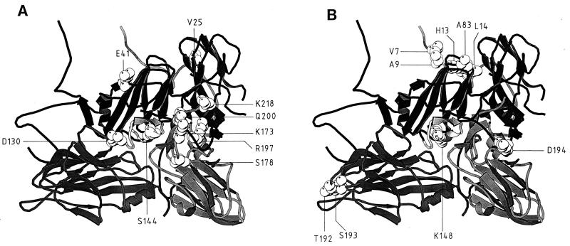
Hypervirulent variants of foot-and-mouth disease virus (FMDV) of serotype C arise upon serial cytolytic or persistent infections in cell culture. A specific mutation in the internal ribosome entry site of persistent FMDV was previously associated with enhanced translation initiation activity that could contribute to the hypervirulent phenotype for BHK-21 cells. Here we report that several hypervirulent FMDV variants arising upon serial cytolytic passage show an invariant internal ribosome entry site but have a number of mutations affecting structural and nonstructural viral proteins. The construction of chimeric type O-type C infectious transcripts has allowed the mapping of a major determinant of hypervirulence to the viral capsid. Tissue culture-adapted FMDV displayed enhanced affinity for heparin, but binding to cell surface heparan sulfate moieties was not required for expression of the hypervirulent phenotype in Chinese hamster ovary (CHO) cells. Virulence was identical or even higher for glycosaminoglycan-deficient CHO cells than for wild-type CHO cells. FMDV variants with decreased affinity for heparin were selected from a high-binding parental population and analyzed. Substitutions associated with decreased heparin binding were located at positions 173 of capsid protein VP3 and 144 of capsid protein VP1. These substitutions had a moderate effect on virulence for BHK-21 cells but completely abrogated infection of CHO cells. The comparative results with several FMDV isolates show that (i) increased affinity for heparin and alterations in cell tropism may be mediated by a number of independent sites on the viral capsid and (ii) the same capsid modifications may have different effects on different cell types.


Figures

FIG. 1
FIG. 1
Passage history of serotype C FMDV variants analyzed in this study. Plaque-purified FMDV C-S8c1 (▪) passaged once cytolytically in BHK-21 cells (○) was further propagated in BHK-21 cells either in cytolytic or in persistent infections. C-S8c1-derived populations p100 and p213 were obtained after 100 and 213 serial cytolytic passages, respectively. Clones c1 and c10 were isolated from C-S8c1 p100 (37). RGG and MARLS are two mutants resistant to MAb SD6 (3, 37). FMDV MARLS was subjected to 10 rounds of selection and amplification (b○) with heparin-Sepharose binding and infection of BHK-21 cells, to eliminate viruses with high affinity for heparin. This led to the isolation of clones MARLS/hs c1, c2, c3, and c4 (bottom right). Procedures for cytolytic infections and isolation of virus from single plaques are described in Materials and Methods. Persistent FMDV infections and the R100 population were described previously (8, 9, 21).
FIG. 2
FIG. 2
Comparison of the virus production upon infection of BHK-21 cell monolayers with (A) FMDV C-S8c1 and the MARLS variant (A) and chimeric FMDVs O1K/C-S8c1, O1K/MARLS, and O1K/MARLST2363, including the capsid of C-S8c1 or its hypervirulent derivatives (described in Materials and Methods) (B). In all cases, BHK-21 cells were infected at a multiplicity of infection of 5 PFU per cell. Virus titers at different times after infection were determined by plaque assays on BHK-21 cells monolayers (12). Each value represents the mean and standard deviation from triplicate assays.
FIG. 3
FIG. 3
Schematic representation of the capsid coding region of FMDV O1K and of C-S8c1 and its variant derivatives, as inserted in the parental type O1 plasmid pFMDV-YEP-poly(C) (60). Type C genomic regions are represented by open boxes. NgoMI restriction sites and their numbering refer to FMDV O1K cDNA (60). NcoI and Hind III sites and their numbering refer to the C-S8c1 genome (20). Amino acid residues differing in the compared chimeric genomes are indicated. The vertical numbers at the bottom refer to the corresponding positions in each protein. Infectious transcripts were produced with plasmid pFMDV-YEP-poly(C), and derived constructs encoding the cDNA of FMDV O1K or type O-type C chimeras under the control of the SP6 promoter. The amino acid sequence of protein VP4 is conserved among the FMDVs of serotypes O and C analyzed here. Procedures for the construction of chimeric viruses and confirmatory sequence analysis of chimeric progeny are described in Materials and Methods. O1K/MARLS includes a capsid-coding region identical to the consensus sequence of FMDV MARLS. T-2363 indicates an Ala-156 → Val mutation in protein VP2 that was accidentally selected during the cloning procedure. Infectious transcripts were produced with plasmid pFMDV-YEP-poly(C) and derived constructs encoding the cDNA of FMDV O1K or type O-type C chimeras under the control of the SP6 promoter. The amino acid sequence of protein VP4 is conserved among the FMDVs of serotypes O and C analyzed here. Procedures for the construction of chimeric viruses and confirmatory sequence analysis of chimeric progeny are described in Materials and Methods.
FIG. 4
FIG. 4
The effect of soluble heparin on the infectivity of FMDV. Mixtures of the indicated FMDV (50 to 300 PFU) and the amount of heparin (Sigma; from bovine lung, sodium salt) were incubated in DMEM for 10 min at 25°C. Then the mixtures were applied to confluent monolayers of either BHK-21, wild-type CHO (WT-CHO), or pgsA-745 CHO cells. After an adsorption period of 1 h at 37°C, the monolayers were washed and overlaid with DMEM containing 1% fetal calf serum and 0.5% agar. Plaques were visualized by staining with crystal violet. Plaque formation by mutant p100RGG required the presence of DEAE-dextran (0.08 mg/ml) in the semisolid medium. Percent plaque reduction was calculated relative to parallel assays with each virus and cell type in the absence of heparin. In addition, virulence assays were performed, as described in Materials and Methods, in the absence or presence of heparin (1 mg/ml) in the virus to be tested. Virulence values are given in parentheses (absence or presence of heparin) and are defined as indicated in Materials and Methods. Some viruses were tested only in BHK-21 cells because they do not infect CHO cells under the assay conditions used (see the text and Table 3).
FIG. 5
FIG. 5
Locations of the amino acid substitutions found in FMDV MARLS (A) and FMDV R100 (B) on a ribbon protein diagram of a crystallographic protomer of C-S8c1 (33). The capsid proteins VP1, VP2, and VP3 are represented as dark, medium, and light grey, respectively. VP1 from a neighboring protomer is shown at the upper right. The position of the G-H loop of VP1 in C-S8c1 corresponding to that found in the complex with MAb SD6 is shown in light grey at the center of each structure (27). The substituted residues are depicted in van der Waals spheres. The amino acids indicated are those listed in Table 5 for MARLS and R100. The minimal side chain-side chain distances measured between critical amino acid residues of MARLS and were 3.6 Å between VP2 D-130 (Cδ2) and VP1 A-145 (N), 4.5 Å between VP3 K-173 (Nζ) and VP1 Q-200 (Oɛ1), 4.7 Å between VP3 K-173 (Cδ) and VP1 R-141 (Nɛ), 5.1 Å between VP1 Q-200 (Cα) and VP1 R-141 (Cζ), 12.7 Å between VP1 R-197 (Cα) and VP1 R-141 (Nη2), and 15.0 Å between VP3 S-178 (Cβ) and VP1 P-156 (Cγ). The corresponding Cα-Cα distances were 6.8, 5.6, 9.5, 9.7, 17.9, and 16.5 Å, respectively. A minimal side chain distance of 13.3 Å was measured between R100 VP1 D-194 (Oδ2) and VP1 R-141 (Nη2). The corresponding Cα-Cα distance was 21.1 Å. The origin of the viruses and the procedures used to locate amino acids on the capsid structure are described in Materials and Methods.

References

    1. Berinstein A, Roivainen M, Hovi T, Mason P W, Baxt B. Antibodies to the vitronectin receptor (integrin αvβ3) inhibit binding and infection of foot-and-mouth disease virus to cultured cells. J Virol. 1995;69:2664–2666. - PMC - PubMed
    1. Borrego B, Novella I S, Giralt E, Andreu D, Domingo E. Distinct repertoire of antigenic variants of foot-and-mouth disease virus in the presence or absence of immune selection. J Virol. 1993;67:6071–6079. - PMC - PubMed
    1. Charpentier N, Dávila M, Domingo E, Escarmís C. Long-term, large-population passage of aphthovirus can generate and amplify defective noninterfering particles deleted in the leader protease gene. Virology. 1996;223:10–18. - PubMed
    1. Clarke D K, Duarte E A, Moya A, Elena S F, Domingo E, Holland J J. Genetic bottlenecks and population passages cause profound fitness differences in RNA viruses. J Virol. 1993;67:222–228. - PMC - PubMed
    1. Costa Giomi M P, Bergmann I E, Scodeller E A, Augé de Mello P, Gómez I, La Torre J L. Heterogeneity of the polyribocytidylic acid tract in aphthoviruses: biochemical and biological studies of viruses carrying polyribocytidylic acid tracts of different lengths. J Virol. 1984;51:799–805. - PMC - PubMed

Publication types

MeSH terms