. 1998 Aug;72(8):6437-41.
doi: 10.1128/JVI.72.8.6437-6441.1998.

Transfectant influenza A viruses with long deletions in the NS1 protein grow efficiently in Vero cells

Affiliations

Transfectant influenza A viruses with long deletions in the NS1 protein grow efficiently in Vero cells

A Egorov et al. J Virol. 1998 Aug.

Abstract

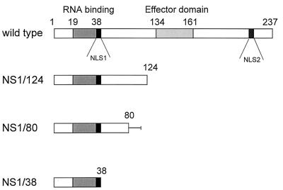
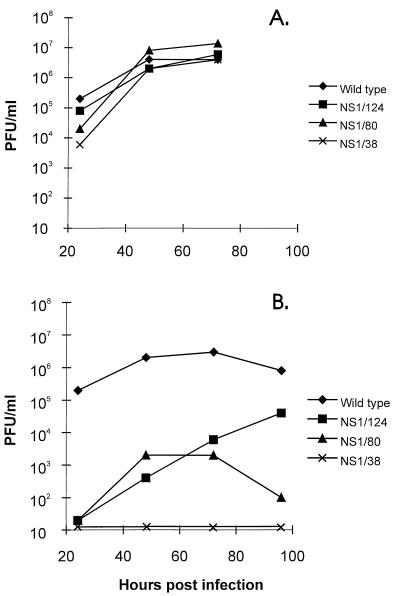
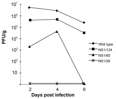
We established a reverse genetics system for the nonstructural (NS) gene segment of influenza A virus. This system is based on the use of the temperature-sensitive (ts) reassortant virus 25A-1. The 25A-1 virus contains the NS gene from influenza A/Leningrad/134/57 virus and the remaining gene segments from A/Puerto Rico (PR)/8/34 virus. This particular gene constellation was found to be responsible for the ts phenotype. For reverse genetics of the NS gene, a plasmid-derived NS gene from influenza A/PR/8/34 virus was ribonucleoprotein transfected into cells that were previously infected with the 25A-1 virus. Two subsequent passages of the transfection supernatant at 40 degreesC selected viruses containing the transfected NS gene derived from A/PR/8/34 virus. The high efficiency of the selection process permitted the rescue of transfectant viruses with large deletions of the C-terminal part of the NS1 protein. Viable transfectant viruses containing the N-terminal 124, 80, or 38 amino acids of the NS1 protein were obtained. Whereas all deletion mutants grew to high titers in Vero cells, growth on Madin-Darby canine kidney (MDCK) cells and replication in mice decreased with increasing length of the deletions. In Vero cells expression levels of viral proteins of the deletion mutants were similar to those of the wild type. In contrast, in MDCK cells the level of the M1 protein was significantly reduced for the deletion mutants.


Figures

FIG. 1
FIG. 1
Rescued transfectant viruses. The bars represent the NS proteins of the generated transfectant viruses; amino acid positions are indicated. The RNA binding domain and the effector domain are represented by shaded bars (28). The two nuclear localization signals (NLS1 and NLS2) described by Greenspan et al. (10) are represented by black bars. The line after amino acid position 80 of NS1/80 corresponds to the amino acids HGLCTCVALPN which resulted from the frameshift at nucleotide position 263. Generation of transfectant viruses is described in Materials and Methods.
FIG. 2
FIG. 2
Growth of transfectant viruses in Vero (A) and MDCK (B) cells. Confluent monolayers of Vero or MDCK cells were infected with viruses at an MOI of 0.05 and incubated at 37°C. At different time points, supernatants were assayed for infectious virus particles in plaque assays on Vero cells as described in Materials and Methods.
FIG. 3
FIG. 3
Viral titers in lung tissue of mice. BALB/c mice were infected intranasally with 105.2 PFU of transfectant viruses. On days 2, 4, and 6 following virus administration, mice were sacrificed and their lungs were removed for virus quantitation. Lungs of four mice were pooled, homogenized, and assayed for infectious virus particles in plaque assays on Vero cells.
FIG. 4
FIG. 4
Synthesis of viral proteins. Confluent monolayers of Vero (lanes 1 to 5) or MDCK (lanes 6 to 10) cells were infected with transfectant viruses at an MOI of 5. Five hours postinfection, cells were labeled with [35S]methionine and [35S]cysteine for 30 min and lysed. Cell extracts were analyzed by SDS-gel electrophoresis on 13% polyacrylamide gels containing 5 M urea. Lane 1, uninfected Vero cell extract; lanes 2 and 6, wild-type-infected cell extracts; lanes 3 and 7, transfectant NS1/124-infected cell extracts; lanes 4 and 8, transfectant NS1/80-infected cell extracts; lanes 5 and 9, transfectant NS1/38-infected cell extracts; line 10, uninfected MDCK cell extract. The positions of the viral proteins are indicated on the left. The faint band migrating between the M1 and NS1 proteins corresponds to the HA2 subunit. For transfectant NS1/38, the NS1 protein is not visible due to the small size and lower number of methionine and cysteine residues.

Similar articles

Cited by

References

    1. Baez M, Taussig R, Zazra J J, Young J F, Palese P, Reisfeld A, Skalka A M. Complete nucleotide sequence of the influenza A/PR/8/34 virus NS gene and comparison with the NS genes of the A/Udorn/72 and A/FPV/Rostock/34 strains. Nucleic Acids Res. 1980;8:5845–5858. - PMC - PubMed
    1. de la Luna S, Fortes P, Beloso A, Ortin J. Influenza virus NS1 protein enhances the rate of translation initiation of viral mRNAs. J Virol. 1995;69:2427–2433. - PMC - PubMed
    1. Diaz M O, Ziemin S, Le Beau M M, Pitha P, Smith S D, Chilcote R R, Rowley J D. Homozygous deletion of the alpha- and beta 1-interferon genes in human leukemia and derived cell lines. Proc Natl Acad Sci USA. 1988;85:5259–5263. - PMC - PubMed
    1. Egorov A, Garmashova L M, Lukashok I V, Nevedomskaia G N, Aleksandrova G I, Klimov A I. The NS gene—a possible determinant of apathogenicity of a cold-adapted donor of attenuation A/Leningrad/134/47/57 and its reassortants. Vopr Virusol. 1994;39:201–205. . (In Russian.) - PubMed
    1. Enami K, Sato T A, Nakada S, Enami M. Influenza virus NS1 protein stimulates translation of the M1 protein. J Virol. 1994;68:1432–1437. - PMC - PubMed

Publication types

MeSH terms

Substances