

Human T-cell leukemia virus type 1 reverse transcriptase (RT) originates from the pro and pol open reading frames and requires the presence of RT-RNase H (RH) and RT-RH-integrase proteins for its activity
- PMID: 9658093
- PMCID: PMC109816
- DOI: 10.1128/JVI.72.8.6504-6510.1998
Human T-cell leukemia virus type 1 reverse transcriptase (RT) originates from the pro and pol open reading frames and requires the presence of RT-RNase H (RH) and RT-RH-integrase proteins for its activity
Abstract
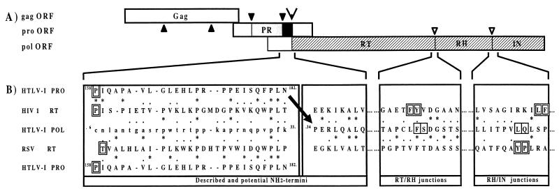
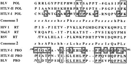
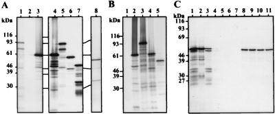
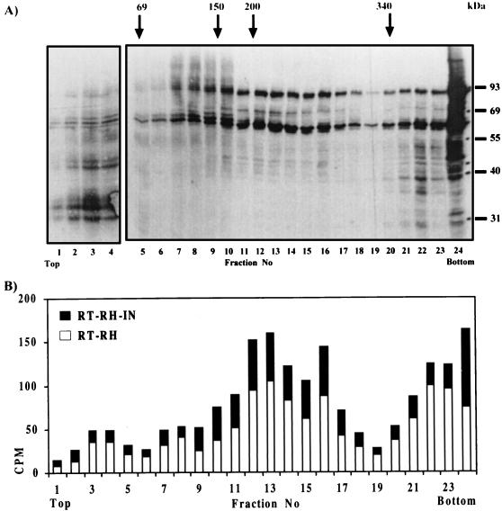
The first description of an active form of a recombinant human T-cell leukemia virus type 1 (HTLV-1) reverse transcriptase (RT) and subsequent predictions of its amino acid sequence and quaternary structure are reported here. By using amino acid alignment methods, the NH2 and COOH termini of the RT, RNase H (RH), and integrase (IN) domains of the Pol polyprotein were determined. The HTLV-1 RT seems to be unique since its NH2 terminus is probably encoded by the pro open reading frame (ORF) fused downstream, via a transframe peptide, to the polypeptide encoded by the pol ORF. The HTLV-1 Pol amino acid sequence was revealed to be highly similar to that of Rous sarcoma virus (RSV), particularly at the RT-RH hinge region. These two domains remain linked for RSV; this may also be the case for HTLV-1. In light of these results, RT, RT-RH, and RT-RH-IN genes were constructed and introduced into His-tagged protein expression vectors. The corresponding proteins were synthesized in vitro, and the DNA polymerase activities of different protein combinations were tested. Solely the RT-RH-RT-RH-IN combination was found to have a significant activity level. Velocity sedimentation analysis suggested that the HTLV-1 RT-RH and RT-RH-IN monomers are likely associated in an oligomeric structure, probably of the alpha3/beta type.
Figures
Similar articles
-
Identification of the RT-RH/IN cleavage site of HTLV-I.Biochem Biophys Res Commun. 2003 Jan 10;300(2):268-70. doi: 10.1016/s0006-291x(02)02848-6. Biochem Biophys Res Commun. 2003. PMID: 12504078
-
Synthesis, processing, and composition of the virion-associated HTLV-1 reverse transcriptase.J Biol Chem. 2006 Feb 17;281(7):3964-71. doi: 10.1074/jbc.M507660200. Epub 2005 Dec 19. J Biol Chem. 2006. PMID: 16368688
-
Expression of an active form of recombinant Ty1 reverse transcriptase in Escherichia coli: a fusion protein containing the C-terminal region of the Ty1 integrase linked to the reverse transcriptase-RNase H domain exhibits polymerase and RNase H activities.Biochem J. 2000 Jun 1;348 Pt 2(Pt 2):337-42. Biochem J. 2000. PMID: 10816427 Free PMC article.
-
Proteolytic processing of the human T-cell lymphotropic virus 1 reverse transcriptase: identification of the N-terminal cleavage site by mass spectrometry.Arch Biochem Biophys. 2001 Aug 1;392(1):93-102. doi: 10.1006/abbi.2001.2432. Arch Biochem Biophys. 2001. PMID: 11469799
-
Purification and characterization of recombinant equine infectious anemia virus reverse transcriptase.J Virol. 1991 Dec;65(12):7004-7. doi: 10.1128/JVI.65.12.7004-7007.1991. J Virol. 1991. PMID: 1719238 Free PMC article.
Cited by
-
The diversity of retrotransposons and the properties of their reverse transcriptases.Virus Res. 2008 Jun;134(1-2):221-34. doi: 10.1016/j.virusres.2007.12.010. Epub 2008 Feb 7. Virus Res. 2008. PMID: 18261821 Free PMC article. Review.
-
Human immunodeficiency virus type 1 integrase protein promotes reverse transcription through specific interactions with the nucleoprotein reverse transcription complex.J Virol. 1999 Mar;73(3):2126-35. doi: 10.1128/JVI.73.3.2126-2135.1999. J Virol. 1999. PMID: 9971795 Free PMC article.
-
Development of a cytotoxic T-cell assay in rabbits to evaluate early immune response to human T-lymphotropic virus type 1 infection.Viral Immunol. 2009 Dec;22(6):397-405. doi: 10.1089/vim.2009.0059. Viral Immunol. 2009. PMID: 19951176 Free PMC article.
-
Cooperation between reverse transcriptase and integrase during reverse transcription and formation of the preintegrative complex of Ty1.Eukaryot Cell. 2006 Oct;5(10):1760-9. doi: 10.1128/EC.00159-06. Eukaryot Cell. 2006. PMID: 17031000 Free PMC article.
-
Current State of Therapeutics for HTLV-1.Viruses. 2024 Oct 15;16(10):1616. doi: 10.3390/v16101616. Viruses. 2024. PMID: 39459949 Free PMC article. Review.
References
-
- Anderson S F, Coleman J E. Conformational changes of HIV reverse transcriptase subunits on formation of the heterodimer: correlation with kcat and Km. Biochemistry. 1992;31:8221–8228. - PubMed
-
- Barber A M, Hizi A, Maizel J V, Jr, Hughes S H. HIV-1 reverse transcriptase: structure predictions for the polymerase domain. AIDS Res Hum Retroviruses. 1990;6:1061–1072. - PubMed
-
- Daenke S, Schramm H J, Bangham C R. Analysis of substrate cleavage by recombinant protease of human T cell leukaemia virus type 1 reveals preferences and specificity of binding. J Gen Virol. 1994;75:2233–2239. - PubMed
-
- Dayhoff M O, Barker W C, Hunt L T. Establishing homologies in protein sequences. Methods Enzymol. 1983;91:524–545. - PubMed
Publication types
MeSH terms
Substances
LinkOut - more resources
Full Text Sources
