. 1998 Aug;72(8):6511-9.
doi: 10.1128/JVI.72.8.6511-6519.1998.

Involvement of aminopeptidase N (CD13) in infection of human neural cells by human coronavirus 229E

Affiliations

Involvement of aminopeptidase N (CD13) in infection of human neural cells by human coronavirus 229E

C Lachance et al. J Virol. 1998 Aug.

Abstract

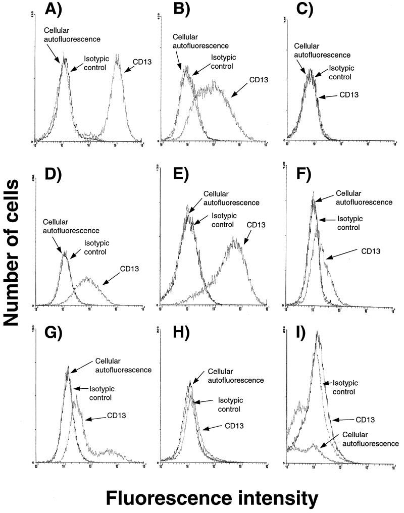
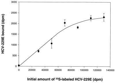
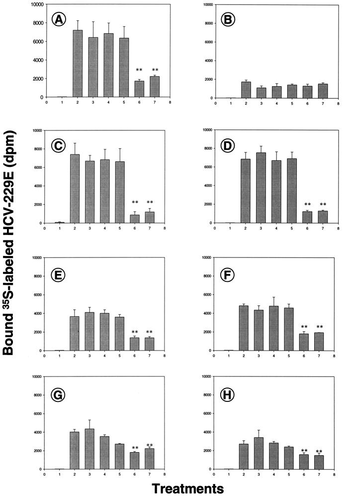
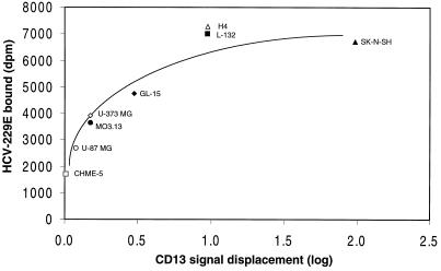
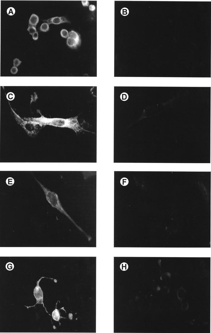
Attachment to a cell surface receptor can be a major determinant of virus tropism. Previous studies have shown that human respiratory coronavirus HCV-229E uses human aminopeptidase N (hAPN [CD13]) as its cellular receptor for infection of lung fibroblasts. Although human coronaviruses are recognized respiratory pathogens, occasional reports have suggested their possible neurotropism. We have previously shown that human neural cells, including glial cells in primary cultures, are susceptible to human coronavirus infection in vitro (A. Bonavia, N. Arbour, V. W. Yong, and P. J. Talbot, J. Virol. 71:800-806, 1997). However, the only reported expression of hAPN in the nervous system is at the level of nerve synapses. Therefore, we asked whether hAPN is utilized as a cellular receptor for infection of these human neural cell lines. Using flow cytometry, we were able to show the expression of hAPN on the surfaces of various human neuronal and glial cell lines that are susceptible to HCV-229E infection. An hAPN-specific monoclonal antibody (WM15), but not control antibody, inhibited the attachment of radiolabeled HCV-229E to astrocytic, neuronal, and oligodendrocytic cell lines. A correlation between the apparent amount of cell surface hAPN and the level of virus attachment was observed. Furthermore, the presence of WM15 inhibited virus infection of these cell lines, as detected by indirect immunofluorescence. These results indicate that hAPN (CD13) is expressed on neuronal and glial cell lines in vitro and serves as the receptor for infection by HCV-229E. This further strengthens the neurotropic potential of this human respiratory virus.


Figures

FIG. 1
FIG. 1
Flow cytometry analysis of cell surface expression of CD13. Cells were labeled with either an anti-CD13 PE-conjugated antibody or a PE-conjugated isotypic control antibody. (A) THP-1 monocytic cell line (2-log-unit signal displacement); (B) L-132 human lung fibroblast cell line (1-log-unit signal displacement); (C) CHME-5 microglial cell line (no signal displacement); (D) H4 neuronal cell line (1-log-unit signal displacement); (E) SK-N-SH neuronal cell line (2-log-unit signal displacement); (F) MO3.13 oligodendrocytic cell line (0.2-log-unit signal displacement); (G) GL-15 astrocytic cell line (0.5-log-unit signal displacement); (H) U-87 MG astrocytic cell line (0.1-log-unit signal displacement); (I) U-373 MG astrocytic cell line (0.1-log-unit signal displacement). These profiles are representative of at least two separate experiments.
FIG. 2
FIG. 2
Specific attachment of 35S-labeled HCV-229E onto L-132 cells. Binding of HCV-229E to L-132 cells was carried out by incubation of increasing amounts of 35S-labeled HCV-229E, in triplicate, on monolayers of L-132 cells for 1 h at room temperature. Cell monolayers were washed four times with DMEM supplemented with 1% (vol/vol) FBS to eliminate unattached virus and were solubilized with 1% (wt/vol) SDS. Cell-bound 35S-labeled HCV-229E was quantitated by liquid scintillation counting. This profile is representative of two separate experiments.
FIG. 3
FIG. 3
Detection of binding of 35S-labeled HCV-229E on cultures of human neural cell lines pretreated with an anti-CD13 MAb or with various controls, all in triplicate. (A) L-132 human lung fibroblast cell line; (B) CHME-5 microglial cell line; (C) H4 neuronal cell line; (D) SK-N-SH neuronal cell line; (E) MO3.13 oligodendrocytic cell line; (F) GL-15 astrocytic cell line; (G) U-373 MG astrocytic cell line; (H) U-87 MG astrocytic cell line. Treatment 1, culture medium; treatment 2, 105 dpm of HCV-229E; treatment 3, 105 dpm of HCV-229E plus HLA-specific antiserum (15 μg); treatment 4, 105 dpm of HCV-229E plus anti-TuMV MAb 6D (15 μg); treatment 5, 105 dpm of HCV-229E plus anti-HCV-229E MAb 5-11H.6 (15 μg); treatment 6, 105 dpm of HCV-229E plus anti-CD13 MAb WM15 (15 μg); treatment 7, 105 dpm of HCV-229E plus unlabeled HCV-229E. These profiles are representative of two separate experiments. ∗∗, statistically significant difference according to Tukey’s multiple comparison test, following an ANOVA test (P < 0.05).
FIG. 4
FIG. 4
Correlation between expression levels of CD13 on human neural cell lines and attachment of 35S-labeled HCV-229E. ▪, L-132 human lung fibroblastic cell line; ▵, H4 neuronal cell line; ▴, SK-N-SH neuronal cell line; ⧫, GL-15 astrocytic cell line; ○, U-87 MG astrocytic cell line; ◊, U-373 MG astrocytic cell line; •, MO3.13 oligodendrocytic cell line; □, CHME-5 microglial cell line.
FIG. 5
FIG. 5
Detection of viral antigens by indirect immunofluorescence in cultures of human neural cells pretreated with an anti-CD13 MAb and inoculated with HCV-229E. (A and B) L-132 human lung fibroblastic cell line; (C and D) SK-N-SH neuronal cell line; (E and F) GL-15 astrocytic cell line; (G and H) MO3.13 oligodendrocytic cell line. Cells were pretreated with 15 μg of an anti-CD13 MAb (B, D, F, and H) or with 15 μg of an isotypic control antibody (A, C, E, and G) and were inoculated with HCV-229E at an MOI of 0.1. After 22 h, the cells were fixed in cold acetone and examined by indirect immunofluorescence. The primary antibody used to detect viral antigens was a guinea pig antiserum against HCV-229E. This was followed by the addition of a fluorescein-conjugated goat affinity-purified anti-guinea pig secondary antibody Magnification, ×100. The results represent several fields, and observations at lower magnification yielded the following percentages of infected cells and qualitative levels of fluorescence (scale, 0 to 3+): 30% and 3+ (A and C), 10% and 1.5+ (E), 10 to 15% and 3+ (G), <5% and 1+ (B), <5% and 0.5+ (D and F), and negative (H).

References

    1. Arbour N, Talbot P J. Persistent infection of neural cell lines by human coronaviruses. Adv Exp Med Biol. 1998;440:575–581. - PubMed
    1. Arbour, N., and P. J. Talbot. 1998. Unpublished data.
    1. Ashmun R A, Look A T. Metalloprotease activity of CD13/aminopeptidase N on the surface of human myeloid cells. Blood. 1990;75:462–469. - PubMed
    1. Barnes K, Kenny A J, Turner A J. Localization of aminopeptidase N and dipeptidyl peptidase IV in pig striatum and in neuronal and glial cell cultures. Eur J Neurosci. 1994;6:531–537. - PubMed
    1. Barnett E M, Perlman S. The olfactory nerve and not the trigeminal nerve is the major site of CNS entry for mouse hepatitis virus, strain JHM. Virology. 1993;194:185–191. - PubMed

Publication types

LinkOut - more resources