. 1998 Aug;72(8):6621-8.
doi: 10.1128/JVI.72.8.6621-6628.1998.

The human homolog of HAVcr-1 codes for a hepatitis A virus cellular receptor

Affiliations

The human homolog of HAVcr-1 codes for a hepatitis A virus cellular receptor

D Feigelstock et al. J Virol. 1998 Aug.

Abstract

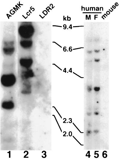
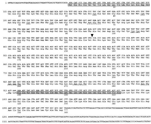
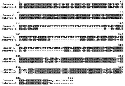
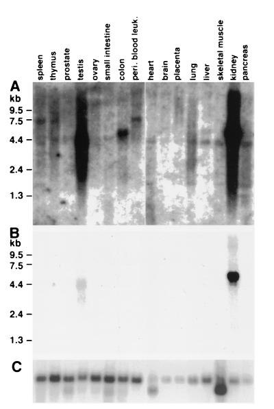
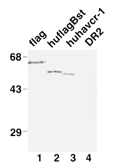
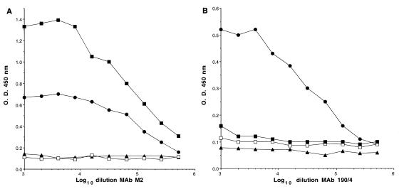
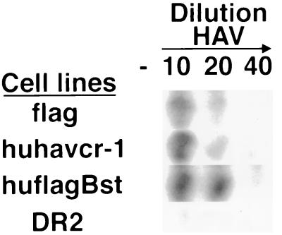
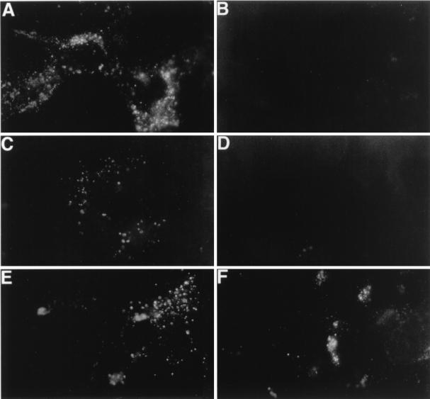
The hepatitis A virus cellular receptor 1 (HAVcr-1) cDNA was isolated from a cDNA expression library of African green monkey kidney (AGMK) cells by using protective monoclonal antibody (MAb) 190/4, which blocks the binding of hepatitis A virus (HAV) to AGMK cells. The HAVcr-1 cDNA codes for havcr-1, a 451-amino-acid class I integral-membrane mucin-like glycoprotein of unknown natural function. To determine the existence of a human homolog(s) of HAVcr-1 (huHAVcr-1), we used HAVcr-1-specific primers to amplify cDNAs from human liver and kidney mRNA by reverse transcription-PCR. Nucleotide sequence analysis revealed that the amplified liver and kidney huHAVcr-1 cDNAs were identical and that they coded for a 359-amino-acid glycoprotein, termed huhavcr-1, which was approximately 79% identical to havcr-1. The six Cys residues of the extracellular domain of havcr-1 and its first N-glycosylation site were conserved in huhavcr-1. However, the number of hexameric repeats of the mucin-like region was reduced from 27 in havcr-1 to 13 in huhavcr-1. In addition, 12 C-terminal amino acids in the cytoplasmic domain of huhavcr-1 were deleted. Northern blot analysis of poly(A) RNA showed that huhavcr-1 is expressed in every organ analyzed, including the liver, small intestine, colon, and spleen, and that it is expressed at higher levels in the kidney and testis. Although dog cells transfected with the huHAVcr-1 cDNA did not express the protective 190/4 epitope, they bound hepatitis A virus (HAV) and gained limited susceptibility to HAV infection. Treatment with MAb 190/4 did not protect AGMK cell transfectants expressing huhavcr-1 against HAV, suggesting that HAV infected these cells via the huhavcr-1 receptor and not the endogenously expressed havcr-1, which was blocked by MAb 190/4. Our data demonstrate that huhavcr-1 is a binding receptor for HAV and suggest that it is also a functional receptor for HAV.


Figures

FIG. 1
FIG. 1
Southern blot analysis of the huhavcr-1 gene. Genomic DNA extracted from simian AGMK cells, mouse Ltk cells transfected with pDR2GL37/5 (Lcr5 cells) or pDR2 (LDR2 cells), human male (M) and female (F) peripheral blood leukocytes, and mouse cells was digested with PstI and examined by Southern blot analysis using a full-length 32P-labeled HAVcr-1 cDNA probe. The positions of size markers in kilobase pairs (kb) are illustrated.
FIG. 2
FIG. 2
cDNA sequence and predicted amino acid composition of huhavcr-1. The nucleotide sequence of the huHAVcr-1 cDNA was determined on both strands by automatic sequencing using specific oligonucleotides. The white box indicates the putative signal sequence. The six cysteines of the Cys-rich region are marked with solid lines. Potential N-linked glycosylation sites are marked with dashed lines. The arrowhead indicates the putative beginning of the TSP-rich region. The putative transmembrane domain is boxed in grey. The termination codon is indicated by asterisks.
FIG. 3
FIG. 3
Alignment of havcr-1 and huhavcr-1. Alignment of amino acid sequences predicted from the HAVcr-1 and huHAVcr-1 cDNAs was done with the Clustal W program. Gaps introduced in the sequences for the alignment are indicated by dashes; numbers of residues starting with the respective initiating methionine codons are indicated. Amino acids identical to those of the havcr-1 sequence are in grey background.
FIG. 4
FIG. 4
Expression of huhavcr-1 in human tissues. MTN blots containing poly(A) RNA from different human tissues were probed with 32P-labeled full-length huHAVcr-1 cDNA at high stringency and autoradiographed for 3 weeks (A) or 1 day (B). (C) The same blots were stripped and rehybridized with a 32P-labeled β-actin probe. The positions of size markers in kilobases are illustrated. peri. blood leuk., peripheral blood leukocytes.
FIG. 5
FIG. 5
Western blot analysis of the expression of huhavcr-1 in dog cell transfectants. Cytoplasmic extracts of dog cell transfectants expressing FLAG-tagged havcr-1 (flag), FLAG-tagged huhavcr-1 (huflagBst), and untagged huhavcr-1 (huhavcr-1) and dog cells transfected with vector pDR2 alone (DR2) were prepared in RSB–1% NP-40, resolved by SDS–10% polyacrylamide gel electrophoresis, transferred to polyvinylidene difluoride membranes, and probed with rabbit anti-GST2 Ab directed against the TSP-rich region of huhavcr-1. The positions of prestained molecular weight markers and their sizes in kilodaltons are shown on the left.
FIG. 6
FIG. 6
Expression of huhavcr-1 at the cell surfaces of dog cell transfectants. Expression of the M2 (A) and 190/4 (B) epitopes at the cell surfaces of dog cell transfectants expressing FLAG-tagged huhavcr-1 (flag cells; solid circles), FLAG-tagged huhavcr-1 (huflagBst cells; solid squares); and untagged huhavcr-1 (huhavcr-1 cells; open squares) and dog cells transfected with vector pDR2 (DR2 cells; solid triangles) was determined by ELISA using twofold dilutions of the MAbs. Absorbance at 450 nm was plotted versus the log10 of the MAb dilution. Data are means of duplicate wells; duplicate values varied by less than 10%. The results correspond to one experiment which was repeated two times with approximately 5 to 10% experimental error.
FIG. 7
FIG. 7
Binding of HAV to dog cells transfected with huHAVcr-1 cDNA. Dog cell transfectants expressing FLAG-tagged huhavcr-1 (flag), FLAG-tagged huhavcr-1 (huflagBst), and untagged huhavcr-1 (huhavcr-1) and dog cells transfected with vector pDR2 (DR2) were grown in 96-well plates and infected with 1:10, 1:20, and 1:40 dilutions of purified HAV or were mock infected (−) for 1 h at 35°C. After being extensive washed, monolayers were fixed, and HAV bound to the cells was detected with 125I-labeled human anti-HAV Ab. Autoradiography of the 96-well plate showing a single well for each treatment is presented.
FIG. 8
FIG. 8
Detection of HAV antigen in the cytoplasm of dog cell transfectants by indirect IF analysis. Monolayers of GL37 (A), DR2 (B), flag (C), d1− (D), huhavcr-1 (E), and huflagBst (F) cells grown in eight-well slides were infected with a MOI of 100 to 1,000 TCID50 of purified HAV per cell for 6 h, washed extensively, and incubated at 35°C for 3 days. Cells were fixed with cold acetone and stained with human anti-HAV Ab and FITC-labeled goat anti-human IgG and IgM Abs. Mock-infected cells did not immunofluoresce (data not shown).
FIG. 9
FIG. 9
Slot blot analysis of MAb 190/4-mediated protection of GL37 cell transfectants against HAV infection. (A) Slot blot analysis of protection of GL37 cells and GL37 cell transfectants expressing FLAG-tagged huhavcr-1 (GL37huflagBst 2 and GL37hflaBst 3 cells) against HAV infection. Confluent monolayers of GL37 cells in six-well plates were treated with MAb 190/4 or anti-human α3 integrin MAb P1B5 for 1 h at 35°C, washed, and inoculated at a MOI of 0.1 TCID50 of HAV per cell for 1 h at 35°C. Monolayers were washed and incubated at 35°C, cytoplasmic extracts were prepared at 3 days postinfection, and total RNA was extracted. HAV-specific RNA was detected with a 32P-labeled HAV cDNA probe. To control for RNA loading, the slot blot was stripped and rehybridized with a 32P-labeled β-actin cDNA probe. (B) Control GL37 cells were infected with HAV or were mock infected (Mock) without prior MAb treatment. RNA samples were prepared in parallel with the samples shown in panel A and hybridized under the same conditions. The autoradiogram was exposed for 2.5 h with intensifying screens.

References

    1. Asher L V, Binn L N, Mensing T L, Marchwicki R H, Vassell R A, Young G D. Pathogenesis of hepatitis A in orally inoculated owl monkeys (Aotus trivirgatus) J Med Virol. 1995;47:260–268. - PubMed
    1. Cohen J I, Feinstone S, Purcell R H. Hepatitis A virus infection in a chimpanzee: duration of viremia and detection of virus in saliva and throat swabs. J Infect Dis. 1989;160:887–890. - PubMed
    1. Cohen J I, Ticehurst J R, Feinstone S M, Rosenblum B, Purcell R H. Hepatitis A virus cDNA and its RNA transcripts are infectious in cell culture. J Virol. 1987;61:3035–3039. - PMC - PubMed
    1. Dotzauer A, Feinstone S M, Kaplan G. Susceptibility of nonprimate cell lines to hepatitis A virus infection. J Virol. 1994;68:6064–6068. - PMC - PubMed
    1. Feigelstock D, Thompson P, Mattoo P, Kaplan G G. Polymorphisms of the hepatitis A virus cellular receptor 1 in African green monkey kidney cells result in antigenic variants that do not react with protective monoclonal antibody 190/4. J Virol. 1998;72:6218–6222. - PMC - PubMed

Publication types

MeSH terms

Associated data

LinkOut - more resources