. 1998 Aug;72(8):6646-56.
doi: 10.1128/JVI.72.8.6646-6656.1998.

Gastrointestinal T lymphocytes retain high potential for cytokine responses but have severe CD4(+) T-cell depletion at all stages of simian immunodeficiency virus infection compared to peripheral lymphocytes

Affiliations

Gastrointestinal T lymphocytes retain high potential for cytokine responses but have severe CD4(+) T-cell depletion at all stages of simian immunodeficiency virus infection compared to peripheral lymphocytes

Z Smit-McBride et al. J Virol. 1998 Aug.

Abstract

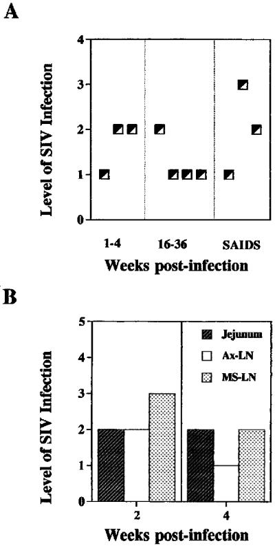
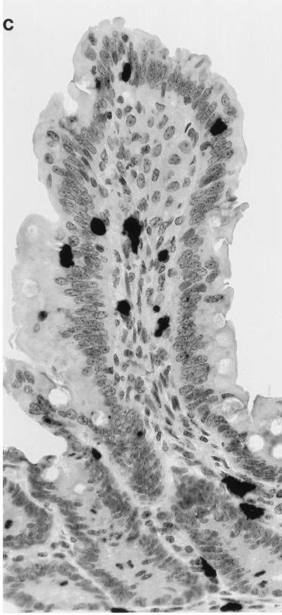
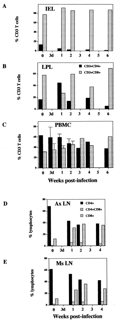
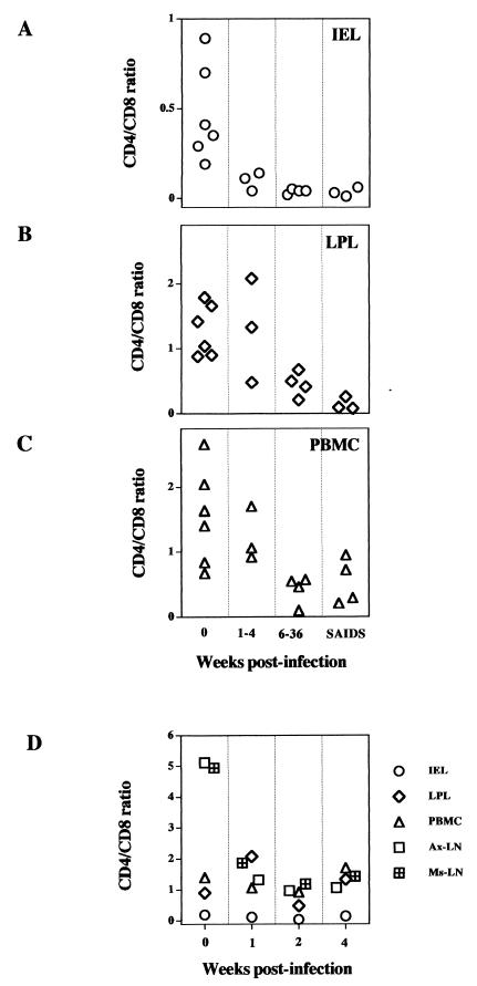
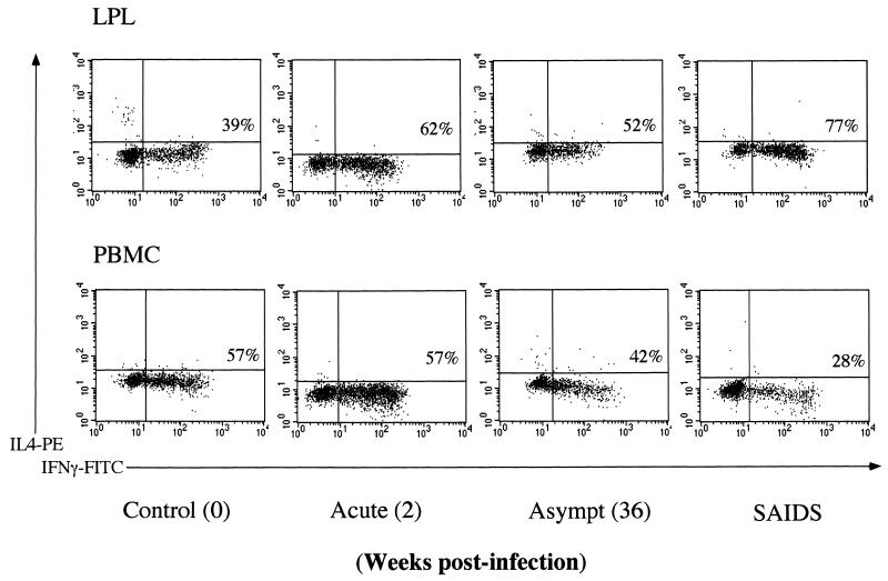
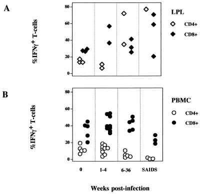
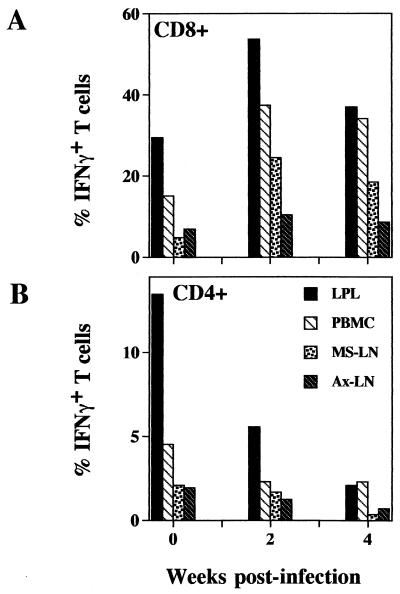
Gastrointestinal complications in human immunodeficiency virus (HIV) infection are indicative of impaired intestinal mucosal immune system. We used simian immunodeficiency virus (SIV)-infected rhesus macaques as an animal model for HIV to determine pathogenic effects of SIV on intestinal T lymphocytes. Intestinal CD4(+) T-cell depletion and the potential for cytokine responses were examined during SIV infection and compared with results for lymphocytes from lymph nodes and blood. Flow cytometric analysis demonstrated severe depletion of CD4(+)CD8(-) single-positive T cells and CD4(+)CD8(+) double-positive T cells in intestinal lamina propria lymphocytes (LPL) and intraepithelial lymphocytes (IEL) during primary SIV infection which persisted through the entire course of SIV infection. In contrast, CD4(+) T-cell depletion was gradual in peripheral lymph nodes and blood. Flow cytometric analysis of intracellular gamma interferon (IFN-gamma) and interleukin-4 (IL-4) production following short-term mitogenic activation revealed that LPL retained same or higher capacity for IFN-gamma production in all stages of SIV infection compared to uninfected controls, whereas peripheral blood mononuclear cells displayed a gradual decline. The CD8(+) T cells were the major producers of IFN-gamma. There was no detectable change in the frequency of IL-4-producing cells in both LPL and peripheral blood mononuclear cells. Thus, severe depletion of CD4(+) LPL and IEL in primary SIV infection accompanied by altered cytokine responses may reflect altered T-cell homeostasis in intestinal mucosa. This could be a mechanism of SIV-associated enteropathy and viral pathogenesis. Dynamic changes in intestinal T lymphocytes were not adequately represented in peripheral lymph nodes or blood.


Figures

FIG. 1
FIG. 1
SIV-infected cells were detected in intestinal mucosa through the entire course of SIV infection. SIV-infected cells in jejunal tissues were detected by in situ hybridization, and the level of SIV infection was scored according to the scale described in the Materials and Methods. (A) Levels of SIV infection in jejunal tissue during the primary acute (1 to 4 weeks p.i.), asymptomatic (16 to 36 weeks p.i.), and SAIDS stages. (B) Comparison of the levels of viral infection in jejunum, Ax-LN, and Ms-LN from the same animal during the primary acute phase of SIV infection. (C) SIV-infected cells in intestinal epithelium and lamina propria.
FIG. 1
FIG. 1
SIV-infected cells were detected in intestinal mucosa through the entire course of SIV infection. SIV-infected cells in jejunal tissues were detected by in situ hybridization, and the level of SIV infection was scored according to the scale described in the Materials and Methods. (A) Levels of SIV infection in jejunal tissue during the primary acute (1 to 4 weeks p.i.), asymptomatic (16 to 36 weeks p.i.), and SAIDS stages. (B) Comparison of the levels of viral infection in jejunum, Ax-LN, and Ms-LN from the same animal during the primary acute phase of SIV infection. (C) SIV-infected cells in intestinal epithelium and lamina propria.
FIG. 2
FIG. 2
Severe depletion of CD4+CD8 and CD4+CD8+ T cells in jejunum occurs in primary SIV infection. The IEL, LPL, and PBMC were isolated from the animals at different stages of SIV infection (primary acute, asymptomatic [Asympt], and SAIDS) and the CD4+CD8, CD4+CD8+, and CD4CD8+ T cells were analyzed by two-color flow cytometry. Freshly isolated cells were stained with PE-conjugated anti-CD4 and FITC-conjugated anti-CD8 and then analyzed on a flow cytometer as described in the text. A lymphocyte gate was set on forward versus side scatter, and percentages of positive cells within this gate are shown. Shown are results for a representative set of animals from different stages of SIV infection, illustrating changes in distribution of CD4+CD8, CD4+CD8+, and CD4CD8+ T-cells subsets between different tissue compartments (IEL, LPL, and PBMC) within the same animal.
FIG. 3
FIG. 3
The severe CD4+ T-cell depletion in intestinal mucosa during primary SIV infection is not reflected in peripheral blood and lymph node lymphocytes. Lymphocytes were isolated from the five sites indicated from rhesus macaques at 3 days, 1, 2, 3, 4, and 6 weeks post-SIV infection. The percentages of CD4+ and CD8+ T cells were determined by flow cytometry.
FIG. 4
FIG. 4
A severe decline in CD4/CD8 T-cell ratio occurred in intestinal mucosa during primary SIV infection compared to peripheral and lymph node lymphocytes and persisted through the SIV infection course. Lymphocytes were isolated from IEL (A), LPL (B), and PBMC (C) during the primary acute (1 to 4 weeks postinfection), asymptomatic (6 to 36 weeks postinfection), and terminal (SAIDS) stages of SIV infection. Ax-LN and Ms-LN lymphocytes (D) were obtained from macaques only at 1, 2, and 3 weeks postinfection. The percentages of CD4+ and CD8+ T cells were determined by flow cytometry.
FIG. 5
FIG. 5
Analysis of intracellular IFN-γ and IL-4 production in CD8+ LPL and PBMC at a single-cell level by three-color flow cytometry. Percentages of CD8+ IFN-γ T cells are shown in the plots. The LPL and PBMC were isolated from rhesus macaques during the primary acute, asymptomatic (Asympt), and terminal stages of SIV infection. Cells were stimulated with PMA-ionomycin, stained with TC-labeled CD8 antibody, fixed, permeabilized, and stained intracellularly with FITC-labeled IFN-γ and PE-labeled IL-4 antibodies. The negative control was stained with an isotype-matched control antibody.
FIG. 6
FIG. 6
The capacity of LPL for intracellular IFN-γ expression was retained through the SIV infection course, while a gradual decline was seen in peripheral blood with disease progression. Frequency of IFN-γ cells was determined among the CD8+ or CD4+ T-cell populations of LPL (A) and PBMC (B) during the course of SIV infection by three-color flow cytometry. Cells were stimulated with PMA-ionomycin, stained for CD8 or CD4, fixed, permeabilized, and stained intracellularly for IFN-γ and IL-4. A negative control was stained with an isotype-matched control antibody.
FIG. 7
FIG. 7
Comparison of the potential of intracellular IFN-γ production by CD8+ (A) and CD4+ (B) T cells among LPL, Ms-LN and Ax-LN lymphocytes, and PBMC at 2 and 4 weeks post-SIV infection by flow cytometric analysis. Variable frequencies of INF-γ cells were found among the cells from different tissues.

References

    1. Adams R B, Planchon S M, Roche J K. IFN-gamma modulation of epithelial barrier function. Time course, reversibility, and site of cytokine binding. J Immunol. 1993;150:2356–2363. - PubMed
    1. Adleman L M, Wofsy D. T-cell homeostasis: implications in HIV infection. J Acquired Immune Defic Syndr. 1993;6:144–152. - PubMed
    1. Badley A D, McElhinny J A, Leibson P J, Lynch D H, Alderson M R, Paya C V. Upregulation of Fas ligand expression by human immunodeficiency virus in human macrophages mediates apoptosis of uninfected T lymphocytes. J Virol. 1996;70:199–206. - PMC - PubMed
    1. Barcellini W, Rizzardi G P, Borghi M O, Fain C, Lazzarin A, Meroni P L. Th1 and Th2 cytokine production by peripheral blood mononuclear cells from HIV-infected patients. AIDS. 1994;8:757. - PubMed
    1. Caruso A, Licenziati S, Canaris A D, Cantalamessa A, Corulli M, Benzoni B, Peroni L, Balsari A, Turano A. Characterization of T cell subsets involved in the production of IFN-gamma in asymptomatic HIV-infected patients. AIDS Res Hum Retroviruses. 1996;12:135–141. - PubMed

Publication types

MeSH terms