Comparative Study
. 1998 Aug;72(8):6678-88.
doi: 10.1128/JVI.72.8.6678-6688.1998.

Comparisons of highly virulent H5N1 influenza A viruses isolated from humans and chickens from Hong Kong

Affiliations
Free PMC article
Comparative Study

Comparisons of highly virulent H5N1 influenza A viruses isolated from humans and chickens from Hong Kong

D L Suarez et al. J Virol. 1998 Aug.
Free PMC article

Abstract

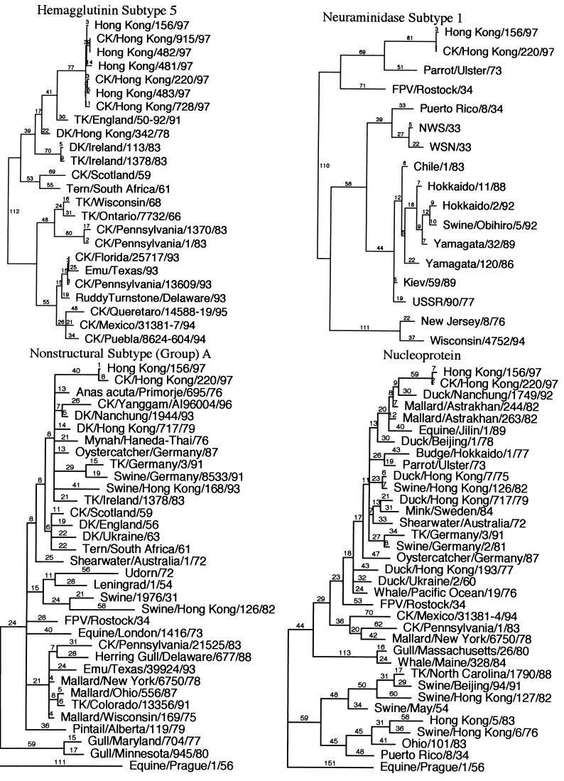
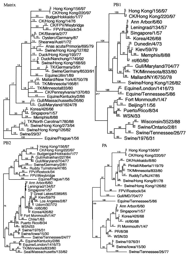
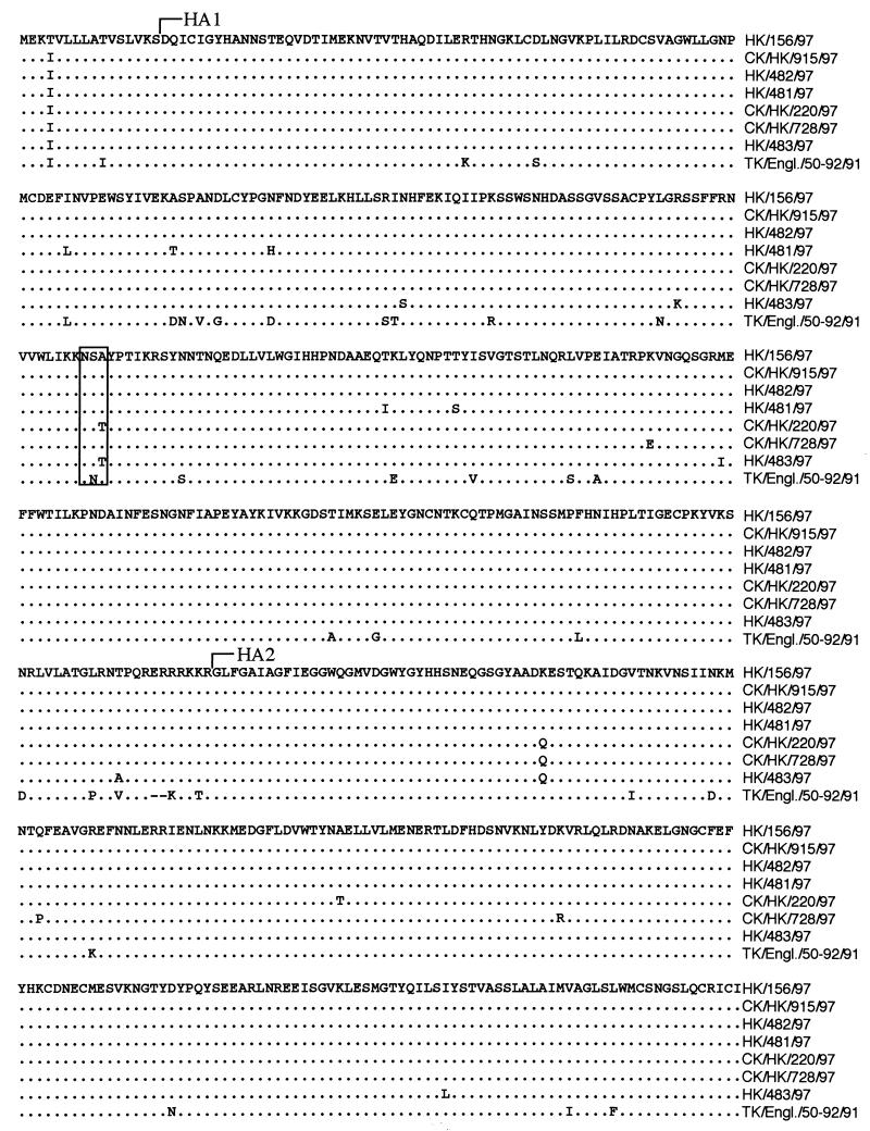
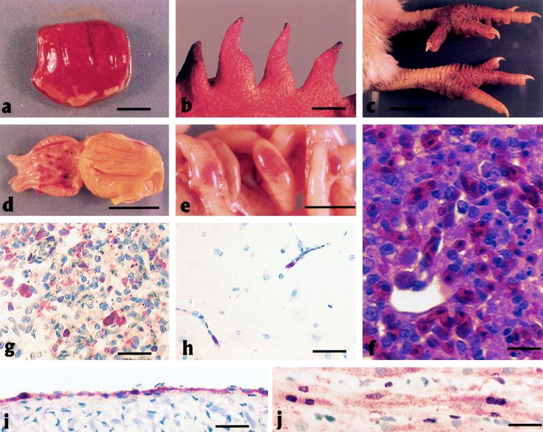
Genes of an influenza A (H5N1) virus from a human in Hong Kong isolated in May 1997 were sequenced and found to be all avian-like (K. Subbarao et al., Science 279:393-395, 1998). Gene sequences of this human isolate were compared to those of a highly pathogenic chicken H5N1 influenza virus isolated from Hong Kong in April 1997. Sequence comparisons of all eight RNA segments from the two viruses show greater than 99% sequence identity between them. However, neither isolate's gene sequence was closely (>95% sequence identity) related to any other gene sequences found in the GenBank database. Phylogenetic analysis demonstrated that the nucleotide sequences of at least four of the eight RNA segments clustered with Eurasian origin avian influenza viruses. The hemagglutinin gene phylogenetic analysis also included the sequences from an additional three human and two chicken H5N1 virus isolates from Hong Kong, and the isolates separated into two closely related groups. However, no single amino acid change separated the chicken origin and human origin isolates, but they all contained multiple basic amino acids at the hemagglutinin cleavage site, which is associated with a highly pathogenic phenotype in poultry. In experimental intravenous inoculation studies with chickens, all seven viruses were highly pathogenic, killing most birds within 24 h. All infected chickens had virtually identical pathologic lesions, including moderate to severe diffuse edema and interstitial pneumonitis. Viral nucleoprotein was most frequently demonstrated in vascular endothelium, macrophages, heterophils, and cardiac myocytes. Asphyxiation from pulmonary edema and generalized cardiovascular collapse were the most likely pathogenic mechanisms responsible for illness and death. In summary, a small number of changes in hemagglutinin gene sequences defined two closely related subgroups, with both subgroups having human and chicken members, among the seven viruses examined from Hong Kong, and all seven viruses were highly pathogenic in chickens and caused similar lesions in experimental inoculations.


Figures

FIG. 1
FIG. 1
Phylogenetic analysis using parsimony for all eight influenza virus gene segments based on nucleotide sequence. HK/156 is used as the reference isolate for all eight trees. All trees were constructed by general bootstrap analysis using 100 replicates, using PAUP 3.1 software (42). Branch lengths are provided in each tree. The NP, M, and NS genes are rooted to A/equine/Prague/1/56, and all other trees are midpoint rooted. All isolates in trees are type A influenza virus isolates and are full-length or close to full-length sequences. The NP and M trees used selected isolates to show the main groups but are concentrated with isolates close to the reference isolate. The NS gene tree also uses selected isolates, but only subtype (group) A isolates are presented. Abbreviations used for identifying isolates: CK (chicken), TK (turkey), DK (duck), FPV (fowl plague virus), WSN (mouse neurovirulent derivative of A/WS/33), Aust (Australia).
FIG. 1
FIG. 1
Phylogenetic analysis using parsimony for all eight influenza virus gene segments based on nucleotide sequence. HK/156 is used as the reference isolate for all eight trees. All trees were constructed by general bootstrap analysis using 100 replicates, using PAUP 3.1 software (42). Branch lengths are provided in each tree. The NP, M, and NS genes are rooted to A/equine/Prague/1/56, and all other trees are midpoint rooted. All isolates in trees are type A influenza virus isolates and are full-length or close to full-length sequences. The NP and M trees used selected isolates to show the main groups but are concentrated with isolates close to the reference isolate. The NS gene tree also uses selected isolates, but only subtype (group) A isolates are presented. Abbreviations used for identifying isolates: CK (chicken), TK (turkey), DK (duck), FPV (fowl plague virus), WSN (mouse neurovirulent derivative of A/WS/33), Aust (Australia).
FIG. 2
FIG. 2
Amino acid sequence alignment of the HA genes from all seven isolates in this study and from A/Turkey/England (TK/Engl.)/50-92/91, the isolate with the closest HA sequence similarity in GenBank. The start sites for the HA1 and HA2 proteins are indicated. Amino acids at positions 154 to 156 are boxed to highlight the glycosylation difference among the different virus isolates.
FIG. 3
FIG. 3
Polyacrylamide gel electrophoresis of purified virus structural proteins. Purified virions were disrupted in sodium dodecyl sulfate and mercaptoethanol and electrophoresed in 10% polyacrylamide gels. Viral polypeptides were identified on the basis of molecular weights. Lane A, CK/HK/220; lane B, HK/156. Po 1–3, polymerase genes PA, PB1, and PB2; NP, nucleoprotein; HA1, hemagglutinin subunit 1; M1, matrix protein 1; HA2, hemagglutinin subunit 2.
FIG. 4
FIG. 4
Experimental studies of chickens inoculated with HK/156 or CK/HK/220. (a to e) Photographs of gross lesions; (f to j) photomicrographs of hematoxylin-and-eosin-stained tissue sections or sections stained immunohistochemically to demonstrate AIV NP. (a) Severe congestion, hemorrhage, and edema of the lung from a 3-week-old WPR chicken that died 2 days after i.v. inoculation with HK/156 (bar = 1.5 cm). (b) Ischemic necrosis at the tips of the comb from a 37-week-old WL chicken that died 3 days after i.n./i.t. inoculation with HK/156 (bar = 1 cm). (c) Severe subcutaneous edema and hemorrhage of the feet and shanks from a 3-week-old WPR chicken that died 5 days after i.n. inoculation with HK/156 (bar = 2 cm). (d) Submucosal hemorrhage surrounding ducts of glands in the proventriculus from a 3-week-old WPR chicken that died 2 days after i.v. inoculation with HK/156 (bar = 2 cm). (e) Prominent hemorrhage in lymphoid tissue of Peyer’s patches in the jejunum from a 4-week-old WPR chicken that died 1 day after i.v. inoculation with CK/HK/220 (bar = 1 cm). (f) Severe diffuse pulmonary edema with congestion, hemorrhage, and interstitial pneumonitis in a 4-week-old WPR chicken that died 2 days after i.n. inoculation with CK/HK/220. Scattered necrotic cellular debris is present in blood capillaries (bar = 15 mm). (g) AIV antigen in cytoplasm and nucleus of blood capillary endothelial cells and macrophages and in necrotic debris from the lung of the chicken in panel f (bar = 50 mm). (h) AIV antigen in the cytoplasm and nuclei of blood capillary endothelial cells in the brain of the chicken in panel f (bar = 30 mm). (i) AIV antigen in the cytoplasm and nuclei of endocardial cells from a 4-week-old WPR chicken that died 1 day after i.v. inoculation with CK/HK/220 (bar = 30 mm). (j) AIV antigen in the cytoplasm and nuclei of cardiac myocytes from a 3-week-old WPR chicken that died on day 3 after inoculation with HK/156 (bar = 15 mm).
FIG. 5
FIG. 5
Lung tissues from a chicken that died 1 day after i.n. inoculation with CK/HK/220 (a and b) and a chicken euthanized 3 days after intranasal sham inoculation (c and d). (a) Hypertrophied blood capillary endothelial cells (E) with luminal heterophils (H) and degeneration of air capillary endothelial cells (A) in the lung of a chicken that died 1 day after i.n. inoculation with CK/HK/220. Note separation (∗) of blood capillary endothelium from air capillary epithelium and proteinic material and inflammatory cells filling the space (bar = 2 nm). (b) Hypertrophied capillary endothelium (E) with luminal heterophil (H) separation of blood capillary endothelium and air capillary epithelium in the lung (∗) and proteinic material in air capillary space (formula image
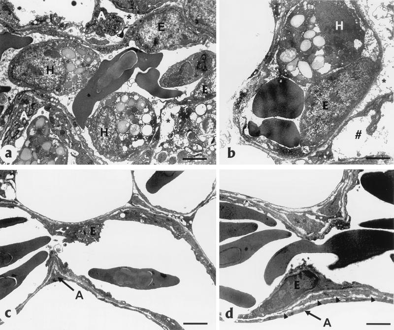
) (bar = 2.5 nm). (c) Normal blood capillary endothelium (E) and air capillary epithelium (A) without inflammatory cells (bar = 2 nm). (d) Thin basal lamina (arrowheads) separating blood capillary endothelium (E) and air capillary epithelium (A) (bar = 2.5 nm).

References

    1. Alexander D J. Avian influenza. Recent developments. Vet Bull. 1982;52:341–359.
    1. Barbeito M S, Abraham G, Best M, Cairns P, Langevin P, Sterritt W G, Barr D, Meulepas W, Sanchez-Vizcaino J M, Saraza M, Requena E, Collado M, Mani P, Breeze R, Brunner H, Mebus C A, Morgan R L, Rusk S, Siegfried L M, Thompson L H. Recommended biocontainment features for research and diagnostic facilities where animal pathogens are used. Rev Sci Tech Off Int Epizoot. 1995;14:873–887. - PubMed
    1. Bashiruddin J B, Gould A R, Westbury H A. Molecular pathotyping of two avian influenza viruses isolated during the Victoria 1976 outbreak. Aust Vet J. 1992;69:140–142. - PubMed
    1. Beare A S, Webster R G. Replication of avian influenza viruses in humans. Arch Virol. 1991;119:37–42. - PubMed
    1. Brown C C, Olander H J, Senne D A. A pathogenesis study of highly pathogenic avian influenza virus H5N2 in chickens, using immunohistochemistry. J Comp Pathol. 1992;107:341–348. - PubMed

Publication types

Substances