. 1998 Aug;72(8):6867-74.
doi: 10.1128/JVI.72.8.6867-6874.1998.

Infectious laryngotracheitis herpesvirus expresses a related pair of unique nuclear proteins which are encoded by split genes located at the right end of the UL genome region

Affiliations

Infectious laryngotracheitis herpesvirus expresses a related pair of unique nuclear proteins which are encoded by split genes located at the right end of the UL genome region

K Ziemann et al. J Virol. 1998 Aug.

Abstract

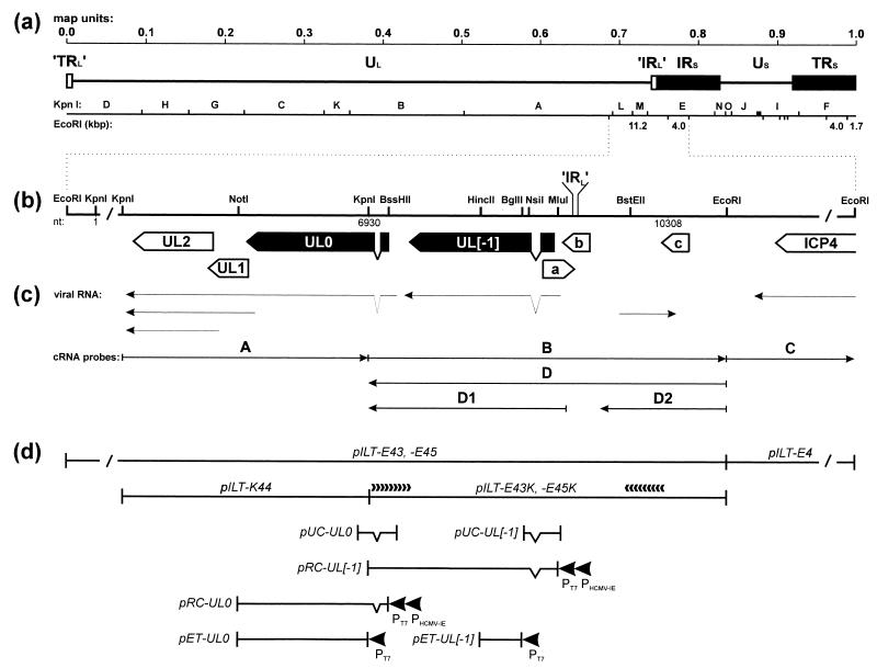
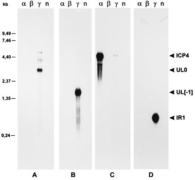
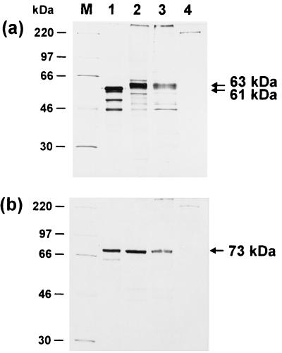
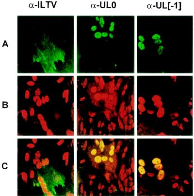
Avian infectious laryngotracheitis virus (ILTV) possesses an alphaherpesvirus type D DNA genome of ca. 155 kbp. Completion of our previous sequence analyses (W. Fuchs and T. C. Mettenleiter, J. Gen. Virol. 77:2221-2229, 1996) of the right end of the unique long (UL) genome region revealed the presence of two adjacent, presumably ILTV-specific genes, which were named UL0 and UL[-1] because of their location upstream of the conserved UL1 (glycoprotein L) gene. Transcriptional analyses showed that both genes are abundantly expressed during the late phase of the viral replication cycle and that both mRNAs are spliced by the removal of short introns close to their 5' ends. Furthermore, the deduced gene products exhibit a moderate but significant homology of 28% to each other. The newly identified ILTV genes encode proteins of 63 kDa (UL0) and 73 kDa (UL[-1]), which both are predominantly localized in the nuclei of virus infected chicken cells. In summary, our results indicate that duplication of a spliced ILTV-specific gene encoding a nuclear protein has occurred during evolution of ILTV.


Figures

FIG. 1
FIG. 1
Genome structure of ILTV. (a) Diagram of the type D genome of ILTV with KpnI restriction fragment map. Locations and sizes of relevant EcoRI restriction fragments are indicated. The US region is flanked by extended inverted repeat sequences (IRS and TRS). Very short (17-bp) inverted repetitions (’IRL’ and ’TRL’) were also found at both ends of the UL region. (b) Enlarged map of the investigated region with relevant restriction sites. Close to either end, the DNA fragments are not plotted to scale (/). Location of the ’IRL’ sequence separating the UL and IRS regions is indicated. Nucleotide numbers refer to the DNA sequence deposited in GenBank (accession no. X97256). Locations and orientations of ORFs are depicted by pointed rectangles, and splice sites are indicated by triangles. Conserved genes are named according to their homologs in HSV-1, and the analyzed ILTV-specific genes are highlighted in black. (c) Arrows indicate identified viral transcripts and the in vitro-transcribed cRNA probes which were used for hybridization as shown in Fig. 2. (d) Plasmid maps. pILT-E43, -E45, -E4, and -K44 contain EcoRI or KpnI fragments of ILTV DNA. Plasmids pILT-E43K and -E45K were used for nested deletion subcloning and directed DNA sequencing as indicated by arrowheads. pUC-UL0 and pUC-UL[−1] are cDNA clones encompassing the 5′ termini of either mRNA. They were used for reconstitution of the processed UL0 and UL[−1] ORFs under control of the bacteriophage T7 (PT7) and HCMV immediate-early (PHCMV-IE) promoters in pRC-UL0 and pRC-UL[−1]. For immunization, bacterial fusion proteins were expressed from pET-UL0 and pET-UL[−1] under the control of the T7 promoter (PT7).
FIG. 2
FIG. 2
Northern analyses of RNA from ILTV-infected and noninfected (n) chicken kidney cells. Cells were infected at an MOI of 5 and incubated for 6 h in the presence of cycloheximide (100 μg/ml) (α), for 16 h in the presence of phosphonoacetic acid (250 μg/ml) (β), or for 16 h without drugs (γ). Total RNA (5 μg per lane) was separated in 1% formaldehyde–0.8% agarose gels, transferred to nylon membranes, and hybridized with the probes depicted in Fig. 1c (A to D). Molecular sizes of RNA markers are indicated on the left. The sizes of the detected ILTV-specific transcripts (ICP4, UL0, UL[−1], IR1) are given in the text.
FIG. 3
FIG. 3
Identification of the UL0 and UL[−1] proteins. Radiolabeled proteins were incubated with monospecific anti-UL0 (a) and anti-UL[−1] sera (b) and immunoprecipitated. The in vitro translation products of pRC-UL0 and pRC-UL[−1] (lanes 1) were compared to proteins from primary chicken kidney cells transfected with the respective plasmid (lanes 2). In addition, lysates of chicken kidney cells were analyzed 24 h after infection with ILTV at an MOI of 5 (lanes 3). Results from precipitation of noninfected cell lysates are shown in lanes 4. In the depicted fluorograms of discontinuous SDS–10% polyacrylamide gels, the molecular masses of marker proteins (M) are indicated on the left, and locations of UL0 (61 and 63 kDa) and UL[−1] (73 kDa) gene products are marked by arrows.
FIG. 4
FIG. 4
Subcellular localization of the UL0 and UL[−1] proteins. ILTV-infected (MOI = 0.01) chicken kidney cells were fixed after 18 h and incubated with a chicken hyperimmune serum (α-ILTV) or monospecific rabbit antisera (α-UL0 or α-UL[−1]). Confocal laser scan microscopy was performed after subsequent incubation with fluorescein-conjugated secondary antibodies (row A) and staining of nuclear DNA with propidium iodide (row B). The combined green and red fluorescence (row C) demonstrates colocalization of the UL0 and UL[−1] proteins with nuclear DNA.

References

    1. Altschul S F, Gish W, Miller W, Myers E W, Lipman D J. Basic local alignment search tool. J Mol Biol. 1990;215:403–410. - PubMed
    1. Bagust T J, Guy J S. Laryngotracheitis. In: Calnek B W, Barnes H J, Beard C W, McDougald L R, Saif Y M, editors. Diseases of poultry. 10th ed. Ames, Iowa: Iowa State University Press; 1997. pp. 527–539.
    1. Baumeister J, Klupp B G, Mettenleiter T C. Pseudorabies virus and equine herpesvirus 1 share a nonessential gene which is absent in other herpesviruses and located adjacent to a highly conserved gene cluster. J Virol. 1995;69:5560–5567. - PMC - PubMed
    1. Ben-Porat T, Veach R A, Ihara S. Localization of the regions of homology between the genomes of herpes simplex virus type 1 and pseudorabies virus. Virology. 1983;127:194–204. - PubMed
    1. Bradley G, Hayashi M, Lancz G, Tanaka A, Nonoyama M. Structure of the Marek’s disease virus BamHI-H gene family: genes of putative importance for tumor induction. J Virol. 1989;63:2534–2542. - PMC - PubMed

Publication types

Associated data

LinkOut - more resources