. 1998 Jul;9(7):1773-86.
doi: 10.1091/mbc.9.7.1773.

Localization of autocrine motility factor receptor to caveolae and clathrin-independent internalization of its ligand to smooth endoplasmic reticulum

Affiliations
Free PMC article

Localization of autocrine motility factor receptor to caveolae and clathrin-independent internalization of its ligand to smooth endoplasmic reticulum

N Benlimame et al. Mol Biol Cell. 1998 Jul.
Free PMC article

Abstract

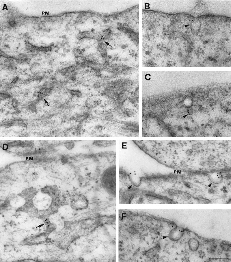
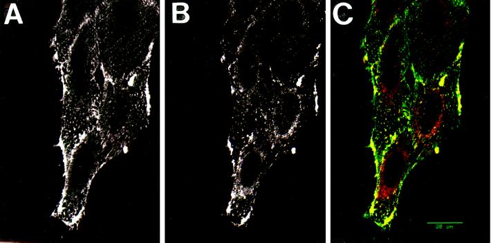
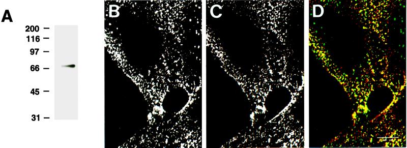
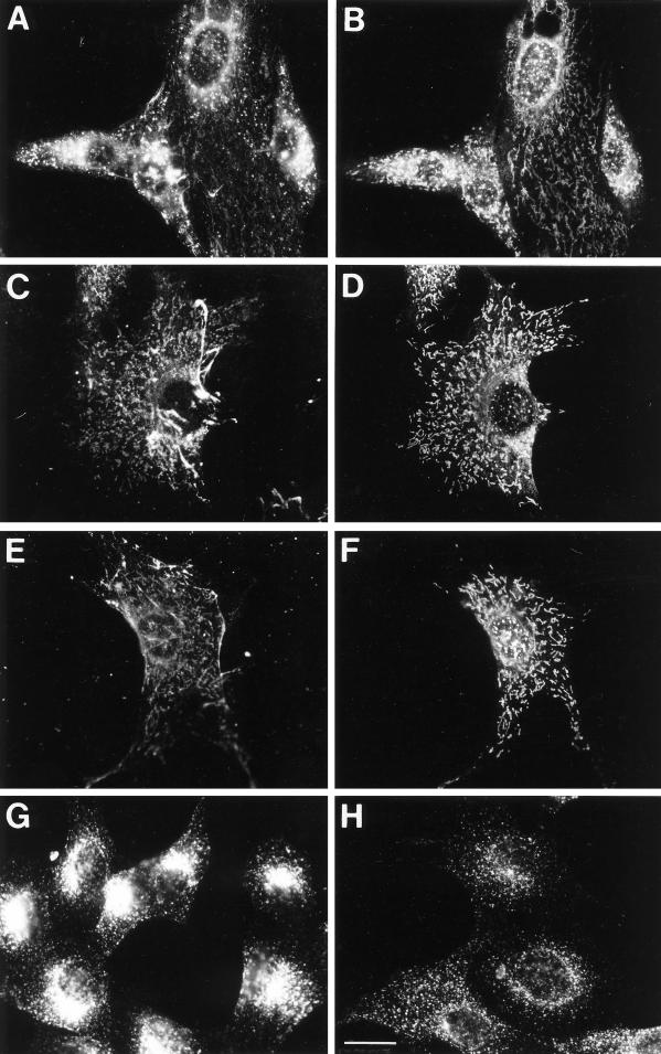
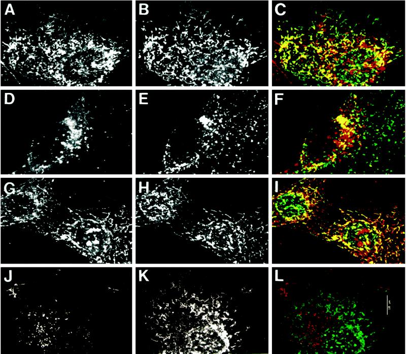
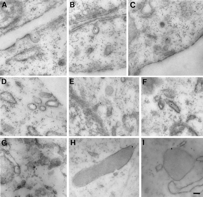
Autocrine motility factor receptor (AMF-R) is a cell surface receptor that is also localized to a smooth subdomain of the endoplasmic reticulum, the AMF-R tubule. By postembedding immunoelectron microscopy, AMF-R concentrates within smooth plasmalemmal vesicles or caveolae in both NIH-3T3 fibroblasts and HeLa cells. By confocal microscopy, cell surface AMF-R labeled by the addition of anti-AMF-R antibody to viable cells at 4 degreesC exhibits partial colocalization with caveolin, confirming the localization of cell surface AMF-R to caveolae. Labeling of cell surface AMF-R by either anti-AMF-R antibody or biotinylated AMF (bAMF) exhibits extensive colocalization and after a pulse of 1-2 h at 37 degreesC, bAMF accumulates in densely labeled perinuclear structures as well as fainter tubular structures that colocalize with AMF-R tubules. After a subsequent 2- to 4-h chase, bAMF is localized predominantly to AMF-R tubules. Cytoplasmic acidification, blocking clathrin-mediated endocytosis, results in the essentially exclusive distribution of internalized bAMF to AMF-R tubules. By confocal microscopy, the tubular structures labeled by internalized bAMF show complete colocalization with AMF-R tubules. bAMF internalized in the presence of a 10-fold excess of unlabeled AMF labels perinuclear punctate structures, which are therefore the product of fluid phase endocytosis, but does not label AMF-R tubules, demonstrating that bAMF targeting to AMF-R tubules occurs via a receptor-mediated pathway. By electron microscopy, bAMF internalized for 10 min is located to cell surface caveolae and after 30 min is present within smooth and rough endoplasmic reticulum tubules. AMF-R is therefore internalized via a receptor-mediated clathrin-independent pathway to smooth ER. The steady state localization of AMF-R to caveolae implicates these cell surface invaginations in AMF-R endocytosis.


Figures

Figure 1
Figure 1
Electron microscopic localization of AMF-R in NIH-3T3 fibroblasts and HeLa cells. HeLa (A, B, and C) and NIH-3T3 (D, E, and F) cells were postembedding immunolabeled with anti-AMF-R and 12-nm gold–conjugated anti-rat IgM secondary antibodies. Typical AMF-R labeling of smooth tubules (A and D, arrows) and cell surface caveolae (B, C, E, F, arrowheads) is shown. PM, Plasma membrane. Bar, 0.2 μm.
Figure 2
Figure 2
Colocalization of AMF-R and caveolin by confocal microscopy. Viable NIH-3T3 cells were labeled for cell surface AMF-R at 4°C (A) and for caveolin after fixation and permeabilization (B). To demonstrate the colocalization of AMF-R and caveolin, confocal images from both fluorescent channels were superimposed (panel C; AMF-R in green and caveolin in red) and colocalization appears in yellow. Bar, 20 μm.
Figure 3
Figure 3
bAMF and anti-AMF-R mAb colocalize on the cell surface. bAMF migrated as a single band in protein blots revealed with HRP-streptavidin (A). Confocal imaging of cell surface labeling of viable NIH-3T3 cells at 4°C with bAMF (B) or anti-AMF-R antibody (C). Confocal images from both fluorescent channels were superimposed (panel D; bAMF in green and AMF-R in red) and revealed a significant degree of colocalization in yellow. Bar, 20 μm.
Figure 4
Figure 4
F). After fixation with methanol/acetone, cells were double labeled with Texas Red-streptavidin to reveal bAMF (A, C, and E) and anti-AMF-R mAb and FITC-conjugated anti-rat secondary antibody to reveal AMF-R labeling (B, D, and F). To ensure that cellular acidification disrupted clathrin-mediated endocytosis of transferrin receptor, NIH-3T3 cells were incubated at 37°C with Texas Red transferrin for 30 min in regular medium (G) or in medium acidified to pH 5.5 (H). Bar, 20 μm.Internalization of bAMF to AMF-R tubules. NIH-3T3 cells were pulse labeled with bAMF at 37°C for 1 h (A and B), for 2 h and chased for 4 h (C and D), or for 1 h in medium acidified to pH 5.5 to disrupt clathrin-mediated endocytosis (E and
Figure 5
Figure 5
Localization of internalized bAMF to AMF-R tubules by confocal microscopy. NIH-3T3 cells were pulse labeled with bAMF at 37°C for 1 h in regular medium (A–F) for 1 h in medium acidified to pH 5.5 to disrupt clathrin-mediated endocytosis (G–I), or in regular medium in the presence of 10-fold excess unlabeled AMF (J–L) before fixation with methanol/acetone. bAMF was revealed with Texas Red-streptavidin (A, D, G, and J) and AMF-R (B, H, and K) or LAMP-1 (E) labeled with the appropriate primary antibodies and FITC-conjugated secondary antibodies. Confocal images from both fluorescent channels were superimposed (panels C, I, and L, bAMF in red and AMF-R in green; panel F, bAMF in red and LAMP-1 in green) and colocalization appears in yellow. Bar, 10 μm.
Figure 6
Figure 6
Electron microscopy of the internalization pathway of bAMF. NIH-3T3 cells were pulsed with bAMF at 37°C for 10 (A, B, and H) or 30 min (C, D, E, F, G, and I). The localization of bAMF was revealed by postembedding labeling with 10-nm gold-conjugated streptavidin. After 10 min, bAMF is localized to cell surface caveolae (A and B). After a 30-min pulse, bAMF is localized to caveolae and smooth vesicles (C and D) and also appears in intracellular membranous tubules (E, F, and G) including distinctive smooth (E) and rough (F) ER elements. bAMF labeling of dense lysosomal structures is also detected (H and I). Bar, 0.1 μm.

References

    1. Altankov G, Grinnell F. Depletion of intracellular potassium disrupts coated pits and reversibly inhibits cell polarization during fibroblast spreading. J Cell Biol. 1993;120:1449–1459. - PMC - PubMed
    1. Anderson HA, Chen Y, Norkin LC. Bound simian virus 40 translocates to caveolin-enriched membrane domains, and its entry is inhibited by drugs that selectively disrupt caveolae. Mol Biol Cell. 1996;7:1825–1834. - PMC - PubMed
    1. Anderson RGW, Kamen BA, Rothberg KG, Lacey SW. Potocytosis: sequestration and transport of small molecules by caveolae. Science. 1992;253:410–411. - PubMed
    1. Bendayan M, Rasio EA. Transport of insulin and albumin by the microvascular endothelium of the rete mirabile. J Cell Sci. 1996;109:1857–1864. - PubMed
    1. Benlimame N, Simard D, Nabi IR. Autocrine motility factor receptor is a marker for a distinct tubular membrane organelle. J Cell Biol. 1995;129:459–471. - PMC - PubMed

Publication types

MeSH terms

LinkOut - more resources