. 1998 Jul;9(7):1803-16.
doi: 10.1091/mbc.9.7.1803.

Serine and threonine phosphorylation of the paxillin LIM domains regulates paxillin focal adhesion localization and cell adhesion to fibronectin

Affiliations
Free PMC article

Serine and threonine phosphorylation of the paxillin LIM domains regulates paxillin focal adhesion localization and cell adhesion to fibronectin

M C Brown et al. Mol Biol Cell. 1998 Jul.
Free PMC article

Abstract

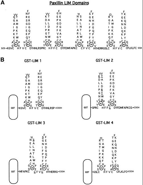
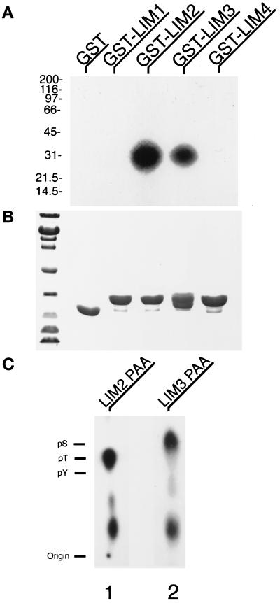
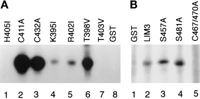
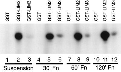
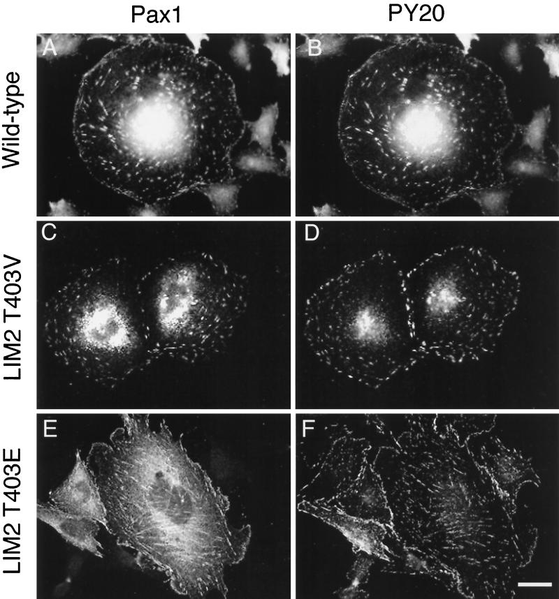
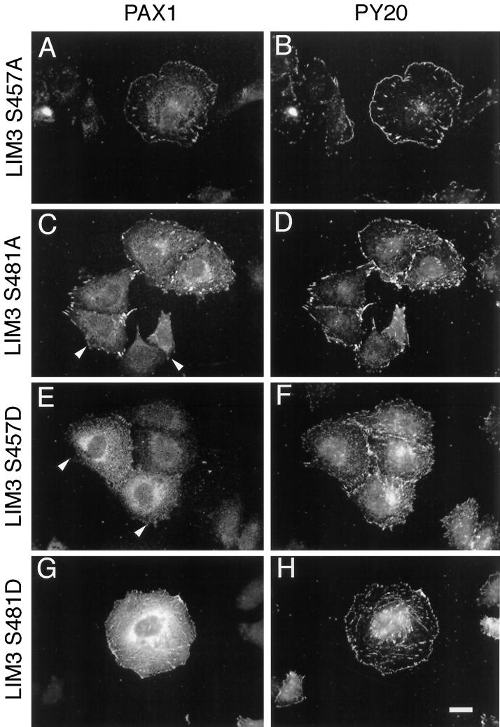
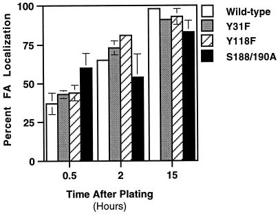
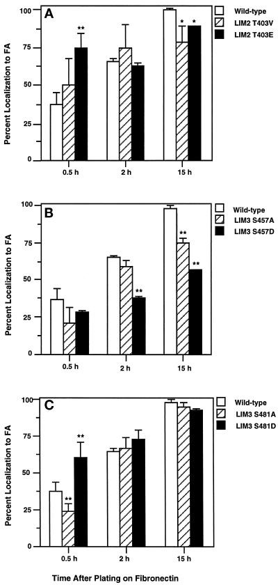
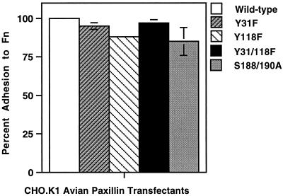
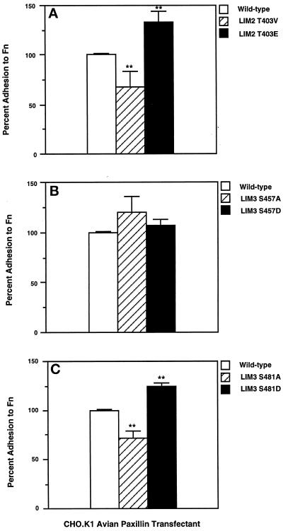
We have previously shown that the LIM domains of paxillin operate as the focal adhesion (FA)-targeting motif of this protein. In the current study, we have identified the capacity of paxillin LIM2 and LIM3 to serve as binding sites for, and substrates of serine/threonine kinases. The activities of the LIM2- and LIM3-associated kinases were stimulated after adhesion of CHO.K1 cells to fibronectin; consequently, a role for LIM domain phosphorylation in regulating the subcellular localization of paxillin after adhesion to fibronectin was investigated. An avian paxillin-CHO.K1 model system was used to explore the role of paxillin phosphorylation in paxillin localization to FAs. We found that mutations of paxillin that mimicked LIM domain phosphorylation accelerated fibronectin-induced localization of paxillin to focal contacts. Further, blocking phosphorylation of the LIM domains reduced cell adhesion to fibronectin, whereas constitutive LIM domain phosphorylation significantly increased the capacity of cells to adhere to fibronectin. The potentiation of FA targeting and cell adhesion to fibronectin was specific to LIM domain phosphorylation as mutation of the amino-terminal tyrosine and serine residues of paxillin that are phosphorylated in response to fibronectin adhesion had no effect on the rate of FA localization or cell adhesion. This represents the first demonstration of the regulation of protein localization through LIM domain phosphorylation and suggests a novel mechanism of regulating LIM domain function. Additionally, these results provide the first evidence that paxillin contributes to "inside-out" integrin-mediated signal transduction.


Figures

Figure 1
Figure 1
A schematic representation of the four LIM domains of paxillin. (A) The four LIM domains of paxillin are found at the carboxyl terminus of the protein and span amino acids 323–559. Each LIM motif is composed of two zinc fingers. (B) Detail of the amino acid residues comprising the individual LIM domain-GST fusion proteins. The residues underlined were targets for mutagenesis in this study.
Figure 2
Figure 2
Phosphorylation of paxillin LIM domains 2 and 3 on threonine and serine. (A) GST fusion proteins comprising the four individual LIM domains of paxillin were incubated with avian smooth muscle lysates and washed free of unbound protein, followed by protein kinase assay as described in MATERIALS AND METHODS. Lane 1, GST alone; lane 2, GST-LIM1; lane 3, GST-LIM2; lane 4, GST-LIM3; and lane 5, GST-LIM4. Only GST-LIM2 and GST-LIM3 specifically precipitated and were phosphorylated by protein kinases. (B) Coomassie brilliant blue staining of the GST precipitation kinase assay SDS-polyacrylamide gel to show equivalent loading of the fusion proteins. (C) PAA was performed on the phosphorylated GST-LIM2 and GST-LIM3 fusion proteins. Comigration of ninhydrin-stained phosphoamino acid standards revealed that the phosphorylation of LIM2 was restricted to the amino acid threonine and paxillin LIM3 on serine. Lower spots are partial hydrolysis products.
Figure 3
Figure 3
Paxillin LIM domain–kinase association is LIM domain structure-dependent. (A) GST-LIM2 fusion proteins were generated containing point mutations of residues potentially involved in kinase binding or phosphorylation and subjected to in vitro kinase assay as in MATERIALS AND METHODS. Lane 1, amino-terminal zinc finger, zinc-chelating histidine 405 to isoleucine; lane 2, carboxyl-terminal zinc finger, zinc-chelating cysteine 411 to alanine; lane 3 carboxyl-terminal zinc finger, zinc-chelating cysteine 432 to alanine; lane 4, lysine 395 to isoleucine; lane 5, arginine 402 to isoleucine; lane 6, threonine 398 to valine; lane 7, threonine 403 to valine; lane 8, GST only. Phosphorylation of C411A was equivalent to wild-type LIM2. GST-LIM3 fusion proteins were generated containing point mutations of residues potentially involved in kinase binding or phosphorylation. Lane 1, GST; lane 2, serine 457 to alanine; lane 3, serine 481 to alanine; lane 4, amino- and carboxyl-terminal zinc finger, zinc-chelating cysteines 467 and 470 to alanine.
Figure 4
Figure 4
Stimulation of LIM2 and LIM3 serine/threonine kinases after adhesion to fibronectin. GST (lanes 1, 4, 7, and 10), GST-LIM2 (lanes 2, 5, 8, and 11), or GST-LIM3 (lanes 3, 6, 9, and 12) fusion proteins were incubated with CHO.K1 lysates, washed free of unbound protein, followed by in vitro kinase assay as in MATERIALS AND METHODS. Lanes 1–3, Lysates from cells maintained in suspension for 60 min; lanes 4–6, cells plated on 10 μg/ml fibronectin-coated Petri dishes for 30 min; lanes 7–9, cells plated for 60 min; lanes 10–12, cells plated for 120 min before harvesting for preparation of lysates for use in in vitro kinase assays. Laser scanning densitometry of the phosphorylated GST-LIM fusion protein autoradiograph signal revealed that CHO.K1 adhesion to fibronectin for 30 min stimulated LIM3 kinase activity 4.5-fold over cells maintained in suspension. After this peak stimulation, the levels decreased over time, to 3.5-fold by 120 min. LIM2 phosphorylation gradually increased 1.2-fold over suspension levels by 120 min.
Figure 5
Figure 5
Immunofluorescence analysis of the capacity and efficiency of paxillin LIM2 phosphorylation mutants to localize to FAs. CHO.K1 cells were transfected with avian paxillin cDNA containing mutations of LIM2. After 24 h of growth on glass coverslips in Ham’s F-12 media containing 10% FBS, ectopically expressed avian paxillin was visualized by immunofluorescence double-labeling with a chicken-specific, polyclonal antiserum Pax1 (A, C, and E) and a monoclonal antibody to phosphotyrosine, PY20 (B, D, and F). (A and B) Wild-type; (C and D) LIM2T403V; (E and F) LIM2T403E are representative of the transfected populations. Bar, 5 μm.
Figure 6
Figure 6
Mutation of paxillin LIM3 phosphorylation sites causes decreased efficiency of FA localization. CHO.K1 avian paxillin transfectants were grown on glass coverslips for 24 h in Ham’s F-12 containing 10% FBS, followed by immunofluorescence double-labeling with a chicken-specific, polyclonal antiserum Pax1 (A, C, E, and G) and a monoclonal antibody to phosphotyrosine, PY20 (B, D, F, and H). (A and B) LIM3S457A; (C and D) LIM3S481A; (E and F) LIM3S457D; (G and H) LIM3S481D are representative of the transfected populations. The LIM3S457D and LIM3S481A mutant molecules showed a reduced colocalization of mutated paxillin and PY20 (see arrows). Bar, 5 μm.
Figure 7
Figure 7
Localization rate of paxillin molecules containing mutations of the fibronectin-inducible phosphorylation sites Y31/118 and S188/190 is comparable to cells expressing wild-type avian paxillin. CHO.K1 fibroblasts expressing avian paxillin were maintained in suspension for 1 h before plating onto duplicate glass coverslips coated with 10 μg/ml fibronectin. At 0.5, 2, and 15 h, the coverslips were processed for immunofluorescence double-labeling using a chicken-specific polyclonal antiserum (Pax1) and either PY20 (Transduction Labs, Lexington, KY) to label FAs, or rhodamine-phalloidin (Molecular Probes, Eugene, OR) to decorate actin stress fibers. At each time point 150–200 transfected cells were counted with the number of avian paxillin transfectants displaying FA localization of the avian paxillin determined and divided by the total number of transfected cells. This is represented in bar graph form as the “Percentage localization to FA.” All experiments were performed in duplicate with at least three independent experiments executed and tabulated to determine mean and SD from the mean. Statistical analyses were performed with Student’s t test; *, p < 0.05; **, p < 0.01.
Figure 8
Figure 8
Increased FA localization rate of paxillin molecules containing phospho-mimetic mutations of phosphorylatable residues within LIM2 or LIM3. (A) LIM2T403V and LIM2T403E. (B) LIM3S457A and LIM3S457D. (C) LIM3S481A and LIM3S481D. After adhesion to 10 μg/ml fibronectin-coated glass coverslips for 0.5, 2, or 15 h, the cells were processed and immunofluorescence double-labeled with Pax1 and PY20 or rhodamine-phalloidin. At each time point 150–200 transfected cells were counted with the number of avian paxillin transfectants displaying FA localization of the avian paxillin determined and divided by the total number of transfected cells. This is represented in bar graph form as the “Percentage localization to FA.” Four independent experiments were executed and tabulated to determine mean and SD from the mean. Statistical analyses were performed with Student’s t test; *, p < 0.05; **, p < 0.01.
Figure 9
Figure 9
Adhesion of CHO.K1 cells expressing avian paxillin containing mutations of Y31/118F and S188/190A is identical to cells expressing wild-type avian paxillin. Avian paxillin transfectants were maintained in suspension for 1 h before adhesion for 30 min on 10 μg/ml fibronectin-coated, 1% BSA-blocked 96-well dishes, 8 wells per transfectant. After extensive washing, absorbance values were obtained by MTT assay, and adhesion relative to wild-type avian paxillin-expressing transfectants was calculated. Four independent experiments were executed, in duplicate, and tabulated to determine mean and SD from the mean. Statistical analyses were performed with Student’s t test; *, p < 0.05; **, p < 0.01.
Figure 10
Figure 10
A role for the LIM domains of paxillin in regulating fibroblast adhesion to fibronectin. Blocking phosphorylation of T403 or S481 reduces cell adhesion by 25% whereas mimicking constitutive phosphorylation of these residues stimulates adhesion by 25%. (A) LIM2T403V and LIM2T403E. (B) LIM3S457A and LIM3S457D. (C) LIM3S481A and LIM3S481D. Avian paxillin transfectants were maintained in suspension for 1 h before adhesion for 30 min on 10 μg/ml fibronectin-coated, 1% BSA-blocked 96-well dishes, 8 wells per transfectant. After extensive washing, absorbance values were obtained by MTT assay and adhesion relative to wild-type avian paxillin-expressing transfectants was calculated. Each graph represents the mean and SD from the mean from three adhesion assays, performed in duplicate. Statistical analyses were performed with Student’s t test; *, p < 0.05; **, p < 0.01.

Similar articles

Cited by

References

    1. Arber S, Caroni P. Specificity of single LIM motifs in targeting and LIM/LIM interactions in situ. Genes Dev. 1996;10:289–300. - PubMed
    1. Aznavoorian S, Stracke ML, Parsons J, McClanahan J, Liotta LA. Integrin αvβ3 mediates chemotactic and haptotactic motility in human melanoma cells through different signaling pathways. J Biol Chem. 1996;271:3247–3254. - PubMed
    1. Barreuther MF, Grabel LB. The role of phosphorylation in modulating β1 integrin localization. Exp Cell Res. 1996;222:10–15. - PubMed
    1. Bellis SL, Miller JT, Turner CE. Characterization of tyrosine phosphorylation of paxillin in vitro by focal adhesion kinase. J Biol Chem. 1995;270:17437–17441. - PubMed
    1. Bellis SL, Perrotta JA, Curtis MS, Turner CE. Adhesion of fibroblasts to fibronectin stimulates both serine and tyrosine phosphorylation of paxillin. Biochem J. 1997;325:375–381. - PMC - PubMed

Publication types

MeSH terms

LinkOut - more resources