. 1998 Jul;9(7):1873-89.
doi: 10.1091/mbc.9.7.1873.

Multiple functions for actin during filamentous growth of Saccharomyces cerevisiae

Affiliations
Free PMC article

Multiple functions for actin during filamentous growth of Saccharomyces cerevisiae

B M Cali et al. Mol Biol Cell. 1998 Jul.
Free PMC article

Abstract

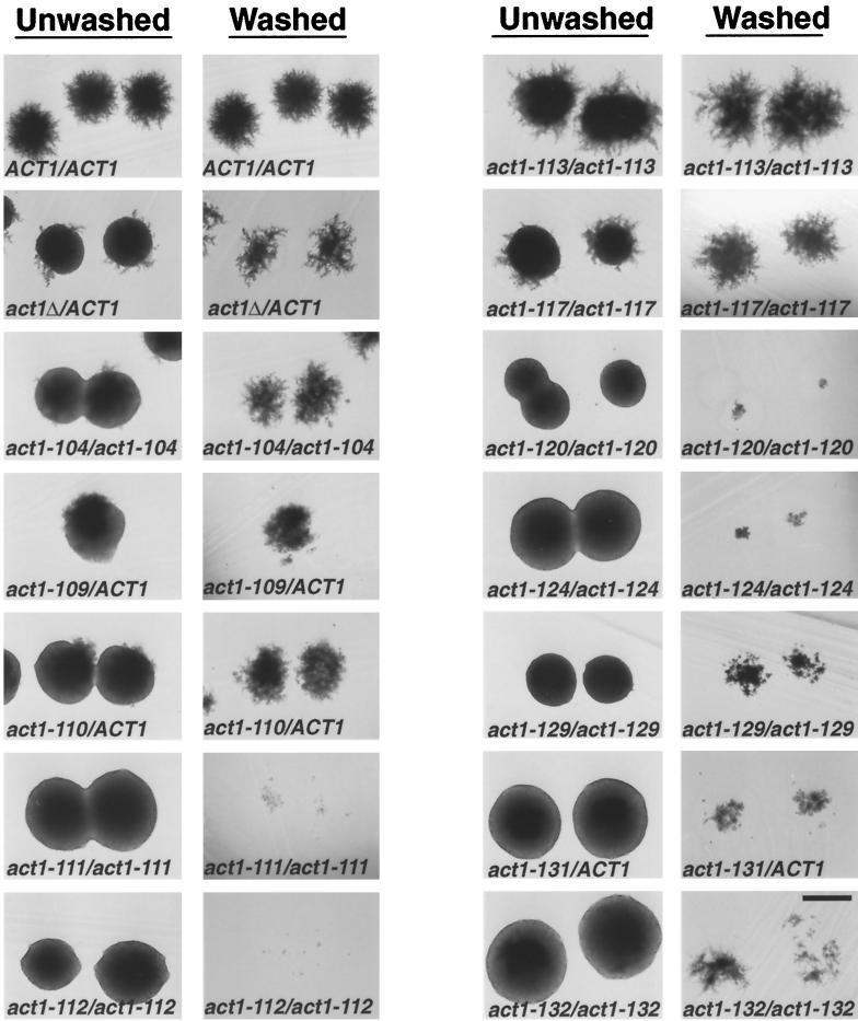
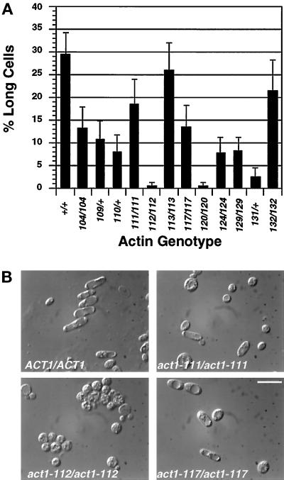
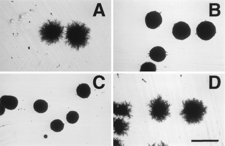
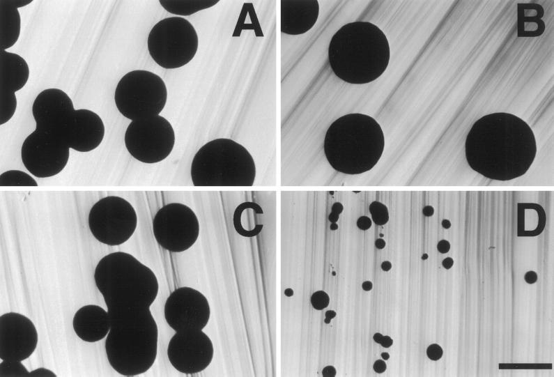
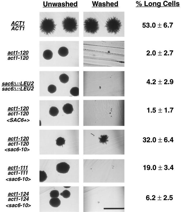
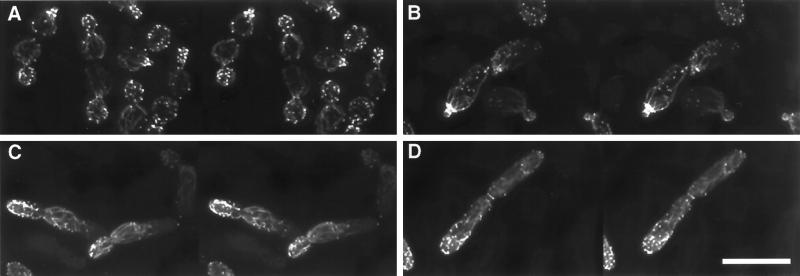
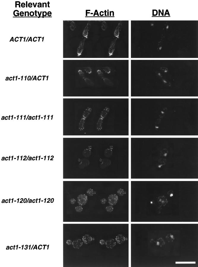
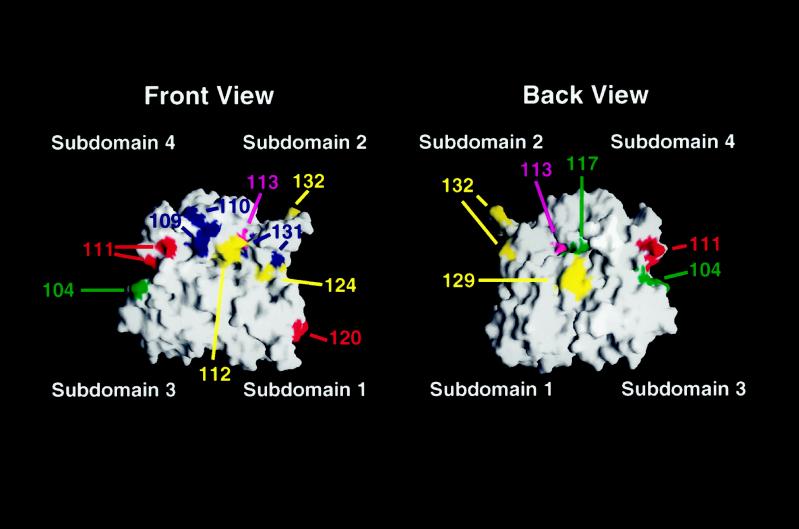
Saccharomyces cerevisiae is dimorphic and switches from a yeast form to a pseudohyphal (PH) form when starved for nitrogen. PH cells are elongated, bud in a unipolar manner, and invade the agar substrate. We assessed the requirements for actin in mediating the dramatic morphogenetic events that accompany the transition to PH growth. Twelve "alanine scan" alleles of the single yeast actin gene (ACT1) were tested for effects on filamentation, unipolar budding, agar invasion, and cell elongation. Some act1 mutations affect all phenotypes, whereas others affect only one or two aspects of PH growth. Tests of intragenic complementation among specific act1 mutations support the phenotypic evidence for multiple actin functions in filamentous growth. We present evidence that interaction between actin and the actin-binding protein fimbrin is important for PH growth and suggest that association of different actin-binding proteins with actin mediates the multiple functions of actin in filamentous growth. Furthermore, characterization of cytoskeletal structure in wild type and act1/act1 mutants indicates that PH cell morphogenesis requires the maintenance of a highly polarized actin cytoskeleton. Collectively, this work demonstrates that actin plays a central role in fungal dimorphism.


Figures

Figure 1
Figure 1
Filamentation and invasion phenotypes of actin mutants. Strains of indicated genotype were streaked for single colonies on SLAD agar and incubated for 4 d at 30°C. Complete genotypes of strains are listed in Table 1. Colonies were photographed before (unwashed column) and after (washed column) washing the surface of the agar to remove noninvaded cells. Bar, 500 μm.
Figure 2
Figure 2
Effect of act1 mutations on cell elongation. (A) Percentage of long cells in each of the actin mutants. Complete genotypes of strains are as indicated in Figure 1. Long cell counts were performed as described in MATERIALS AND METHODS. Bars show the SE of two independent experiments. (B) Examples of different cell elongation phenotypes. Images shown are differential interference contrast images of cells scraped from the agar as above. Bar, 10 μm.
Figure 3
Figure 3
act1–111 and act1–120 exhibit intragenic complementation for filamentation defects. Shown is the growth on SLAD agar after four days at 30°C. (A) BCY209 (ACT1/ACT1); (B) BCY204 (act1–111/act1–111); (C) BCY210 (act1–120/act1–120); (D) BCY310 (act1–111/act1–120). Bar, 500 μm.
Figure 4
Figure 4
act1–111 and act1–112 exhibit a negative growth interaction. Shown are colonies after growth for 3 d at 30°C on YNB. (A) BCY209 (ACT1/ACT1); (B) BCY204 (act1–111/act1–111); (C) BCY202 (act1–112/act1–112); (D) BCY302 (act1–111/act1–112). Bar, 1 mm.
Figure 5
Figure 5
Expression of a mutant fimbrin (Sac6p) with an altered actin-binding domain suppresses the invasion and cell elongation defects of an act1–120/act1–120 diploid. Strains BCY209 (ACT1/ACT1), BCY210 (act1–120/act1–120), BCY372 (sac6/sac6), BCY412 (act1–120/act1–120SAC6+〉), BCY411 (act1–120/act1–120sac6–10〉), BCY432 (act1–111/act1–111sac6–10〉), and BCY433 (act1–124/act1–124sac6–10〉), where genotypes in angle brackets refer to relevant genotypes of plasmids AAB117 (SAC6 URA3 CEN) and AAB289 (sac6–10 URA3 CEN) (Brower et al., 1995), were streaked to SLAD agar and incubated at 30°C for 4 d. The resultant colonies were photographed before and after washing the agar surface. Long cells were quantified as described in MATERIALS AND METHODS and are reported with SE. Bar, 500 μm.
Figure 6
Figure 6
Comparison of the YF and PH actin cytoskeletons. Stereo images of the filamentous actin cytoskeleton (stained with rhodamine–phalloidin) in strain BCY209 grown either in YPD (A) or in SLAD alginate (B–D). (A) YF cells: all stages of cell cycle. (B) PH cell, early bud emergence; note prominent ring of actin in mother cell at site of bud emergence and at the neck of the very small emerging bud. (C) PH cell, midbud emergence; image shows the pronounced actin cables and patch polarization to the distal end of the emerging bud. (D) PH cell shortly before cytokinesis; a ring of actin has formed at the septation site between mother and daughter, whereas some patches remain near the distal end of the daughter cell. Bar, 10 μm.
Figure 7
Figure 7
The actin cytoskeleton in actin mutants grown under conditions that induce PH growth. Shown are wild-type and actin mutant strains grown in SLAD alginate and stained with rhodamine–phalloidin (to detect F-actin) and DAPI (to detect DNA) as described in MATERIALS AND METHODS. F-actin images are in stereo. Strains shown are BCY209 (ACT1/ACT1), BCY157 (act1–110/ACT1), BCY204 (act1–111/act1–111), BCY202 (act1–112/act1–112), BCY210 (act1–120/act1–120), and BCY168 (act1–131/ACT1). Bar, 10 μm.
Figure 8
Figure 8
Modeling of the mutations analyzed in this study onto the solvent-exposed surface of an actin monomer. Shown is the calculated surface of the actin monomer based on the coordinates of the rabbit muscle actin filament structure (Lorenz et al., 1993). Actin subdomains are indicated. Red, locations of residues altered by viable, temperature-sensitive alleles with recessive effects on PH growth; yellow, location of residues altered by viable, temperature-sensitive alleles with dominant effects on PH growth; blue, positions of residues affected by recessive lethal alleles with dominant effects on PH growth; green, locations of residues affected by mutations that have no effect on viability but disrupt PH growth; magenta, positions of residues altered by mutations with no effect on PH growth. Surfaces were generated using GRASP (Nicholls et al., 1991) on a Silicon Graphics Iris computer.
Figure 9
Figure 9
Model for mechanism of intragenic complementation between act1 alleles (after Ayscough and Drubin, 1996). Actin monomers encoded by ACT1, act1-x, and act1-y are represented by white, black, and gray circles, respectively. The ability of hypothetical actin-binding proteins A and B to interact with actin filaments comprising wild-type actin, each mutant actin singly, or a combination of mutant actins is schematized. The PH growth phenotype of strains containing such actin filaments is also indicated.

References

    1. Adams AE, Botstein D. Dominant suppressors of yeast actin mutations that are reciprocally suppressed. Genetics. 1989;121:675–683. - PMC - PubMed
    1. Adams AE, Botstein D, Drubin DG. A yeast actin-binding protein is encoded by SAC6, a gene found by suppression of an actin mutation. Science. 1989;243:231–233. - PubMed
    1. Adams AE, Pringle JR. Relationship of actin and tubulin distribution to bud growth in wild-type and morphogenetic-mutant Saccharomyces cerevisiae. J Cell Biol. 1984;98:934–945. - PMC - PubMed
    1. Adams AE, Shen W, Lin CS, Leavitt J, Matsudaira P. Isoform-specific complementation of the yeast sac6 null mutation by human fimbrin. Mol Cell Biol. 1995;15:69–75. - PMC - PubMed
    1. Agard DA, Hiraoka Y, Shaw P, Sedat JW. Fluorescence microscopy in three dimensions. Methods Cell Biol. 1989;30:353–377. - PubMed

Publication types

MeSH terms

LinkOut - more resources