. 1998 Jul;9(7):1891-902.
doi: 10.1091/mbc.9.7.1891.

RhoA GTPase and serum response factor control selectively the expression of MyoD without affecting Myf5 in mouse myoblasts

Affiliations
Free PMC article

RhoA GTPase and serum response factor control selectively the expression of MyoD without affecting Myf5 in mouse myoblasts

G Carnac et al. Mol Biol Cell. 1998 Jul.
Free PMC article

Abstract

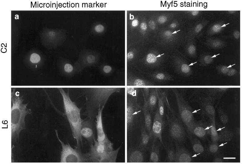
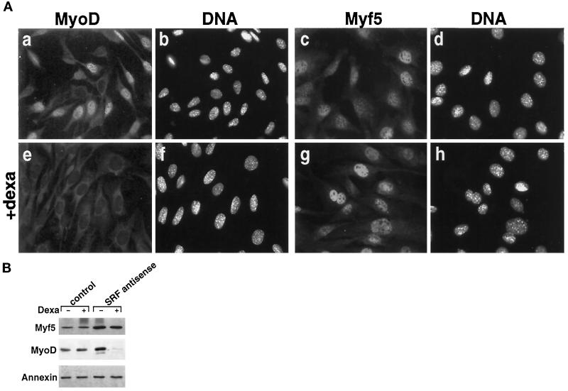
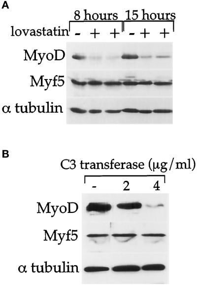
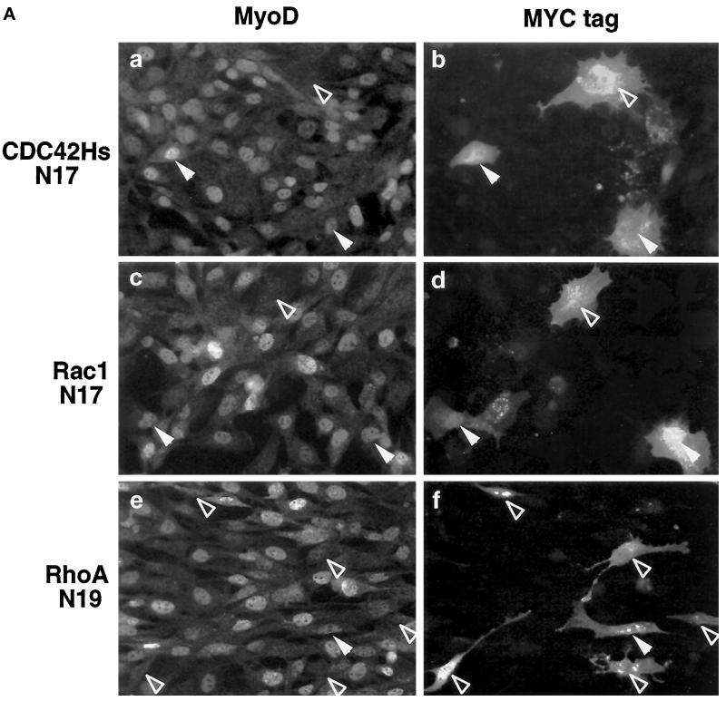
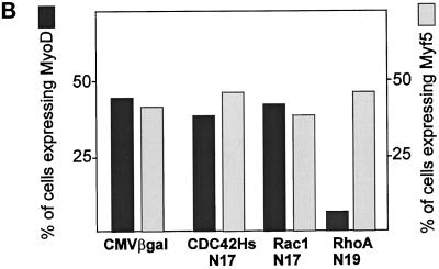
MyoD and Myf5 belong to the family of basic helix-loop-helix transcription factors that are key operators in skeletal muscle differentiation. MyoD and Myf5 genes are selectively activated during development in a time and region-specific manner and in response to different stimuli. However, molecules that specifically regulate the expression of these two genes and the pathways involved remain to be determined. We have recently shown that the serum response factor (SRF), a transcription factor involved in activation of both mitogenic response and muscle differentiation, is required for MyoD gene expression. We have investigated here whether SRF is also involved in the control of Myf5 gene expression, and the potential role of upstream regulators of SRF activity, the Rho family G-proteins including Rho, Rac, and CDC42, in the regulation of MyoD and Myf5. We show that inactivation of SRF does not alter Myf5 gene expression, whereas it causes a rapid extinction of MyoD gene expression. Furthermore, we show that RhoA, but not Rac or CDC42, is also required for the expression of MyoD. Indeed, blocking the activity of G-proteins using the general inhibitor lovastatin, or more specific antagonists of Rho proteins such as C3-transferase or dominant negative RhoA protein, resulted in a dramatic decrease of MyoD protein levels and promoter activity without any effects on Myf5 expression. We further show that RhoA-dependent transcriptional activation required functional SRF in C2 muscle cells. These data illustrate that MyoD and Myf5 are regulated by different upstream activation pathways in which MyoD expression is specifically modulated by a RhoA/SRF signaling cascade. In addition, our results establish the first link between RhoA protein activity and the expression of a key muscle regulator.


Figures

Figure 1
Figure 1
Inhibition of SRF through microinjection of purified SRF-DB does not modify Myf5 expression in muscle cell lines. Mouse C2 (upper panels) and rat L6 (lower panels) cells were cultured in proliferation medium. They were injected with a solution containing mouse marker antibodies and purified SRF-DB proteins, a dominant negative form of SRF corresponding to the DNA-binding region of SRF but lacking the transactivation domain (Gauthier-Rouvière et al., 1993). Six hours after microinjection, cells were fixed and double stained for microinjected markers with biotinylated anti-mouse IgGs followed by streptavidin-Texas red (both from Amersham) (panels a and c) and Myf5 expression with rabbit anti-Myf5 polyclonal antibody followed by fluorescein-conjugated anti-rabbit IgGs (panels b and d). In both cases, injected cells (marked by arrows) present a level of Myf5 protein comparable to noninjected control surrounding cells.
Figure 2
Figure 2
Myf5 has a short half-life. C2 cells were cultured for 48 h in proliferation medium before being treated with 15 μg/ml CHX added to the medium. Myf5 and α-tubulin protein levels were followed by immunoblot analysis at the indicated time after CHX addition. Immunoblots were quantified by densitometric scanning, and Myf5 protein levels were expressed as the ratio of Myf5/α-tubulin signals.
Figure 3
Figure 3
Inhibition of SRF expression by SRF antisense does not inhibit Myf5 expression while blocking efficiently MyoD expression. Control C2 cells (stably transfected with the glucocorticoid receptor only) and SRF antisense C2 cells (stably transfected with the glucocorticoid receptor and dexamethasone-inducible antisense SRF) (see Soulez et al., 1996) were cultured in proliferation medium for 3 d in the presence or absence of 10−6 M dexamethasone to induce the production of SRF antisense. (A) cells were fixed and stained for MyoD (a and e) or Myf5 (c and g) and for DNA with Hoechst dye (b, d, f, and h) after 3 d of culture in the absence (a, b, c, and d) or presence (e, f, g, and h) of 10−6 M dexamethasone (Dexa). (B) Culture conditions are the same as the one described above. Three days after plating, proteins were extracted and immunoblot analyses were performed with rabbit anti-MyoD antibodies, rabbit anti-Myf5 antibodies, and rabbit anti-annexin antibodies as described in MATERIALS AND METHODS.
Figure 4
Figure 4
A lovastatin-/C3 transferase-sensitive G-protein is required for MyoD, but not for Myf5, gene expression. C2 cells were cultured for 48 h in proliferation medium and 50 μM lovastatin (A) or 2–4 μg/ml C3 transferase (B) was added to the medium. (A) Eight and 15 h after lovastatin treatment, proteins were analyzed by Western blot for MyoD, Myf5, and α-tubulin expression. Two different protein samples of lovastatin-treated cells were loaded. (B) Twenty-four hours after C3 transferase treatment, proteins were analyzed by Western blot for MyoD, Myf5, and α-tubulin expression.
Figure 5
Figure 5
Only RhoA-dominant negative mutant selectively inhibits MyoD expression with no effect on Myf5. C2 cells were plated in proliferation medium 16 h before transfection: 1 μg of CMVβgal, CMV Myc-tagged CDC42HsN17, Rac1N17, or RhoAN19 was transiently transfected with DOSPER lipids. Twenty four hours after transfections, cells were fixed and analyzed by coimmunofluorescence for the expression of either βgal or Myc-tagged proteins together with either MyoD or Myf5. (A) The staining for Myc-tagged dominant negative G proteins is shown as indicated (b, d, and f) and for MyoD protein (a, c, and e). Open arrows, MyoD-negative cells; solid arrows, MyoD-positive cells. (B) Summary of the quantification for both MyoD and Myf5 expression in cells transfected with either βgal or Myc-tagged CDC42HsN17-, Rac1N17-, and RhoAN19-encoding plasmids.
Figure 5
Figure 5
Only RhoA-dominant negative mutant selectively inhibits MyoD expression with no effect on Myf5. C2 cells were plated in proliferation medium 16 h before transfection: 1 μg of CMVβgal, CMV Myc-tagged CDC42HsN17, Rac1N17, or RhoAN19 was transiently transfected with DOSPER lipids. Twenty four hours after transfections, cells were fixed and analyzed by coimmunofluorescence for the expression of either βgal or Myc-tagged proteins together with either MyoD or Myf5. (A) The staining for Myc-tagged dominant negative G proteins is shown as indicated (b, d, and f) and for MyoD protein (a, c, and e). Open arrows, MyoD-negative cells; solid arrows, MyoD-positive cells. (B) Summary of the quantification for both MyoD and Myf5 expression in cells transfected with either βgal or Myc-tagged CDC42HsN17-, Rac1N17-, and RhoAN19-encoding plasmids.
Figure 6
Figure 6
RhoA requires a functional SRF-binding site to regulate the activity of a reporter construct containing the MLC1A gene promoter in C2 myoblasts. (A) C2 cells plated in 60-mm dishes were transfected with 0.8 μg of the reporter constructs, either MLC1AWT-CAT or MLC1A(mSRF)-CAT, together with either 0.8 μg of empty vector (CMV), CMVRhoA-WT or CMVRhoA-Val14, and 0.4 μg of CMVβgal. Forty-eight hours after transfection, CAT activity was measured and corrected with respect to βgal activity. (B) C2 cells plated in 60-mm dishes were transfected with 1 μg of MLC1AWT-CAT or MLC1A(mSRF)-CAT together with 1 μg of CMVβgal. Twenty-four hours after transfection, cells were treated with C3 transferase at 4 μg/ml (or not, as indicated) for 24 h. CAT activity was then determined as in panel A. CAT activities are expressed relative to that of MLC1AWT-CAT transfected with the empty vector set as 100%.
Figure 7
Figure 7
C3 transferase represses MyoD promoter activity. MyoD promoter (DRR and PRR regions driving βgal reporter gene, Tapscott et al., 1992) was stably transfected in mouse C2 myoblasts and mouse 10T1/2 fibroblasts in the presence of PSV2neo encoding a gene for resistance to geneticin (G418). After selection with G418, a pool of clones was cultured as a permanent cell line. (A) Shown is βgal activity in myoblasts (Myob.), in fibroblasts (Fibr.), and after the onset of differentiation (Myot.). βgal values are expressed relative to that of myoblasts set as 100%. (B) To determine the effect of C3 transferase on MyoD promoter activity, cells stably transfected with MyoD promoter were grown for 48 h in proliferation medium and then treated for 24 h with 4 μg/ml C3 transferase. As a control, CMVβgal was transiently transfected in myoblasts cells in the presence or not of C3 transferase. βgal values determined in nontreated cells were fixed at 100 for each case, and those obtained after C3 treatment were expressed relative to their respective control.

Similar articles

Cited by

References

    1. Afti A, Djelloul S, Chastre E, Davis R, Gespach C. Evidence for a role of Rho-like GTPases and stress-activated protein kinase/c-jun N-terminal kinase (SAPK/JNK) in transforming Growth factor β-mediated signalling. J Biol Chem. 1997;272:1429–1432. - PubMed
    1. Aktories K, Hall A. Botulinum ADP-ribosyl transferase: a new tool to study low molecular weight GTP-binding proteins. Trends Pharmacol Sci. 1989;10:415–418. - PubMed
    1. Alberts AS, Geneste O, Treisman R. Activation of SRF-regulated chromosomal templates by Rho-family GTPases requires a signal that also induces H4 hyperacetylation. Cell. 1998;92:475–487. - PubMed
    1. Auradé F, Pfarr CM, Lindon C, Garcia A, Primig M, Montarras D, Pinset C. The glucocorticoid receptor and AP-1 are involved in a positive regulation of the muscle regulatory gene Myf5 in cultured myoblasts. J Cell Sci. 1997;110:2771–2779. - PubMed
    1. Brennan TJ, Edmonson DG, Olson EN. Aberrant regulation of MyoD1 contributes to the partially defective myogenic phenotype of BC3H1 cells. J Cell Biol. 1990;110:929–937. - PMC - PubMed

Publication types

MeSH terms

LinkOut - more resources