. 1998 Jul;9(7):1919-38.
doi: 10.1091/mbc.9.7.1919.

Isolation and contraction of the stress fiber

Affiliations
Free PMC article

Isolation and contraction of the stress fiber

K Katoh et al. Mol Biol Cell. 1998 Jul.
Free PMC article

Abstract

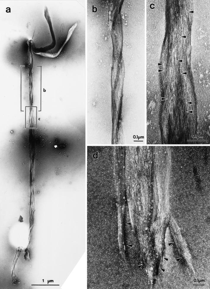
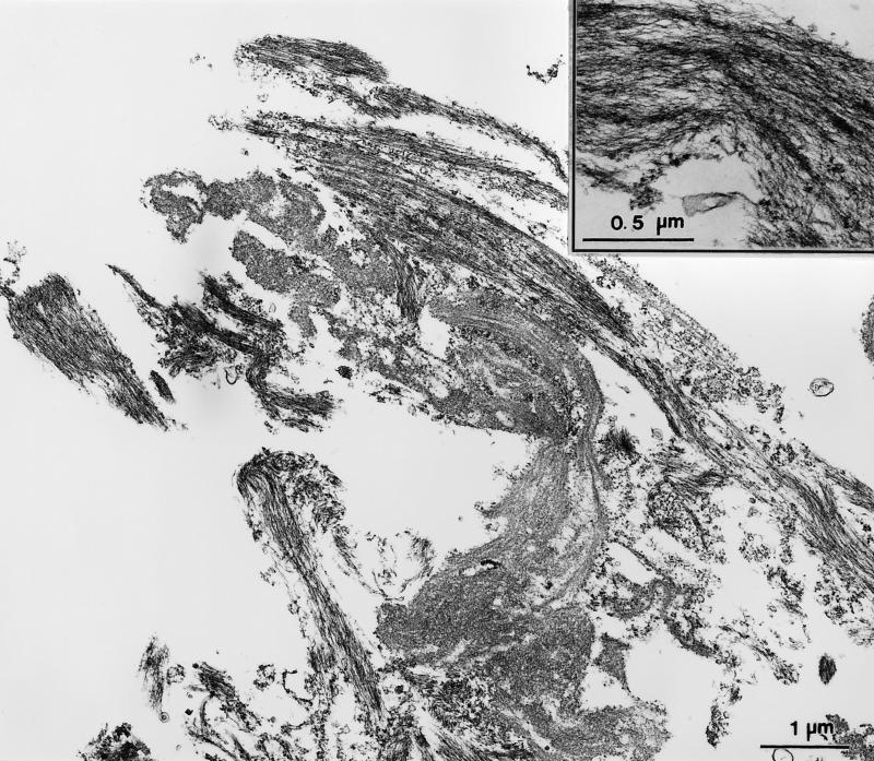
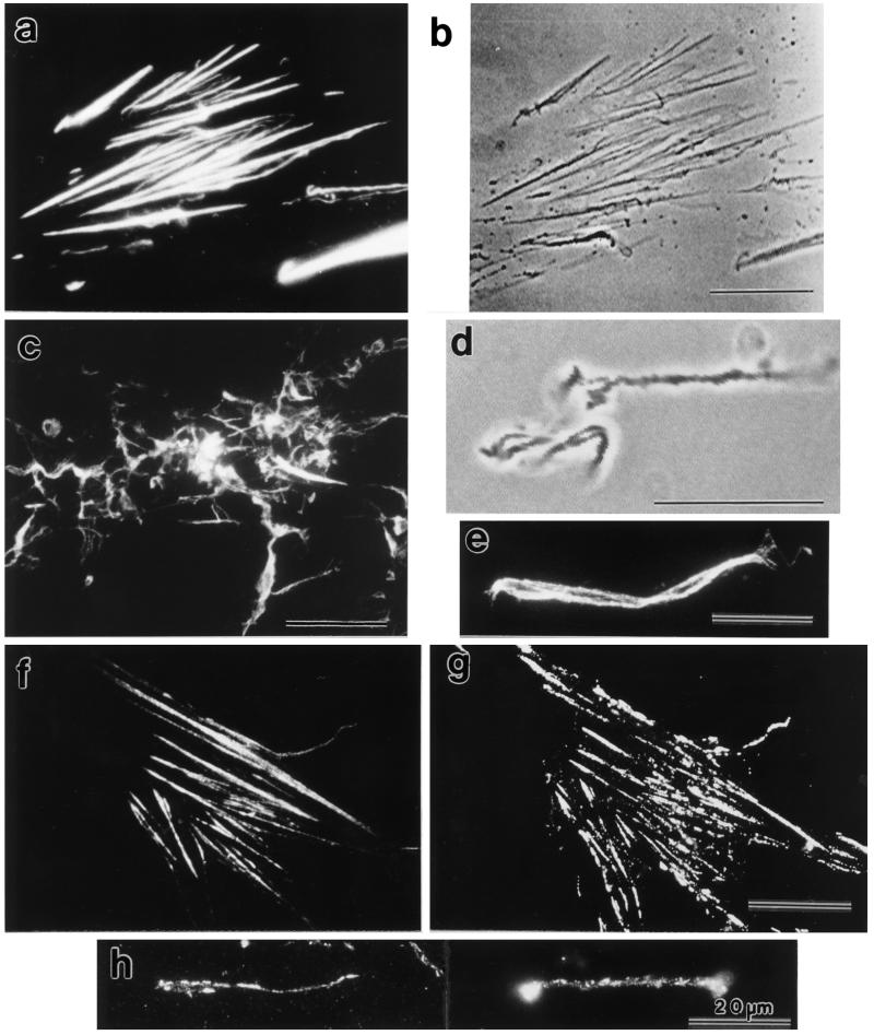
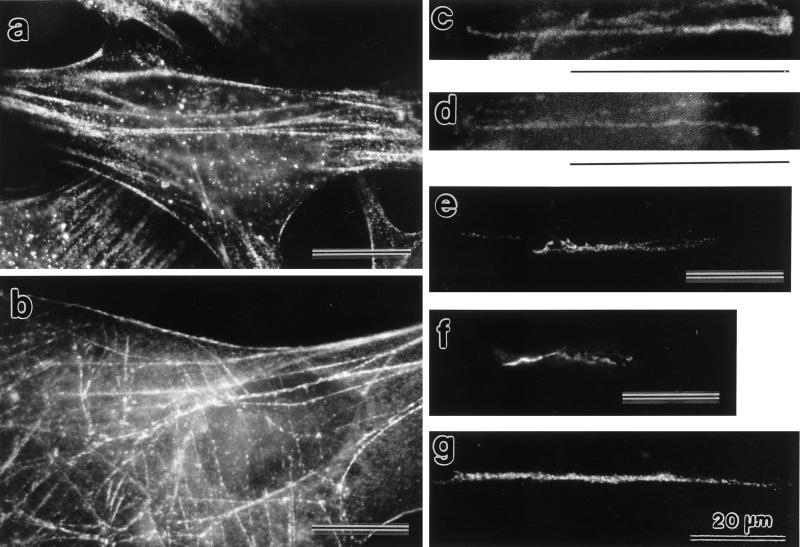
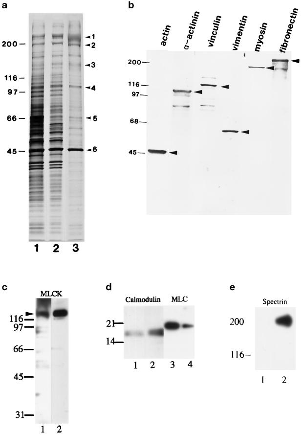
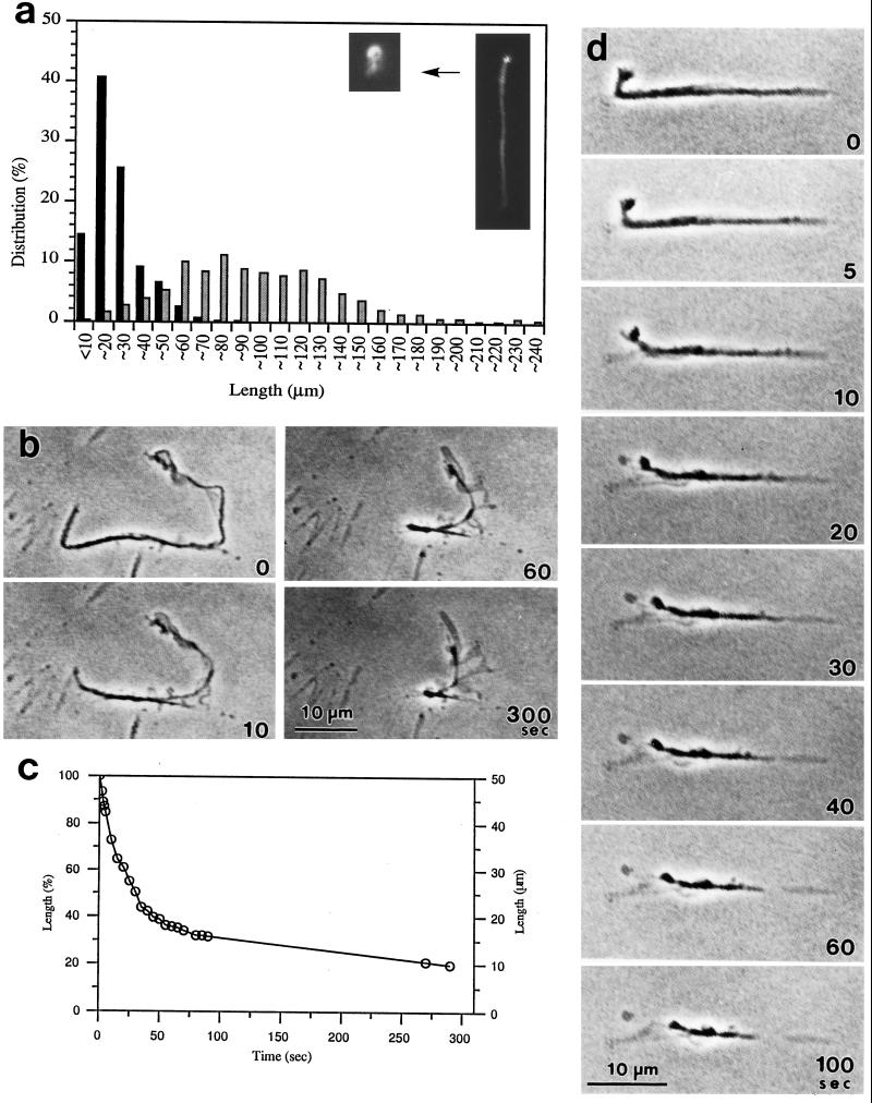
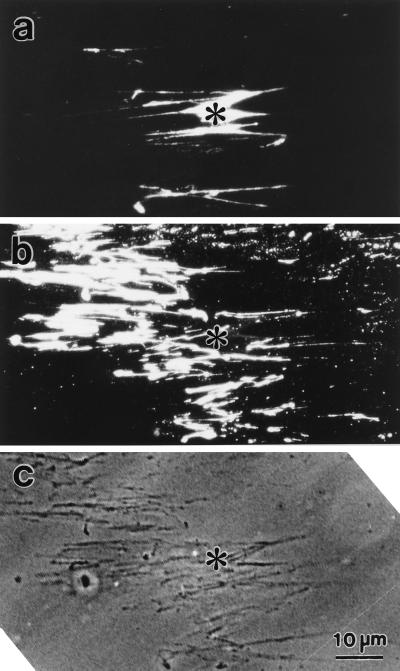
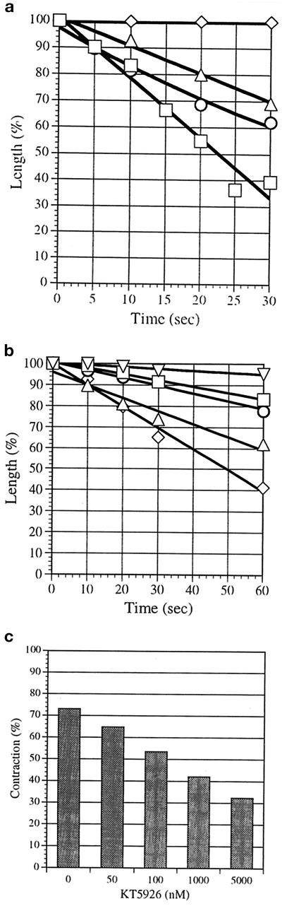
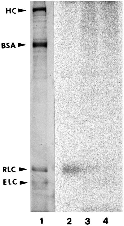
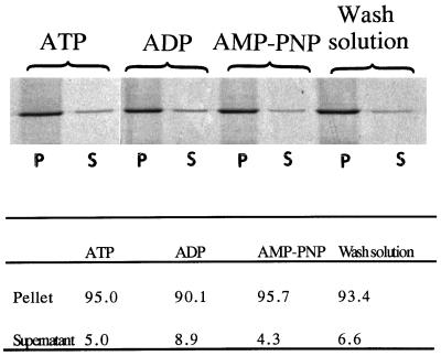
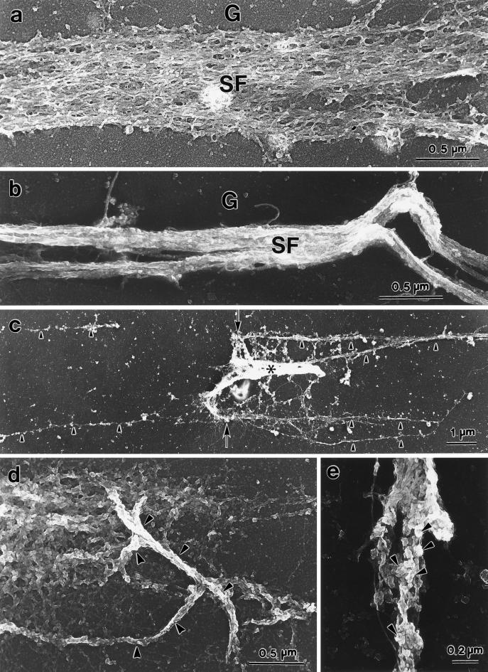
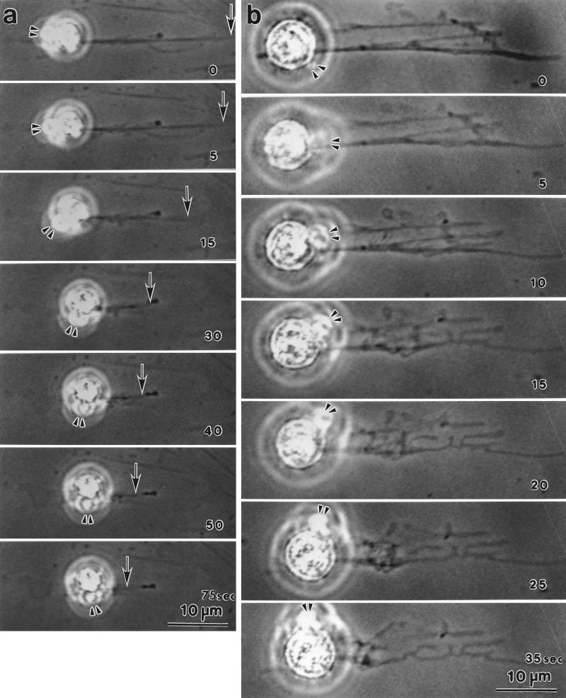
Stress fibers were isolated from cultured human foreskin fibroblasts and bovine endothelial cells, and their contraction was demonstrated in vitro. Cells in culture dishes were first treated with a low-ionic-strength extraction solution and then further extracted using detergents. With gentle washes by pipetting, the nucleus and the apical part of cells were removed. The material on the culture dish was scraped, and the freed material was forced through a hypodermic needle and fractionated by sucrose gradient centrifugation. Isolated, free-floating stress fibers stained brightly with fluorescently labeled phalloidin. When stained with anti-alpha-actinin or anti-myosin, isolated stress fibers showed banded staining patterns. By electron microscopy, they consisted of bundles of microfilaments, and electron-dense areas were associated with them in a semiperiodic manner. By negative staining, isolated stress fibers often exhibited gentle twisting of microfilament bundles. Focal adhesion-associated proteins were also detected in the isolated stress fiber by both immunocytochemical and biochemical means. In the presence of Mg-ATP, isolated stress fibers shortened, on the average, to 23% of the initial length. The maximum velocity of shortening was several micrometers per second. Polystyrene beads on shortening isolated stress fibers rotated, indicating spiral contraction of stress fibers. Myosin regulatory light chain phosphorylation was detected in contracting stress fibers, and a myosin light chain kinase inhibitor, KT5926, inhibited isolated stress fiber contraction. Our study demonstrates that stress fibers can be isolated with no apparent loss of morphological features and that they are truly contractile organelle.


Figures

Figure 1
Figure 1
Electron micrographs of negatively stained stress fibers isolated from bovine endothelial cells (a–c) or human foreskin fibroblasts (d). In a, one whole isolated stress fiber is shown. Two boxed areas in a are enlarged and shown in b and c. Isolated stress fibers consisted of bundles of thin microfilaments. Globular actin monomers in individual filaments are clearly seen (c, arrowheads). Note that the stress fiber in a shows gentle twisting (better shown in b). The ends of isolated stress fibers showed distinct specialization, such as enlargement (a) and finger-like projections (d, arrowheads).
Figure 2
Figure 2
Conventional transmission electron micrographs of stress fibers isolated from human foreskin fibroblasts. A low-power view shows bundles of filaments cut in both longitudinal and cross-sectional directions. The morphology of isolated stress fibers is similar to that of in vivo stress fibers. In a high-power view, semiperiodic electron-dense regions within a microfilament bundle are visible (inset). This structure is one of the morphological characteristics of stress fibers.
Figure 3
Figure 3
stress fibers with fluorescein-labeled phalloidin (f) and anti-vinculin (g) is shown. Fractionated stress fibers stained with anti-vinculin are shown (h). Note the dotty anti-vinculin staining pattern along the stress fibers (h). F-actin and vinculin distribution in isolated stress fibers. Attached stress fibers stained with fluorescein-labeled phalloidin (a) and a video-enhanced phase-contrast image of the same sample (b) are shown. Fractionated stress fibers stained with fluorescein-labeled phalloidin (c and e) and a video-enhanced phase-contrast image of a small tangle of isolated stress fibers in the same fraction as in c (d) are shown. Double staining of attached
Figure 4
Figure 4
Myosin, α-actinin, myosin light chain kinase, calmodulin, and vimentin distribution in isolated stress fibers. Paraformaldehyde-fixed cells stained with anti-myosin (a) or anti-α-actinin (b) show a dotty pattern along the stress fiber. Fractionated stress fibers stained with anti-myosin (c) or anti-α-actinin (d) are shown. Note that isolated stress fibers also show dotty staining patterns. Anti-myosin light chain kinase (e), anti-calmodulin (f), and anti-vimentin (g) staining were also detected in isolated stress fibers with some dotty patterns.
Figure 5
Figure 5
Gel electrophoresis and immunoblotting of isolated stress fibers. SDS-PAGE gels of a crude extract of fibroblasts (a, lane 1), a TEA-treated cell fraction (a, lane 2), and fractionated stress fibers (a, lane 3) are shown after silver staining. The identity of six major bands (1, fibronectin; 2, myosin; 3, vinculin; 4, α-actinin; 5, vimentin; and 6, actin) in the isolated stress fiber (a, lane 3, arrowheads) was determined by immunoblotting (b). Antibodies used in the immunoblotting analyses in b were anti-α-smooth muscle actin, anti-α-actinin, anti-vinculin, anti-vimentin, anti-myosin, and anti-fibronectin. The presence of myosin light chain kinase (c), calmodulin (d, lanes 1 and 2) and myosin regulatory light chain (d, lanes 3 and 4) was also detected by immunoblotting. Fractionated stress fibers (c, lane 1; d, lanes 1 and 3) and a crude extract of guinea pig aorta as a positive control (c, lane 2; d, lanes 2 and 4) were immunoblotted with anti-myosin light chain kinase (c), anti-calmodulin (d, lanes 1 and 2) and anti-myosin regulatory light chain (d, lanes 3 and 4). Fractionated stress fibers (e, lane 1) and a crude extract of fibroblasts (e, lane 2) were immunoblotted with anti-α-spectrin. Note that no immunoreactivity was detected in the stress fiber sample. Arrowheads in b and c indicate the position of the intact antigen bands.
Figure 6
Figure 6
Shortening of free-floating and attached stress fibers. Length distribution (percent) of free-floating isolated stress fibers before (shaded bars) and 10 min after (black bars) the addition of Mg-ATP were plotted (a). Length measurements were done using stress fibers fixed and stained with rhodamine-labeled phalloidin. The total number of isolated stress fibers counted for each type is 500. A typical example of free-floating isolated stress fiber either before or after shortening is shown in the inset (a). Isolated stress fibers still attached to glass surface were treated with Mg-ATP (b and d). The number in each frame indicates time in seconds after the addition of Mg-ATP solution. Note that the thickness of stress fibers decreases as they shorten. Note also some material left behind shortening stress fibers. The length change (expressed in percent of the initial length and also in actual length) of the stress fiber shown in b is plotted against time in c. The initial maximum speed of shortening was 2.4 μm/s, and the degree of shortening was 80% after 5 min.
Figure 7
Figure 7
Identification of the material left behind shortening stress fibers. Attached stress fibers were treated with Mg-ATP, fixed, and stained doubly with rhodamine-labeled phalloidin (a) and anti-fibronectin (b). A video-enhanced phase-contrast image of the same field of view is shown in c. Phalloidin staining shows contracted stress fibers in the center of the micrograph (a), but the fibrous anti-fibronectin pattern occupies a wide portion of the field (b). Note that the anti-fibronectin staining structures are detectable in the phase-contrast micrograph (c). Asterisks indicate the same position in the three micrographs.
Figure 8
Figure 8
The extent of stress fiber shortening depends on Mg-ATP concentration, and the effect of a myosin light chain kinase inhibitor, KT5926, inhibits stress fiber shortening. Isolated stress fibers still attached to glass surface were incubated with 0.1 mM (□), 0.05 mM (○), 0.01 mM (▵), or 0.005 mM (⋄) Mg-ATP solution, and their lengths were measured during shortening (a). These measurements were made from video images. Length is expressed in percent of the initial length. Mg-ATP at 0.1 mM induced >50% shortening within 30 s. Mg-ATP at 0.01 but not 0.005 mM was sufficient to induce shortening. Attached stress fibers were preincubated with no (⋄), 50 nM (▵), 100 nM (○), 1000 nM (□), or 5000 nM (▿) KT5926 for 30 min and then treated with 0.01 mM Mg-ATP. The length change measured from video recording is shown in b. The length is expressed as percent of the initial length. For each datum point, 20 stress fibers were measured. As the KT5926 concentration increased, stress fibers shortened more slowly. The extent of stress fiber shortening after 5 min of treatment with various concentrations of KT5926 is shown (c). Attached stress fibers were incubated with the indicated concentrations of KT5926 for 30 min and then with 0.01 mM Mg-ATP. After 5 min, the extent of shortening was measured. For each datum point, 20 stress fibers were measured. In this figure, the amount of shortening in percentage of the original length is shown.
Figure 9
Figure 9
Phosphorylation of the myosin regulatory light chain in the isolated stress fiber. Fractionated stress fibers were preincubated with 0 (lane 2), 100 (lane 3), or 5000 nM (lane 4) KT5926 for 30 min on ice and then incubated with Mg-[γ-32P]ATP. Purified chicken gizzard heavy meromyosin (HMM) mixed with BSA was used as a molecular marker (lane 1). Phosphorylation of the myosin regulatory light chain is inhibited by KT5926 in a concentration-dependent manner. HC, HMM heavy chain; RLC, myosin regulatory light chain; ELC, myosin essential light chain.
Figure 10
Figure 10
Absence of F-actin dissolution during shortening of isolated stress fibers. Fractionated stress fibers were treated with Mg-ATP, ADP, AMP-PNP, or wash solution. Centrifuged pellets (P) and supernatants (S) were electrophoresed and stained with SYPRO-orange. Actin bands are shown here. Relative amounts (in percent) of actin in the supernatants and the pellets were calculated from intensities of actin bands (table). Note that no dissolution of F-actin is caused by shortening.
Figure 11
Figure 11
Replica electron microscopy of isolated stress fibers before and after Mg-ATP treatment. A loose bundle of microfilaments of an isolated stress fiber before Mg-ATP addition is shown (a). Individual microfilaments can be observed. After Mg-ATP addition, microfilaments in a bundle were no longer loose but were tightly packed (b). (c) Low-power view of an attached stress fiber at the end of shortening. The stress fiber (asterisk) is short and dense. It appears to have “contracted” from the left side of the micrograph and flipped over at the end about an imaginary line indicated by the two arrows. Note the extracellular matrix materials on the glass surface (c, arrowheads). Tangled stress fibers (d, arrowheads) and stress fibers exhibiting small, knot-like structures on the surface (e, arrowheads) are illustrated. G, glass surface; SF, stress fiber.
Figure 12
Figure 12
Rotation of microbeads during contraction of stress fibers. Microbeads were attached to isolated stress fibers to analyze the motion of shortening stress fibers. These video-enhanced phase-contrast images are from time-lapse recording using a disk recorder. The number at the right bottom corner in each plate is time in seconds after Mg-ATP addition. Microbeads attached to the vicinity of one end of isolated stress fibers were selected for observation. Two examples are shown (a and b). The double arrowheads (a and b) indicate the same position on the bead clusters. The number in each panel indicates time in seconds after Mg-ATP addition.

References

    1. Avnur Z, Geiger B. Substrate attached membranes of cultured cells. Isolation and characterization of ventral membranes and the associated cytoskeleton. J Mol Biol. 1981;153:361–379. - PubMed
    1. Avnur Z, Small V, Geiger B. Actin-independent association of vinculin with the cytoplasmic aspect of the plasma membrane in cell-contact areas. J Cell Biol. 1983;96:1622–1630. - PMC - PubMed
    1. Badley RA, Lloyd CW, Woods A, Carruthers L, Allcock C. Mechanisms of cellular adhesion. Exp Cell Res. 1978;117:231–244. - PubMed
    1. Ball EH, Freitag C, Gurofsky S. Vinculin interaction with permeabilized cells: disruption and reconstruction of a binding site. J Cell Biol. 1986;103:641–648. - PMC - PubMed
    1. Brands R, Feltkamp CA. Wet cleaving of cell: a method to introduce macromolecules into the cytoplasm. Exp Cell Res. 1988;176:309–318. - PubMed

Publication types

MeSH terms