

Characterisation of coronary atherosclerotic morphology by spectral analysis of radiofrequency signal: in vitro intravascular ultrasound study with histological and radiological validation
- PMID: 9659192
- PMCID: PMC1728682
- DOI: 10.1136/hrt.79.5.459
Characterisation of coronary atherosclerotic morphology by spectral analysis of radiofrequency signal: in vitro intravascular ultrasound study with histological and radiological validation
Abstract
Objective: To determine whether spectral analysis of unprocessed radiofrequency (RF) signal offers advantages over standard videodensitometric analysis in identifying the morphology of coronary atherosclerotic plaques.
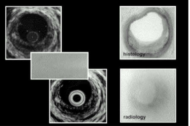
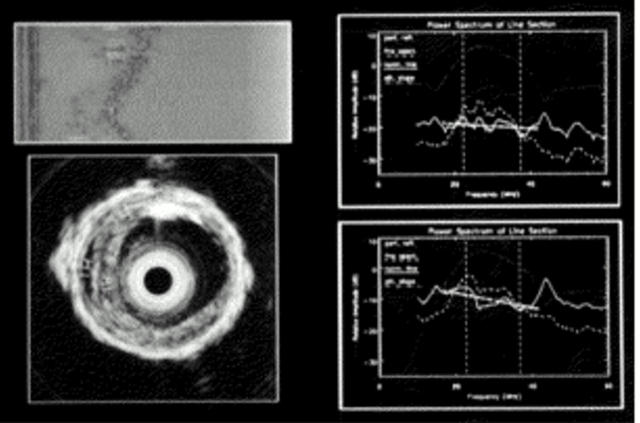
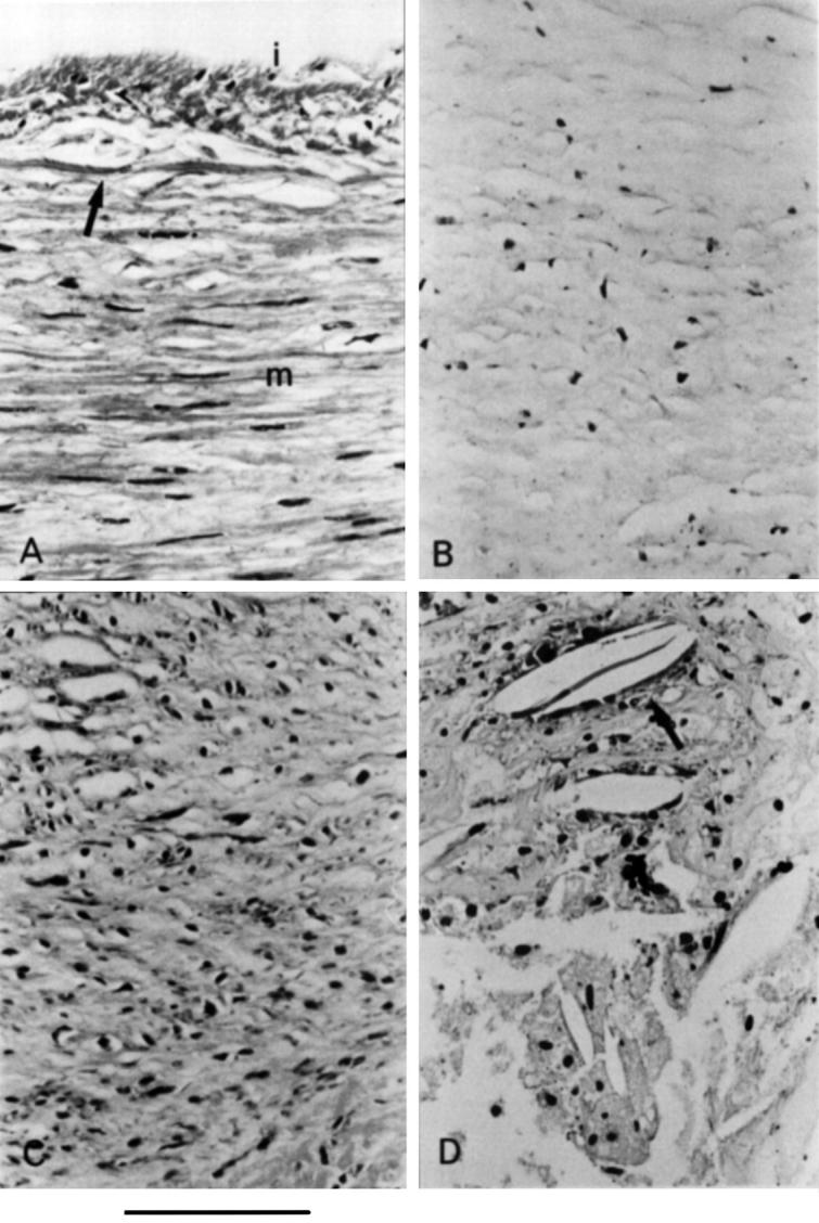
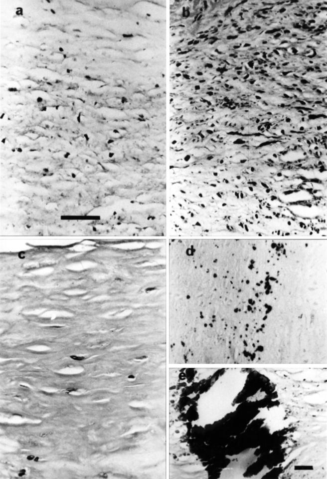
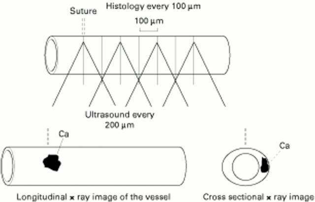
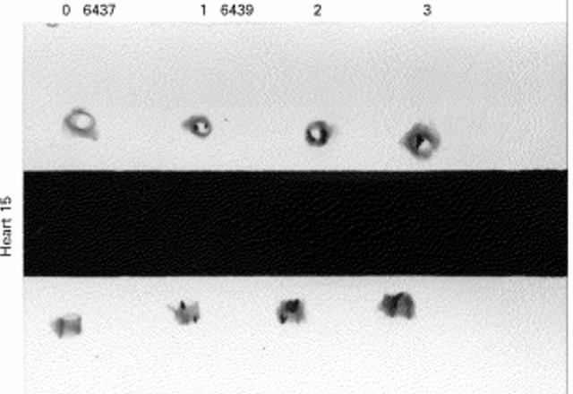
Methods: 97 regions of interest (ROI) were imaged at 30 MHz from postmortem, pressure perfused (80 mm Hg) coronary arteries in saline baths. RF data were digitised at 250 MHz. Two different sizes of ROI were identified from scan converted images, and relative amplitudes of different frequency components were analysed from raw data. Normalised spectra was used to calculate spectral slope (dB/MHz), y-axis intercept (dB), mean power (dB), and maximum power (dB) over a given bandwidth (17-42 MHz). RF images were constructed and compared with comparative histology derived from microscopy and radiological techniques in three dimensions.
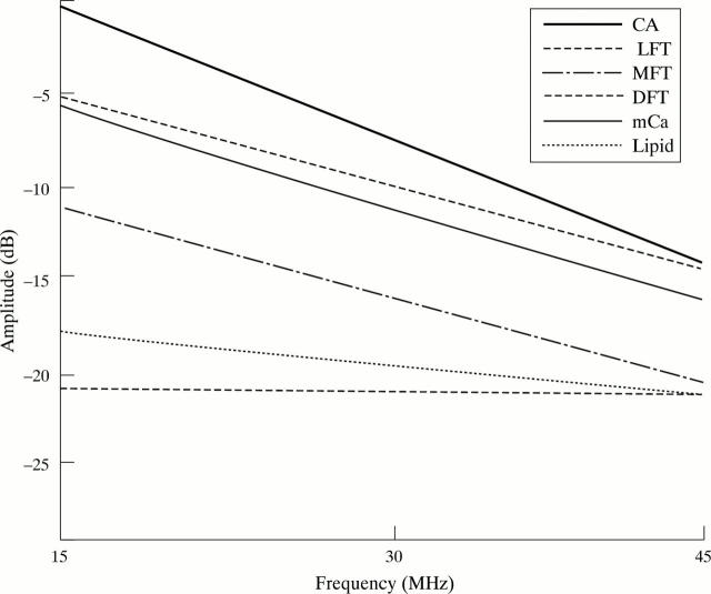
Results: Mean power was similar from dense fibrotic tissue and heavy calcium, but spectral slope was steeper in heavy calcium (-0.45 (0.1)) than in dense fibrotic tissue (-0.31 (0.1)), and maximum power was higher for heavy calcium (-7.7 (2.0)) than for dense fibrotic tissue (-10.2 (3.9)). Maximum power was significantly higher in heavy calcium (-7.7 (2.0) dB) and dense fibrotic tissue (-10.2 (3.9) dB) than in microcalcification (-13.9 (3.8) dB). Y-axis intercept was higher in microcalcification (-5.8 (1.1) dB) than in moderately fibrotic tissue (-11.9 (2.0) dB). Moderate and dense fibrotic tissue were discriminated with mean power: moderate -20.2 (1.1) dB, dense -14.7 (3.7) dB; and y-axis intercept: moderate -11.9 (2.0) dB, dense -5.5 (5.4) dB. Different densities of fibrosis, loose, moderate, and dense, were discriminated with both y-axis intercept, spectral slope, and mean power. Lipid could be differentiated from other types of plaque tissue on the basis of spectral slope, lipid -0.17 (0.08). Also y-axis intercept from lipid (-17.6 (3.9)) differed significantly from moderately fibrotic tissue, dense fibrotic tissue, microcalcification, and heavy calcium. No significant differences in any of the measured parameters were seen between the results obtained from small and large ROIs.
Conclusion: Frequency based spectral analysis of unprocessed ultrasound signal may lead to accurate identification of atherosclerotic plaque morphology.
Figures
Similar articles
-
Characterisation of atherosclerotic plaque by spectral analysis of intravascular ultrasound: an in vitro methodology.Ultrasound Med Biol. 1997;23(2):191-203. doi: 10.1016/s0301-5629(96)00199-8. Ultrasound Med Biol. 1997. PMID: 9140178
-
Classification of arterial plaque by spectral analysis of in vitro radio frequency intravascular ultrasound data.Ultrasound Med Biol. 2000 Jan;26(1):73-80. doi: 10.1016/s0301-5629(99)00112-x. Ultrasound Med Biol. 2000. PMID: 10687795
-
Assessing spectral algorithms to predict atherosclerotic plaque composition with normalized and raw intravascular ultrasound data.Ultrasound Med Biol. 2001 Oct;27(10):1319-31. doi: 10.1016/s0301-5629(01)00436-7. Ultrasound Med Biol. 2001. PMID: 11731045
-
[Intravascular ultrasound investigation with virtual hystology in the study of coronary artery lesions].Kardiologiia. 2009;49(12):58-61. Kardiologiia. 2009. PMID: 20038284 Review. Russian.
-
Discrimination of early/intermediate and advanced/complicated coronary plaque types by radiofrequency intravascular ultrasound analysis.Am J Cardiol. 2002 Jul 1;90(1):19-23. doi: 10.1016/s0002-9149(02)02379-2. Am J Cardiol. 2002. PMID: 12088773
Cited by
-
Rationale and methods of the integrated biomarker and imaging study (IBIS): combining invasive and non-invasive imaging with biomarkers to detect subclinical atherosclerosis and assess coronary lesion biology.Int J Cardiovasc Imaging. 2005 Aug;21(4):425-41. doi: 10.1007/s10554-004-7986-y. Int J Cardiovasc Imaging. 2005. PMID: 16047125
-
Virtual histology.Heart. 2007 Aug;93(8):977-82. doi: 10.1136/hrt.2007.116384. Epub 2007 May 13. Heart. 2007. PMID: 17502324 Free PMC article.
-
Spectral analysis of ultrasound radiofrequency backscatter for the identification of epicardial adipose tissue.J Med Imaging (Bellingham). 2022 Jan;9(1):017001. doi: 10.1117/1.JMI.9.1.017001. Epub 2022 Jan 6. J Med Imaging (Bellingham). 2022. PMID: 35005059 Free PMC article.
-
Recent trends in coronary intravascular ultrasound: tracking atherosclerosis, pursuit of vulnerable plaques, and beyond.J Nucl Cardiol. 2006 Jan-Feb;13(1):91-6. doi: 10.1016/j.nuclcard.2005.11.001. J Nucl Cardiol. 2006. PMID: 16464722 Review.
-
Coronary plaque classification using intravascular ultrasound -- radiofrequency analysis in a patient with severe coronary vasospasm.Clin Res Cardiol. 2007 Jul;96(7):514-8. doi: 10.1007/s00392-007-0520-1. Epub 2007 Apr 26. Clin Res Cardiol. 2007. PMID: 17453131 No abstract available.
References
Publication types
MeSH terms
Substances
LinkOut - more resources
Full Text Sources
Other Literature Sources
Medical
Miscellaneous