. 1998 Jul 13;142(1):1-11.
doi: 10.1083/jcb.142.1.1.

The human homologue of Bub3 is required for kinetochore localization of Bub1 and a Mad3/Bub1-related protein kinase

Affiliations

The human homologue of Bub3 is required for kinetochore localization of Bub1 and a Mad3/Bub1-related protein kinase

S S Taylor et al. J Cell Biol. .

Abstract

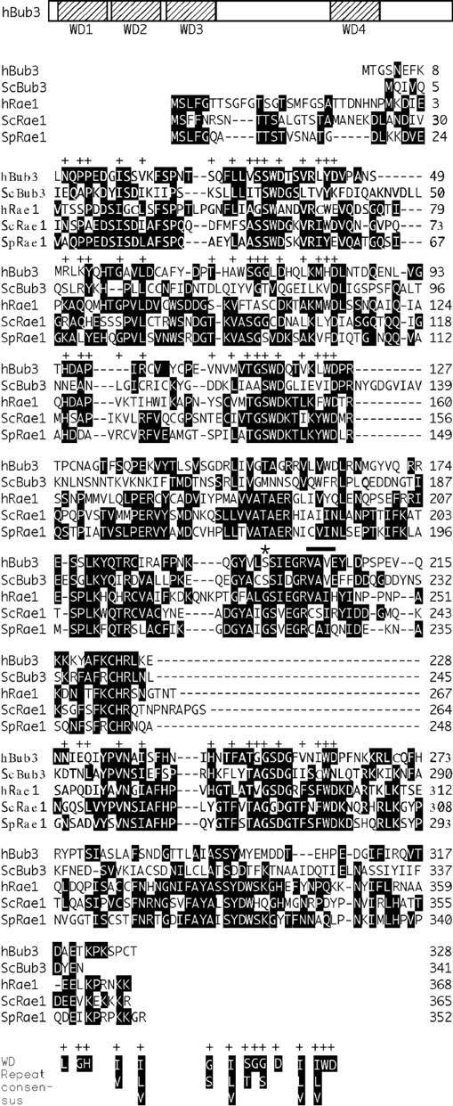
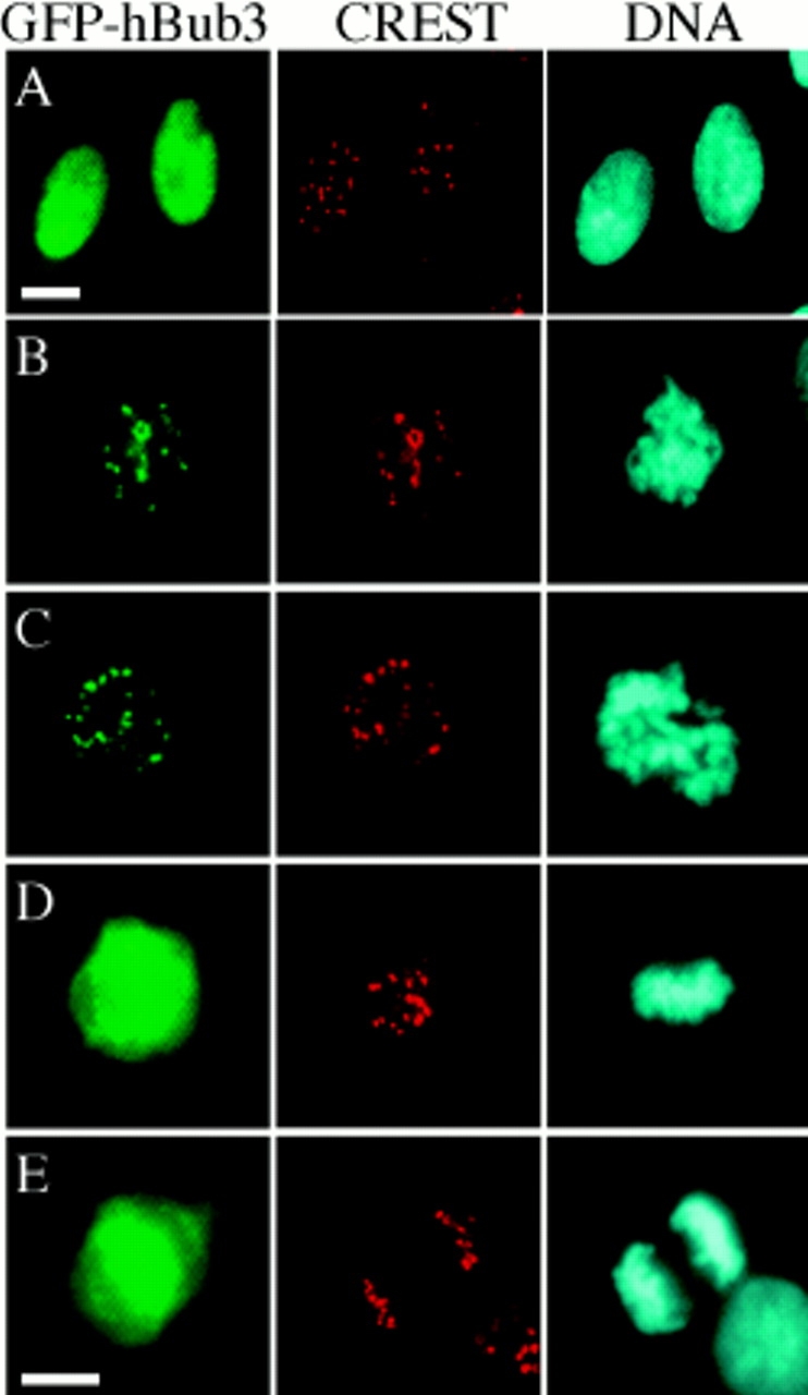
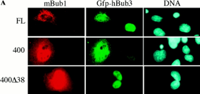
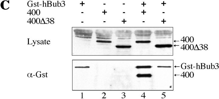
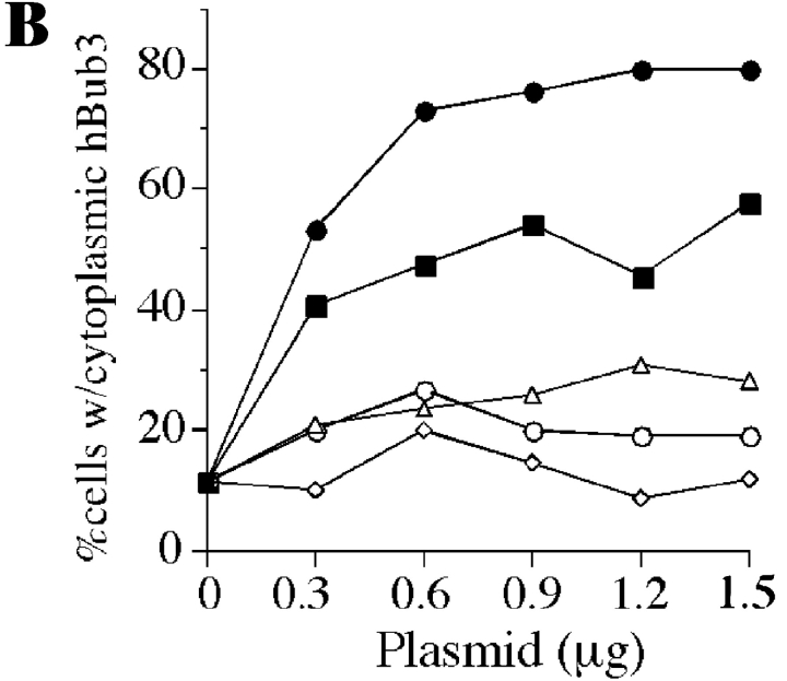
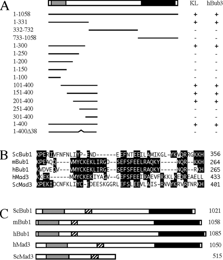
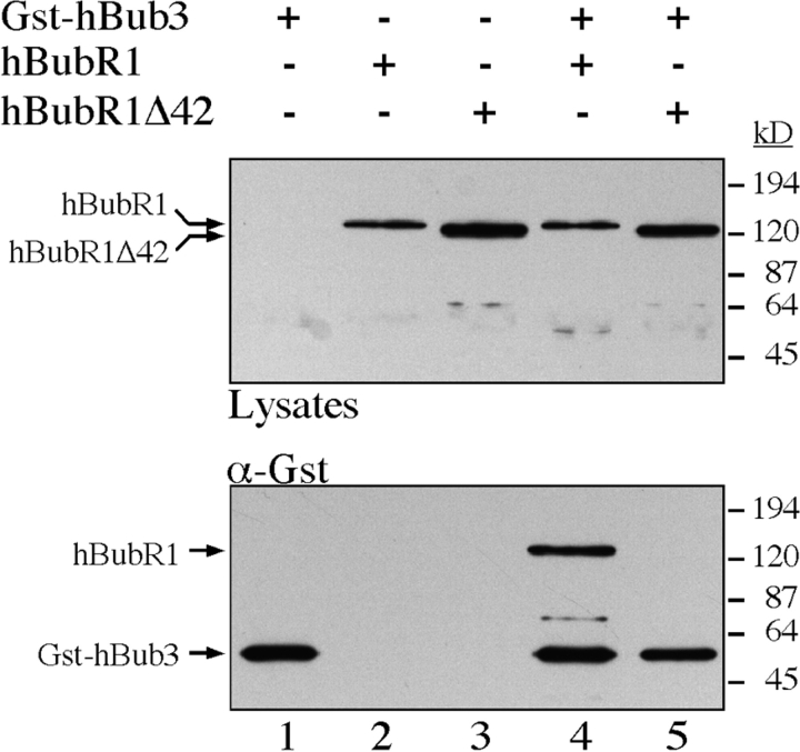
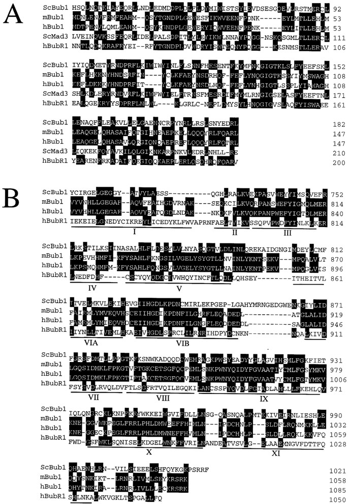
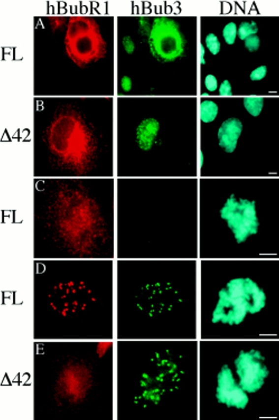
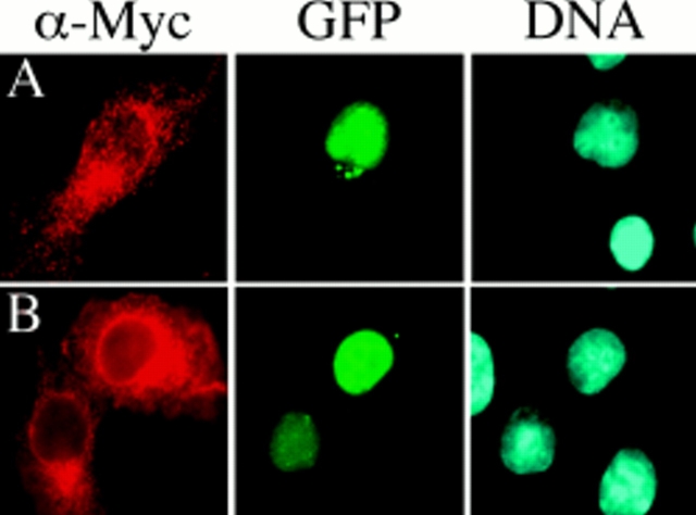
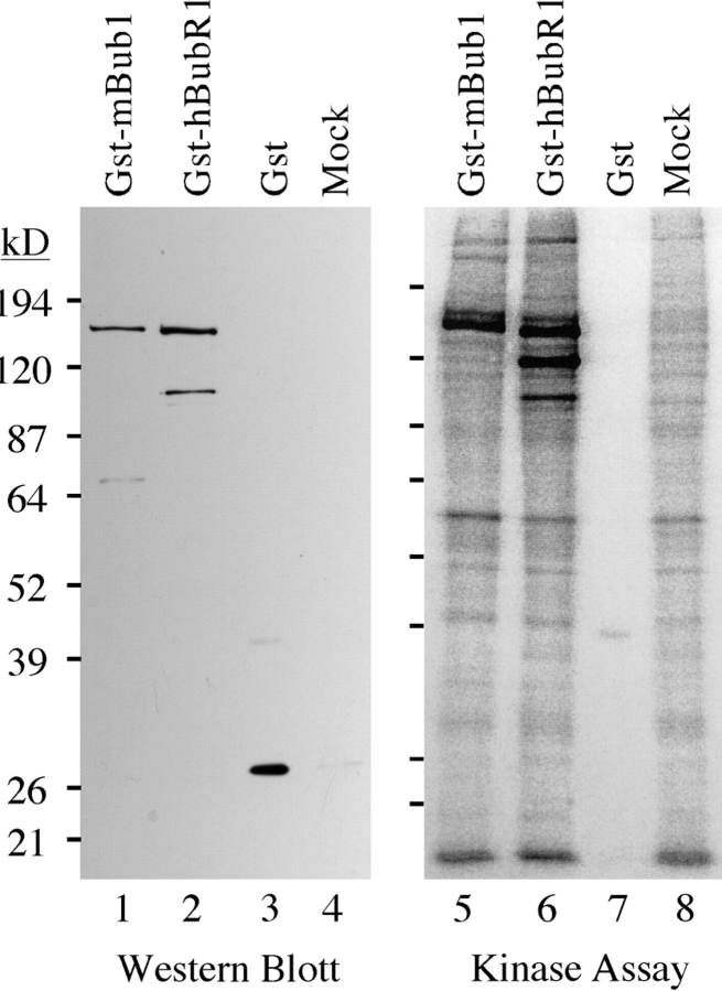
A feedback control mechanism, or cell cycle checkpoint, delays the onset of anaphase until all the chromosomes are correctly aligned on the mitotic spindle. Previously, we showed that the murine homologue of Bub1 is not only required for checkpoint response to spindle damage, but also restrains progression through a normal mitosis (Taylor, S.S., and F. McKeon. 1997. Cell. 89:727-735). Here, we describe the identification of a human homologue of Bub3, a 37-kD protein with four WD repeats. Like Bub1, Bub3 localizes to kinetochores before chromosome alignment. In addition, Bub3 and Bub1 interact in mammalian cells. Deletion mapping was used to identify the domain of Bub1 required for binding Bub3. Significantly, this same domain is required for kinetochore localization of Bub1, suggesting that the role of Bub3 is to localize Bub1 to the kinetochore, thereby activating the checkpoint in response to unattached kinetochores. The identification of a human Mad3/Bub1-related protein kinase, hBubR1, which can also bind Bub3 in mammalian cells, is described. Ectopically expressed hBubR1 also localizes to kinetochores during prometaphase, but only when hBub3 is overexpressed. We discuss the implications of the common interaction between Bub1 and hBubR1 with hBub3 for checkpoint control.


Figures

Figure 1
Figure 1
Sequence comparison of Bub3 homologues. Schematic representation of hBub3 showing the location of the four WD repeats and an alignment of the Bub3 and Rae1 homologues from human (h), S. cerevisiae (Sc) and S. pombe (Sp). The sequences are arranged such that the WD repeats are aligned with each other and with the WD repeat consensus (Dalrymple et al., 1989). The positions of WD repeat consensus residues are indicated with a +. Note the conserved glycine in the Rae1 homologues, marked with an *, that is not conserved in the Bub3 homologues. The rae1-1 loss of function allele in S. pombe results from a substitution of this glycine with a glutamic acid (Brown et al., 1995). The four amino acids deleted to generate the hBub3ΔVAVE mutant are identified with a solid bar.
Figure 2
Figure 2
hBub3 localizes to kinetochores during prometaphase. BHK cells were transfected with a GFP–hBub3 expression construct, fixed, and then stained with a CREST antiserum to identify the kinetochores (red) and Hoechst dye to visualize the chromatin (blue). GFP fluorescence is shown in green. (A) Transfected cells showing that hBub3 is diffusely nuclear during interphase. (B) Transfected prophase and (C) prometaphase cells showing colocalization of GFP–hBub3 with kinetochores. (D) Transfected metaphase and (E) anaphase cells showing GFP–hBub3 diffusely distributed throughout the cell. Bars, 10 μm. B–D are to the same scale as E.
Figure 3
Figure 3
A domain in the NH2-terminus of mBub1 binds hBub3. (A) BHK cells were cotransfected with GFP–hBub3 and Myc-tagged mBub1 expression constructs. After fixation, cells were stained with the 9E10 monoclonal antibody (red) to localize mBub1 and Hoechst dye to visualize the chromatin (blue). GFP fluorescence is shown in green. Coexpression of both full-length mBub1 (top row) and the NH2-terminal domain (NH2-mBub1[400], middle row) results in a cytoplasmic sequestration of GFP–hBub3. In contrast, NH2-mBub1(400Δ38) does not sequester GFP–hBub3 in the cytoplasm (bottom row). (B) BHK cells were cotransfected with 0.5 μg of GFP–hBub3 and increasing amounts of full-length mBub1 (solid squares), the NH2-terminal domain (amino acids 1–331) (solid circles), the central domain (amino acids 332–731) (open triangles), the COOH-terminal kinase domain (amino acids 732–1,058) (open circles) or a control plasmid (open diamonds). The total amount of DNA used in each transfection was maintained at 2.0 μg by addition of carrier DNA where necessary. The percentage of cells with cytoplasmic GFP–hBub3 was scored and plotted against the amount of cotransfected mBub1. Only coexpression of full-length mBub1 and the NH2-terminal domain resulted in a dose-dependent, cytoplasmic sequestration of GFP–hBub3. (C) BHK cells were transfected with GST–hBub3 (lanes 1, 4, and 5) and/or Myc-tagged NH2-mBub1(400) (lanes 2 and 4) and/or Myc-tagged NH2-mBub1(400Δ38) (lanes 3 and 5). Proteins from cell lysates (top) and purified GST–hBub3 complexes (bottom) were resolved by SDS-PAGE, transferred to a membrane, and then probed with anti-GST and anti-Myc antibodies. In the absence of GST–hBub3, neither NH2-mBub1(400) nor NH2-mBub1(400Δ38) purify with the glutathione beads (lanes 2 and 3). In the presence of GST–hBub3, NH2-mBub1(400) does purify with the glutathione beads (lane 4) but NH2-mBub1(400Δ38) does not (lane 5). Note that GST–hBub3 cannot be identified in the cell lysates due to several comigrating background bands.
Figure 3
Figure 3
A domain in the NH2-terminus of mBub1 binds hBub3. (A) BHK cells were cotransfected with GFP–hBub3 and Myc-tagged mBub1 expression constructs. After fixation, cells were stained with the 9E10 monoclonal antibody (red) to localize mBub1 and Hoechst dye to visualize the chromatin (blue). GFP fluorescence is shown in green. Coexpression of both full-length mBub1 (top row) and the NH2-terminal domain (NH2-mBub1[400], middle row) results in a cytoplasmic sequestration of GFP–hBub3. In contrast, NH2-mBub1(400Δ38) does not sequester GFP–hBub3 in the cytoplasm (bottom row). (B) BHK cells were cotransfected with 0.5 μg of GFP–hBub3 and increasing amounts of full-length mBub1 (solid squares), the NH2-terminal domain (amino acids 1–331) (solid circles), the central domain (amino acids 332–731) (open triangles), the COOH-terminal kinase domain (amino acids 732–1,058) (open circles) or a control plasmid (open diamonds). The total amount of DNA used in each transfection was maintained at 2.0 μg by addition of carrier DNA where necessary. The percentage of cells with cytoplasmic GFP–hBub3 was scored and plotted against the amount of cotransfected mBub1. Only coexpression of full-length mBub1 and the NH2-terminal domain resulted in a dose-dependent, cytoplasmic sequestration of GFP–hBub3. (C) BHK cells were transfected with GST–hBub3 (lanes 1, 4, and 5) and/or Myc-tagged NH2-mBub1(400) (lanes 2 and 4) and/or Myc-tagged NH2-mBub1(400Δ38) (lanes 3 and 5). Proteins from cell lysates (top) and purified GST–hBub3 complexes (bottom) were resolved by SDS-PAGE, transferred to a membrane, and then probed with anti-GST and anti-Myc antibodies. In the absence of GST–hBub3, neither NH2-mBub1(400) nor NH2-mBub1(400Δ38) purify with the glutathione beads (lanes 2 and 3). In the presence of GST–hBub3, NH2-mBub1(400) does purify with the glutathione beads (lane 4) but NH2-mBub1(400Δ38) does not (lane 5). Note that GST–hBub3 cannot be identified in the cell lysates due to several comigrating background bands.
Figure 4
Figure 4
The hBub3-binding domain and kinetochore localization domain of mBub1 overlap. (A) A schematic representation of mBub1 and the Myc-tagged deletion mutants tested for kinetochore localization and GFP–hBub3 interaction after transient transfection of BHK cells. The lightly shaded box represents the NH2-terminal homology domain defined previously (Taylor and McKeon, 1997), whereas the darkly shaded box represents the COOH-terminal kinase domain. A + under KL indicates that kinetochore localization of the Myc epitope was observed during prometaphase. A + under hBub3 indicates that cytoplasmic sequestration of GFP–hBub3 was observed in interphase cells. The numbers on the right correspond to the amino acids expressed by each construct. This analysis shows that amino acids 201–300 are required for both kinetochore localization and hBub3 binding. (B) A sequence alignment of the Bub1 and Mad3-related proteins within the Bub3-binding domain. Deletion of this domain from mBub1 and hBubR1 abolishes hBub3 binding (refer to Figs. 3 and 7). (C) Schematic representations of the Bub1 and Mad3-related proteins showing the relative positions of the NH2-terminal homology domains (lightly shaded boxes), the kinase domains (darkly shaded boxes), and the Bub3-binding domains (hatched boxes).
Figure 7
Figure 7
The Bub3 binding domain of mBub1 and hBubR1 is conserved. BHK cells were transfected with GST–hBub3 (lanes 1, 4, and 5) and/or Myc-tagged hBubR1 (lanes 2 and 4), and/or Myc-tagged hBubR1Δ42 (lanes 3 and 5). Proteins from cell lysates (top) and purified GST– hBub3 complexes (bottom) were resolved by SDS-PAGE, transferred to a membrane, and then probed with anti-GST and anti-Myc antibodies. In the absence of GST–hBub3, neither hBubR1 nor hBubR1Δ42 purify with the glutathione beads (lanes 2 and 3). In the presence of GST–hBub3, hBubR1 purifies with the glutathione beads (lane 4) but hBubR1Δ42 does not (lane 5). Note that GST-hBub3 cannot be identified in the cell lysates.
Figure 5
Figure 5
Sequence comparison of Bub1 and Mad3-related proteins. (A) Sequence alignment of the NH2-terminal domains of the Bub1 and Mad3-related proteins from S. cerevisiae (Sc), mouse (m), and human (h). (B) Sequence alignment of the COOH-terminal kinase domains of the Bub1-related proteins. The 12 subdomains conserved among protein kinases (Hanks and Hunter, 1995) are underlined.
Figure 6
Figure 6
hBubR1 localizes to kinetochores when hBub3 is coexpressed. BHK cells were transiently transfected with either a full-length Myc-tagged hBubR1 expression construct (FL) or an hBubR1 mutant lacking the domain shown in Fig. 4 B (Δ42), fixed, and then stained with the 9E10 monoclonal antibody to determine the subcellular localization of hBubR1 (red). Cells were also stained with Hoechst to visualize the chromatin (blue). In A, B, D, and E, cells were cotransfected with GFP–hBub3 (green). (A) Transfected cells showing that ectopically expressed hBubR1 is predominantly cytoplasmic during interphase. In addition, coexpression of hBubR1 results in cytoplasmic sequestration of GFP–hBub3. (B) Expression of hBubR1Δ42 does not result in cytoplasmic sequestration of GFP–hBub3. (C) Transfected prometaphase cell showing ectopically expressed hBubR1 diffusely localized throughout the cell. (D) A prometaphase cell showing localization of hBubR1 at kinetochores when GFP– hBub3 is coexpressed. (E) Two transfected prometaphase cells showing diffuse localization of hBubR1Δ42 despite coexpression of GFP–hBub3. Bars, 5 μm.
Figure 8
Figure 8
hBub3ΔVAVE does not bind mBub1 or hBubR1. BHK cells were cotransfected with GFP–hBub3ΔVAVE and either (A) Myc-tagged mBub1 or (B) Myc-tagged hBubR1. After transfection, the cells were fixed and then stained with the 9E10 monoclonal antibody to determine the subcellular localization of the Myc-tagged fusion proteins (red). The cells were also stained with Hoechst to visualize the chromatin (blue). GFP fluorescence is shown in green. Despite coexpression of mBub1 and hBubR1, hBub3ΔVAVE remains in the nucleus, suggesting that this hBub3 mutant does not interact with mBub1 or hBubR1.
Figure 9
Figure 9
In vitro phosphorylation of mBub1 and hBubR1. BHK cells were transiently transfected with GST–mBub1, GST–hBubR1, and GST alone or mock-transfected as indicated. After transfection, cell lysates were prepared and incubated with glutathione Sepharose beads. Bound proteins were then analyzed by Western blotting to normalize the amounts of GST-fusion proteins used in the subsequent kinase assay. To assay for associated kinase activity, the beads were incubated with 32[P] γ-ATP. Bound proteins were then resolved by SDS-PAGE and analyzed by autoradiography. Lanes 5 and 6 show that there are several phosphorylated proteins associated with the beads incubated in lysates from cells expressing either GST–mBub1 or GST–hBubR1. The Western blot shows that the major phosphorylated proteins correspond to GST–mBub1 (lane 1) and GST– hBubR1 (lane 2), suggesting that the observed kinase activity is autophosphorylation. The major phosphorylated proteins observed in lanes 5 and 6 are not associated with beads incubated in cell lysates expressing GST alone (lane 7) or from mock-transfected cells (lane 8).

References

    1. Adams MD, Kerlavage AR, Fleischmann RD, Fuldner RA, Bult CJ, Lee NH, Kirkness EF, Weinstock KG, Gocayne JD, White O, et al. Initial assessment of human gene diversity and expression patterns based upon 83 million nucleotides of cDNA sequence. Nature. 1995;377(Suppl.):3–174. - PubMed
    1. Altschul SF, Gish W, Miller W, Myers EW, Lipman DJ. Basic local alignment search tool. J Mol Biol. 1990;215:403–410. - PubMed
    1. Bharathi A, Ghosh A, Whalen WA, Yoon JH, Pu R, Dasso M, Dhar R. The human RAE1 gene is a functional homologue of Schizosaccharomyces pombe rae1gene involved in nuclear export of Poly(A)+ RNA. Gene. 1997;198:251–258. - PubMed
    1. Brown JA, Bharathi A, Ghosh A, Whalen W, Fitzgerald E, Dhar R. A mutation in the Schizosaccharomyces pombe rae1gene causes defects in poly(A)+ RNA export and in the cytoskeleton. J Biol Chem. 1995;270:7411–7419. - PubMed
    1. Cahill DP, Lengauer C, Yu J, Riggins GJ, Willson JK, Markowitz SD, Kinzler KW, Vogelstein B. Mutations of mitotic checkpoint genes in human cancers. Nature. 1998;392:300–303. - PubMed

Publication types

MeSH terms

Associated data