. 1998 Jul 13;142(1):117-27.
doi: 10.1083/jcb.142.1.117.

Junctional adhesion molecule, a novel member of the immunoglobulin superfamily that distributes at intercellular junctions and modulates monocyte transmigration

Affiliations

Junctional adhesion molecule, a novel member of the immunoglobulin superfamily that distributes at intercellular junctions and modulates monocyte transmigration

I Martìn-Padura et al. J Cell Biol. .

Abstract

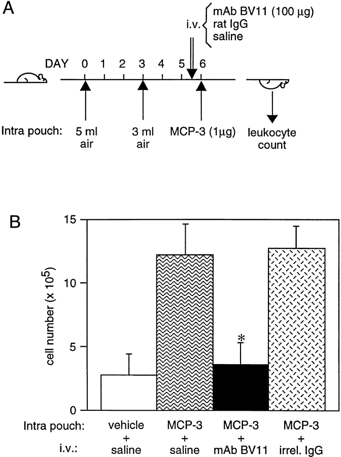
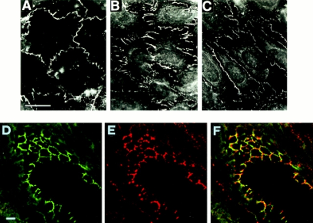
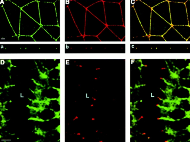
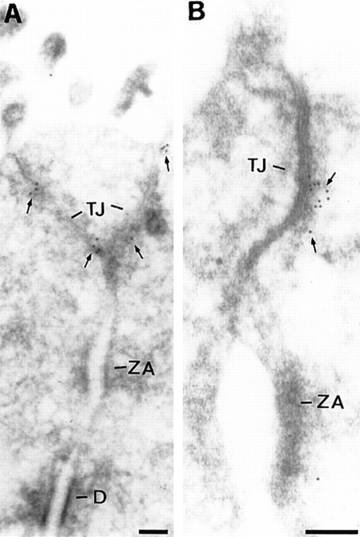
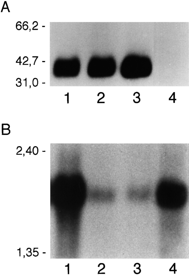
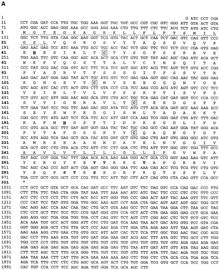
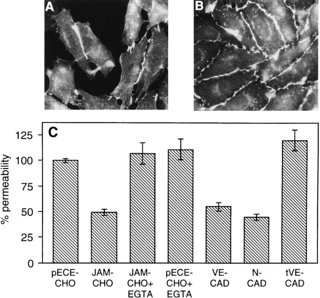
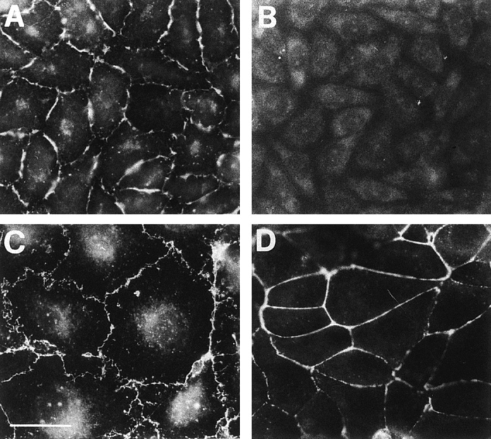
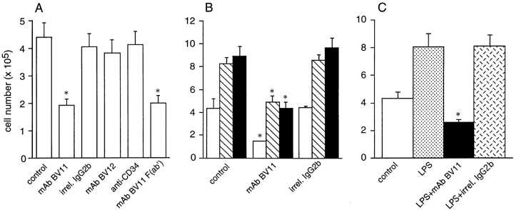
Tight junctions are the most apical components of endothelial and epithelial intercellular cleft. In the endothelium these structures play an important role in the control of paracellular permeability to circulating cells and solutes. The only known integral membrane protein localized at sites of membrane-membrane interaction of tight junctions is occludin, which is linked inside the cells to a complex network of cytoskeletal and signaling proteins. We report here the identification of a novel protein (junctional adhesion molecule [JAM]) that is selectively concentrated at intercellular junctions of endothelial and epithelial cells of different origins. Confocal and immunoelectron microscopy shows that JAM codistributes with tight junction components at the apical region of the intercellular cleft. A cDNA clone encoding JAM defines a novel immunoglobulin gene superfamily member that consists of two V-type Ig domains. An mAb directed to JAM (BV11) was found to inhibit spontaneous and chemokine-induced monocyte transmigration through an endothelial cell monolayer in vitro. Systemic treatment of mice with BV11 mAb blocked monocyte infiltration upon chemokine administration in subcutaneous air pouches. Thus, JAM is a new component of endothelial and epithelial junctions that play a role in regulating monocyte transmigration.


Figures

Figure 9
Figure 9
(A) Treatment scheme of the air pouch model of skin inflammation in mice. Two different volumes of air were injected subcutaneously in the animals at 3-d intervals. BV11 or an irrelevant purified mAb (HB151) of the same isotype or saline were administered intravenously (i.v.) 60 h after the second air injection. 12 h later MCP-3 was injected intrapouch. After 1 h mice were killed, and the pouches were washed with 1 ml of saline. Mononuclear cells present were stained with erythrosin and counted. (B) Effect of BV11 mAb on MCP-3–induced monocyte emigration in the air pouch model in vivo. The figure reports the results of typical experiments out of at least three performed. Error bars, SD. *P < 0.001 by analysis of variance and Dunnett's test in comparison to values obtained in the absence of antibodies.
Figure 1
Figure 1
Immunofluores-cence analysis of the cellular distribution of BV11 antigen, ZO-1, and VE-cadherin in cultured EC (H5V) monolayers (A C), and of BV11 and cingulin in a section of a small artery of the liver (D– F). In cultured EC, BV11 antigen distribution (A) shows a cell–cell contact pattern similar to that shown with ZO-1 (B) and VE-cadherin (C). In artery cryosections, at confocal double-label immunofluorescence microscopy, BV11 antigen (red fluorescence; E) distributed at cell– cell contacts in a way similar to cingulin (green fluorescence; D). In merging (F), BV11 antigen staining showed colocalization with cingulin in few areas (yellow fluorescence; F). Bar, 5 μm.
Figure 2
Figure 2
Immunofluores-cence analysis of the cellular BV11 antigen distribution in cultured epithelial cells (A C, a c) and in a tissue section of a mouse duodenum (D F). Cultured epithelial cells (PDV) were costained with cingulin (green fluorescence; A, a) and BV11 mAb (red fluorescence; B, b). Confocal laser-scanning micrographs of horizontal (A, B) and vertical (a, b) focal planes, and merging of the two staining patterns (yellow fluorescence; C and c) are shown. BV11 and cingulin codistributed at the immediate subapical level of the intercellular cleft. Thickness of cultured epithelial cells is 5–8 μm. Cryosections of the epithelium of the mouse duodenum were costained with β-catenin (D) and BV11 mAb (E). The merging of the two staining patterns is shown in F. BV11 was restricted at the apical region of the cell junctions, and did not colocalize with β-catenin, which was more diffusely distributed along the lateral side of the membrane. L, lumen. Bars, 5 μm.
Figure 3
Figure 3
(A and B) Electron microscopy localization of BV11 antigen (JAM) on ultrathin cryosections of the epithelium of mouse duodenum. The gold particles decorated the tight junction (TJ) area (arrows). The immunogold was absent from adherens junction (zonula adhaerentes, ZA) and desmosomes (D). Bars, 0.1 μM.
Figure 4
Figure 4
(A) Immunoprecipitation analysis of BV11 antigen in different cultured cells. Biotinylated whole-cell extracts were obtained from confluent EC (H5V, lane 1), epithelial cells (PDV, lane 2), COS cells transfected with the full length cDNA of mouse JAM (lane 3), and COS cells transfected with the empty pCDM8 plasmid (lane 4) and immunoprecipitated as described in Materials and Methods, showing a single band of 36–41 kD under reduced conditions. Migration of molecular weight markers is reported on the left. (B) Northern blot analysis of mRNA expression in different murine organs: lung (lane 1), spleen (lane 2), brain (lane 3), and heart (lane 4). Migration of the molecular mass markers is indicated on the left.
Figure 5
Figure 5
(A) Nucleotide and deduced amino acid sequence of murine BV11 antigen (JAM) cDNA. The putative hydrophobic signal peptide (dotted underlined) and transmembrane sequences (underlined) are marked. Potential N-linked glycosylation sites are marked in bold and underlined at positions 42 and 185. Putative phosphorylation sites are marked in bold. Cysteines likely to form disulfide bonds in the two Ig domains (V-type) are boxed. These sequence data are available from GenBank, European Molecular Biology Laboratory, and DNA Data Base of Japan under the accession code U89915. (B) Structural model for murine JAM. The extracellular portion contains two domains with intrachain disulfide bonds typical of immunoglobulin-like loops of the V-type. Two putative N-linked glycosylation sites (—•) are shown.
Figure 5
Figure 5
(A) Nucleotide and deduced amino acid sequence of murine BV11 antigen (JAM) cDNA. The putative hydrophobic signal peptide (dotted underlined) and transmembrane sequences (underlined) are marked. Potential N-linked glycosylation sites are marked in bold and underlined at positions 42 and 185. Putative phosphorylation sites are marked in bold. Cysteines likely to form disulfide bonds in the two Ig domains (V-type) are boxed. These sequence data are available from GenBank, European Molecular Biology Laboratory, and DNA Data Base of Japan under the accession code U89915. (B) Structural model for murine JAM. The extracellular portion contains two domains with intrachain disulfide bonds typical of immunoglobulin-like loops of the V-type. Two putative N-linked glycosylation sites (—•) are shown.
Figure 6
Figure 6
Immunofluorescence analysis of BV11 mAb distribution in CHO cells transfected with JAM, and evaluation of paracellular permeability of transfectant monolayers. In sparse JAM-CHO cells the BV11 staining pattern is restricted to the areas of cell–cell contacts (A), while in confluent monolayers BV11 mAb distributed all along the intercellular junctions (B). Permeability of control CHO cell monolayers (pECE-CHO) to FITC-dextran was reduced by JAM transfection (JAM-CHO), and adding EGTA (JAM-CHO+EGTA) abolished this difference (C). EGTA did not modify permeability of control CHO cells (pECE-CHO+EGTA). Transfection of VE-cadherin (VE-CAD) and N-cadherin (N-CAD) reduced paracellular permeability in a way similar to JAM transfection. In contrast, transfection of truncated VE-cadherin (tVE-CAD) was ineffective. Samples from three different wells from three independent experiments were grouped. Overlapping results were obtained when similar experiments were performed with three other independent clones of control and transfectant cells.
Figure 7
Figure 7
Immunofluorescence analysis of BV12 mAb distribution in CHO cells transfected with JAM (A), control CHO cells (B), H5V endothelial cells (C), and PDV epithelial cells (D). Similarly to BV11 (see Figs. 1, 2, and 6), BV12 staining pattern is restricted to the areas of cell–cell contacts in all the cells expressing JAM. Bar, 5 μm.
Figure 8
Figure 8
Effect of JAM antibodies on transendothelial migration of monocytes in vitro. (A) Spontaneous monocyte migration across EC monolayers in vitro is inhibited by mAb BV11 (IgG and F(ab′) fragments), but not by anti-JAM mAb BV12 nor anti-CD34 (MEC 14) mAb. An irrelevant mAb (MK 2.7) of the same isotype (IgG2b) of BV11 was also inactive. (B) Anti-JAM mAb BV11 blocks spontaneous (open bars), MCP-1 (100 ng/ ml; hatched bars), and MCP-3 (100 ng/ml)–induced migration (solid bars) of monocytes across EC monolayers in vitro. (C) mAb BV11 inhibited monocyte transmigration through LPS-treated EC monolayers. EC were incubated with LPS (50 ng/ml) for 24 h, and were then washed before monocyte addition as described. Data are mean of at least three experiments performed in quadruplicates. Error bars, SD. *P < 0.001 by analysis of variance and Dunnett's test in comparison to values obtained in absence of antibodies.

References

    1. Allavena P, Paganin C, Martin-Padura I, Peri G, Gaboli M, Dejana E, Marchisio PC, Mantovani A. Molecules and structures involved in the adhesion of natural killer cells to vascular entothelium. J Exp Med. 1991;173:439–448. - PMC - PubMed
    1. Anderson AO, Shaw S. T cell adhesion to endothelium: the FRC conduit system and other anatomic and molecular features which facilitate the adhesion cascade in lymph node. Semin Immunol. 1993;5:271–282. - PubMed
    1. Anderson JM, Balda MS, Fanning AS. The structure and regulation of tight junctions. Curr Opin Cell Biol. 1993;5:772–776. - PubMed
    1. Anderson JM, Van Itallie CM. Tight junctions and the molecular basis for regulation of paracellular permeability. Am J Physiol. 1995;269:G465–G475. - PubMed
    1. Ayalon O, Sabanai H, Lampugnani M-G, Dejana E, Geiger B. Spatial and temporal relationships between cadherins and PECAM-1 in cell-cell junctions of human endothelial cells. J Cell Biol. 1994;126:247–258. - PMC - PubMed

Publication types

MeSH terms

Associated data