. 1998 Jul 13;142(1):167-79.
doi: 10.1083/jcb.142.1.167.

STOP proteins are responsible for the high degree of microtubule stabilization observed in neuronal cells

Affiliations

STOP proteins are responsible for the high degree of microtubule stabilization observed in neuronal cells

L Guillaud et al. J Cell Biol. .

Abstract

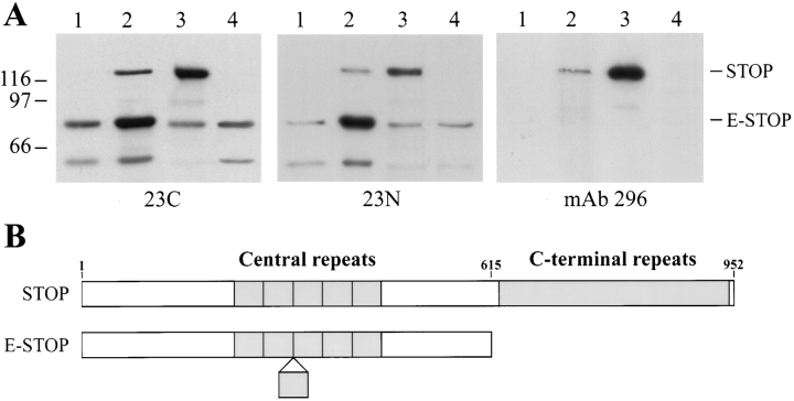
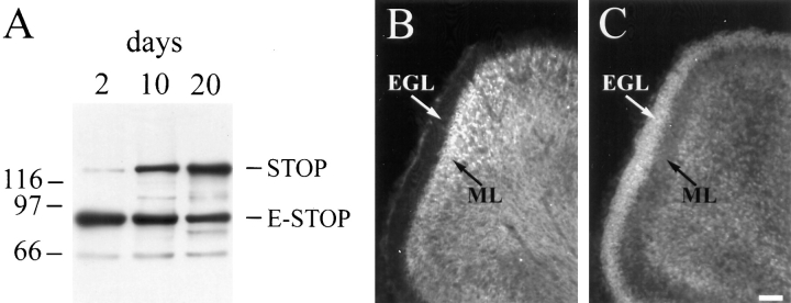
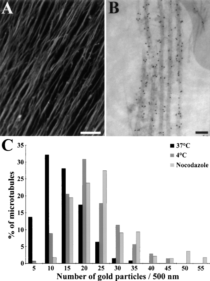
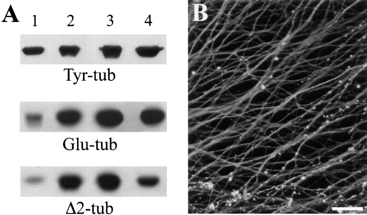
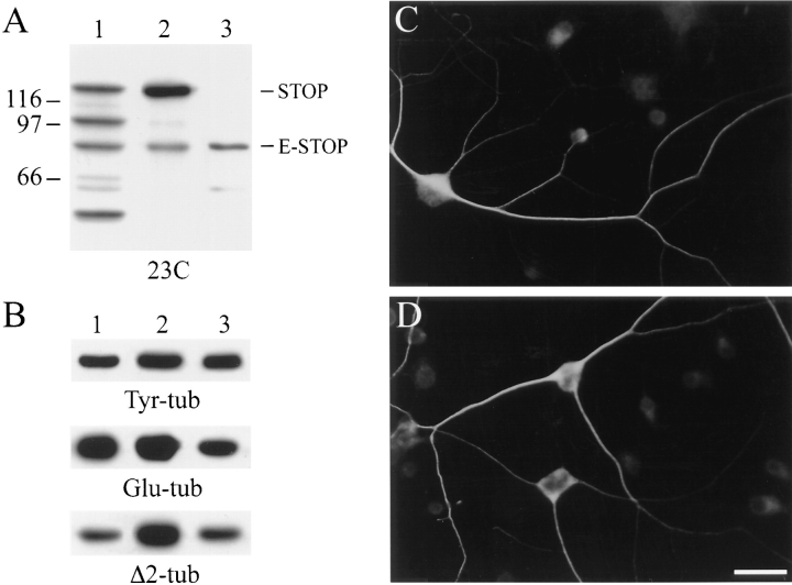
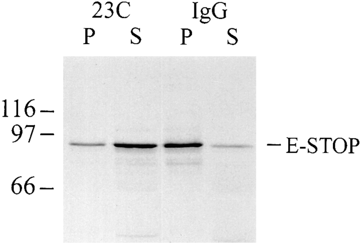
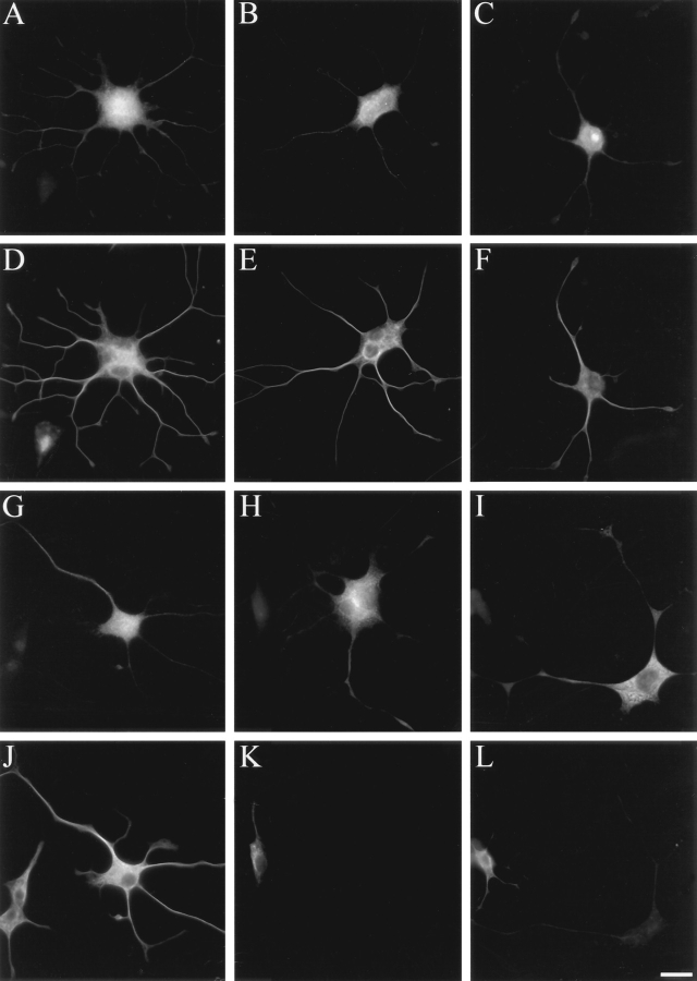
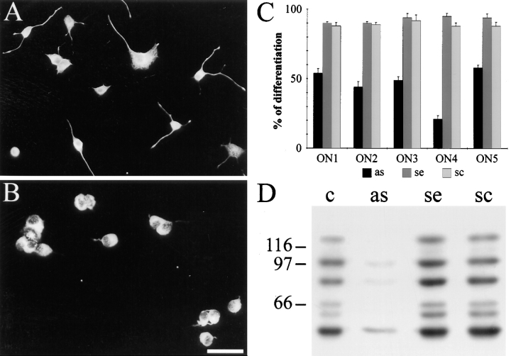
Neuronal differentiation and function require extensive stabilization of the microtubule cytoskeleton. Neurons contain a large proportion of microtubules that resist the cold and depolymerizing drugs and exhibit slow subunit turnover. The origin of this stabilization is unclear. Here we have examined the role of STOP, a calmodulin-regulated protein previously isolated from cold-stable brain microtubules. We find that neuronal cells express increasing levels of STOP and of STOP variants during differentiation. These STOP proteins are associated with a large proportion of microtubules in neuronal cells, and are concentrated on cold-stable, drug-resistant, and long-lived polymers. STOP inhibition abolishes microtubule cold and drug stability in established neurites and impairs neurite formation. Thus, STOP proteins are responsible for microtubule stabilization in neurons, and are apparently required for normal neurite formation.


Figures

Figure 1
Figure 1
STOP expression in DRG cells. (A) Immunoblot analysis of STOP expression. Proteins from 3-d differentiated DRG cells (lane 1), 10-d differentiated DRG cells (lane 2), adult rat brains (lane 3), and embryonic rat brains (lane 4) were run on 7.5% SDS gels. 20 μg of DRG cell extract proteins were loaded onto the gel. Amounts of loaded brain proteins were adjusted to equilibrate brain and DRG cell STOP signals. Proteins were immunoblotted with polyclonal STOP antibodies 23C and 23N (central repeat antibodies), and with monoclonal STOP antibody 296 (COOH-terminal repeat antibody) as indicated. The bands corresponding to STOP and E-STOP are indicated. Size markers are in kD. (B) Schematic representation of STOP and E-STOP showing domain structure of the two proteins. Both proteins contain a central domain composed of 5 (STOP) or 6 (E-STOP) repeats. STOP also contains a COOH-terminal repeat domain that is lacking in E-STOP. The E-STOP sequence ends at a position corresponding to aa 614 in the STOP sequence (Bosc et al., 1996). The E-STOP sequence data are available from GenBank/EMBL/DDBJ under accession no. AJ002556.
Figure 2
Figure 2
STOP expression during postnatal development of the rat cerebellum. (A) Immunoblot analysis of STOP expression in the cerebellum. Proteins from 2-d, 10-d, and 20-d-old rat were run on 7.5% SDS gels. 2 μg of cerebellum extract proteins were loaded onto the gel. Proteins were immunoblotted with polyclonal STOP antibody 23C. The bands corresponding to STOP and E-STOP are indicated. Size markers are in kD. (B and C) Immunofluorescence staining of the cerebellar cortex. Sections of 2-d aged rat cerebellum were stained with polyclonal STOP antibody 23C (B); (C) corresponding DNA staining with Hoechst. EGL, external germinal layer, ML, molecular layer. Bar, 40 μm.
Figure 3
Figure 3
Immunolocalization of STOP proteins in DRG cell axons. After 10 d of culture, DRG cells were stained using the affinity-purified 23C STOP antibody and a Cy3- (A) or gold-labeled (B) secondary antibody. (A) Immunofluorescence analysis of STOP localization in DRG cell axons. Bar, 25 μm. (B) Immunoelectron microscopy of a DRG cell axon: microtubules were specifically decorated with gold particles. Bar, 50 nm. (C) Gold particle distribution along axonal microtubules in the distal part of DRG cell axons. (Black bars) total microtubules; (dark gray bars) cold stable microtubules; (light gray bars) nocodazole-resistant microtubules. In control experiments with secondary antibody alone, microtubules were never decorated with more than four gold particles.
Figure 4
Figure 4
Tubulin modification in DRG cells. (A) Immunoblot analysis of tubulin modification in DRG cells: proteins from DRG cells cultured for 3 d (lane 1) or 10 d (lane 2), adult rat brains (lane 3), and embryonic rat brains (lane 4) were run on 7.5% SDS gels. Amounts of loaded proteins were as indicated in the legend to Fig. 1 A. Proteins were immunoblotted with isoform-specific tubulin antibodies as indicated. (B) Immunofluorescence analysis of Δ2-tubulin localization in 10-d cultured DRG cell axons. Cells were reacted with a specific Δ2-tubulin antibody. Axons showed bright and apparently homogeneous Δ2-tubulin staining along their length. Bar, 25 μm.
Figure 5
Figure 5
STOP expression and tubulin modification in PC12 cells. (A) Immunoblot analysis of STOP expression in PC12 cells: proteins from PC12 cells after 10 d of differentiation (lane 1), adult rat brains (lane 2), and embryonic rat brains (lane 3) were run on 7.5% SDS gels. 20 μg of PC12 cell proteins were loaded onto the gel. Amounts of loaded brain proteins were adjusted to equilibrate brain and PC12 cell STOP signals. Proteins were immunoblotted with polyclonal STOP antibody 23C. Superimposable results were observed with the 23N STOP antibody (data not shown). The bands corresponding to STOP and E-STOP are indicated. Size markers are in kD. (B) Immunoblot analysis of tubulin modification in PC12 cells: proteins from PC12 cells after 10 d of differentiation (lane 1), adult rat brains (lane 2), and embryonic rat brains (lane 3) were run on 7.5% SDS gels. Amounts of loaded proteins were as indicated in A. Proteins were immunoblotted with isoform-specific tubulin antibodies as indicated. (C and D) Immunofluorescence analysis of mixed populations of differentiated and undifferentiated PC12 cells with 23C STOP antibody (C) or Δ2-tubulin antibody (D). Neurite extensions in differentiated cells showed bright STOP or Δ2-tubulin staining while undifferentiated cells showed low or background levels of staining. Bar, 50 μm.
Figure 6
Figure 6
In vitro inhibition of E-STOP binding to microtubules by STOP antibody. In vitro–translated E-STOP (35S-labeled) was sedimented in the presence of bovine brain–polymerized microtubules (for details see Materials and Methods) after preincubation (5 min, 37°C) with polyclonal STOP antibody 23C (0.20 mg/ml) or with naive IgGs at the same concentration, as indicated. Equal amounts of the pellet (P) and supernatant (S) were separated by 7.5% SDS/ PAGE and exposed to autoradiography. In the presence of STOP antibody 23C, extensive inhibition of E-STOP binding to microtubules was observed. Results were similar when E-STOP solutions were preincubated with microtubules before adding STOP antibody 23C, and when STOP antibody 23C was replaced by STOP antibody 23N (data not shown). Size markers are in kD.
Figure 7
Figure 7
STOP inhibition suppresses microtubule cold and drug stability in fused PC 12 cells Fused PC12 cells injected with control IgGs (A–F) showing IgG (A–C) and microtubule (D–F) staining. (A and D) Untreated cells; (B and E) cold-treated cells; (C and F) nocodazole-treated cells. Fused PC12 cells injected with affinity-purified 23C STOP antibody (G–L) showing STOP antibody (G–I) and microtubule (J–L) staining. (G and J) Untreated cells; (H and K) cold-treated cells; (I and L) nocodazole-treated cells. Note that after cell lysis, enough microinjected IgGs remain associated with insoluble cell structures to allow clear identification of injected cells. STOP antibody injection suppressed microtubule cold and drug stability. Bar, 50 μm.
Figure 8
Figure 8
Inhibition of neurite formation in PC12 cells treated with STOP antisense oligonucleotides. (A and B) Immunofluorescence analysis of PC12 cells after a 36-h NGF treatment in the presence of sense ON4 (A) or antisense ON4 (B). For visualization of neurite extensions, cells were stained with mAb TUB 2.1 tubulin antibody. Bar, 100 μm. (C) Effect of five different STOP antisense oligonucleotides on neurite outgrowth. Each antisense oligonucleotide (as) was tested in parallel with the corresponding sense (se) and scrambled (sc) oligonucleotides. ON1–ON5 designate the five groups of oligonucleotides tested. In each condition, the proportion of differentiated cells among the total cell population was determined. Results are expressed as percent of control values determined in the presence of DOTAP alone (for details see Materials and Methods). (D) Immunoblot analysis of STOP proteins in PC12 cells after a 3-d NGF treatment in the presence of DOTAP alone (c), antisense (as), sense (se), or scrambled (sc) oligonucleotide ON4. 20 μg of PC12 cell proteins were loaded on 7.5% SDS gels. Proteins were immunoblotted with polyclonal STOP antibody 23C. Results marked diminution of STOP expression in cells treated with antisense oligonucleotide ON4. Size markers are in kD.

Similar articles

Cited by

References

    1. Baas PW, Ahmad FJ. The plus ends of stable microtubules are the exclusive nucleating structures for microtubules in the axon. J Cell Biol. 1992;116:1231–1241. - PMC - PubMed
    1. Baas PW, Black MM. Individual microtubules in the axon consist of domains that differ in both composition and stability. J Cell Biol. 1990;111:495–509. - PMC - PubMed
    1. Baas PW, Heidemann SR. Microtubule reassembly from nucleating fragments during the regrowth of amputated neurites. J Cell Biol. 1986;103:917–927. - PMC - PubMed
    1. Baas PW, Pienkowski TP, Cimbalnik KA, Toyama K, Bakalis S, Ahmad FJ, Kosik KS. Tau confers drug stability but not cold stability to microtubules in living cells. J Cell Sci. 1994;107:135–143. - PubMed
    1. Barra HS, Arce CA, Argaraña CE. Posttranslational tyrosination/detyrosination of tubulin. Mol Neurobiol. 1988;2:133–153. - PubMed

Publication types

MeSH terms

Associated data