. 1998 Jul 13;142(1):181-90.
doi: 10.1083/jcb.142.1.181.

Targeting, capture, and stabilization of microtubules at early focal adhesions

Affiliations

Targeting, capture, and stabilization of microtubules at early focal adhesions

I Kaverina et al. J Cell Biol. .

Abstract

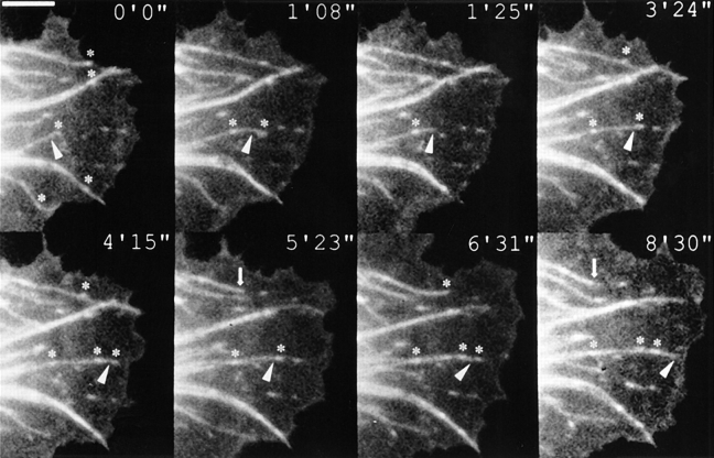
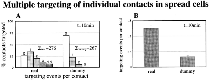
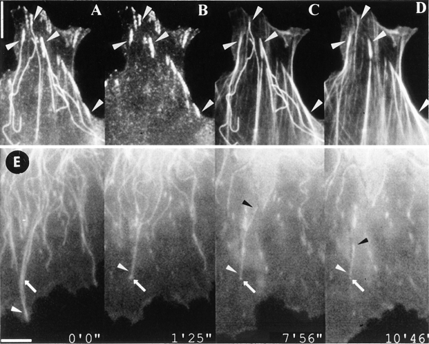
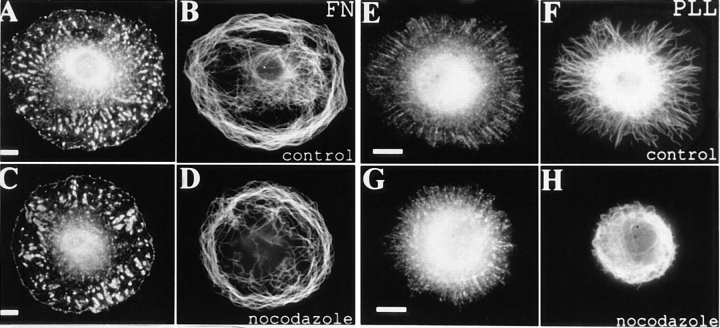
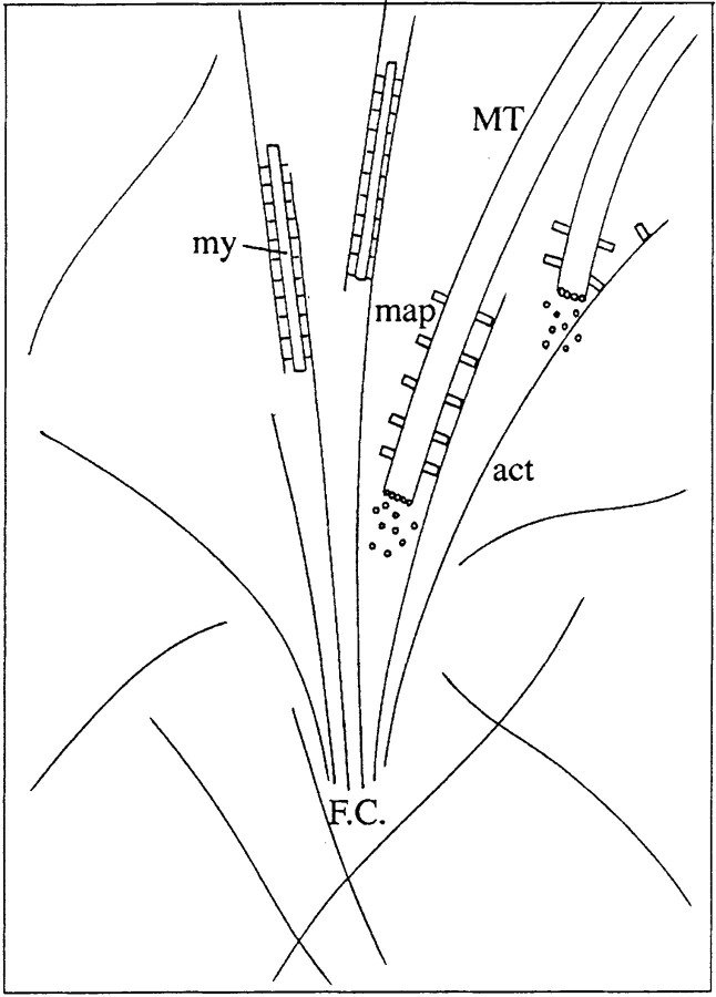
By co-injecting fluorescent tubulin and vinculin into fish fibroblasts we have revealed a "cross talk" between microtubules and early sites of substrate contact. This mutuality was first indicated by the targeting of vinculin-rich foci by microtubules during their growth towards the cell periphery. In addition to passing directly over contact sites, the ends of single microtubules could be observed to target several contacts in succession or the same contact repetitively, with intermittent withdrawals. Targeting sometimes involved side-stepping, or the major re-routing of a microtubule, indicative of a guided, rather than a random process. The paths that microtubules followed into contacts were unrelated to the orientation of stress fiber assemblies and targeting occurred also in mouse fibroblasts that lacked a system of intermediate filaments. Further experiments with microtubule inhibitors showed that adhesion foci can: (a) capture microtubules and stabilize them against disassembly by nocodazole; and (b), act as preferred sites of microtubule polymerization, during either early recovery from nocodazole, or brief treatment with taxol. From these and other findings we speculate that microtubules are guided into substrate contact sites and through the motor-dependent delivery of signaling molecules serve to modulate their development. It is further proposed this modulation provides the route whereby microtubules exert their influence on cell shape and polarity.


Figures

Figure 1
Figure 1
Microtubule targeting of focal adhesions. Figure shows selected frames from a video sequence of a goldfish fibroblast co-injected with rhodamine tubulin and TAMRA vinculin. Some of the focal contacts crossed by microtubules are indicated by asterisks. The ends of two microtubules (arrowhead and arrow) are highlighted. One (arrowhead) targeted successive adhesion sites on its way to the cell periphery, and the other (arrow, 5′23′′–8′30′′) targeted and withdrew from a focal adhesion. For further details, see text. The inset times are given in minutes and seconds in this and subsequent figures of video sequences. Bar, 5 μm.
Figure 2
Figure 2
Progressive targeting in advancing lamellae. (A) Plots for six cells of the percentage, over time, of vinculin-containing contacts targeted by microtubules in a pre-selected, rectangular area positioned close to the cell front in the first frame of the video sequence. Solid lines show data for real contacts and broken lines for dummy contacts (see B and text for further details). (B) Graphic illustration of targeting analysis. A rectangle 4.5 μm in width was placed behind the cell front in the first video frame to define the area of analysis of contact targeting. The dummy contact pattern was created by rotating the real contact pattern by 180 degrees about a line through the center and perpendicular to the long axis of the rectangle.
Figure 3
Figure 3
Re-routing of microtubules from one contact to another and multiple targeting. Selected video frames of three lamella regions of fish fibroblasts co-injected with fluorescent tubulin and vinculin, as for Fig. 1. Times are given in minutes and seconds. (A) Two contacts on the cell edge (open and closed arrowhead) were targeted by microtubules 1, 2, and 3. Microtubule 2 targeted one contact three times (open arrowhead at 0′44′′, 2′12′′, and 4′24′′), and the other contact once (closed arrowhead, 3′40′′). This involved a dramatic re-routing from one contact to the other and back (2′34′′–4′24′′). (B) Two peripheral contacts (open and closed arrowhead) situated on either side of a retracted cell edge were targeted by the same microtubule. This involved shrinkage to a more proximal contact (asterisk at 2′50′′) followed by growth into the second contact in a direction normal to the first. (C) Example of one contact (solid arrowhead) that was targeted successively by three microtubules (1, 2, and 3) that approached from different directions. Bar, 10 μm.
Figure 4
Figure 4
Multiple targeting of individual contacts in spread cells. (A) Histogram of the number of targeting events of individual contacts by microtubules in a period of 10 min. The data is presented for real contacts (total 276) and for dummy contacts (total 267). (B) Data in A summarized in terms of the average number of targeting events per contact. See text for further details.
Figure 5
Figure 5
Stabilization of microtubules at focal adhesions. (A–D) Figure shows a 3T3 fibroblast that was fixed and triple labeled for actin (D), paxillin (B), and tubulin (A and C) after treatment with 1.5 μg/ml nocodazole for 10 min. All peripheral microtubules disassembled, except those whose ends targeted focal adhesions (arrowheads). (E) Video sequence showing the stabilization of a shrinking microtubule at a focal adhesion. Goldfish fibroblast co- injected with vinculin and tubulin. Frames are taken from a video sequence for which nocodazole (1.5 μg/ ml) was added at time 0. One of a pair of microtubules that extended to the periphery at the beginning of the sequence (white arrowhead) was prevented from shrinking beyond an adhesion site over which it passed (arrow). Eventually, it shrank into this adhesion site via depolymerization at its minus end (black arrowhead). Bars, 5 μm.
Figure 6
Figure 6
Capture and stabilization of a microtubule at a focal adhesion that was remote from the contact site at the time of addition of nocodazole. Conditions as for Fig. 4, except that negative and positive times signify before and after nocodazole addition, respectively. Before nocodazole treatment, the microtubule marked with an arrowhead grew and moved laterally and became positioned over a focal adhesion (arrow, 0′44′′). Nocodazole caused rapid shrinkage down to the contact (+1′42′′ –2′16′′), where the end then remained stable for a further 3 min (2′16′′ –5′06′′) before finally shrinking into the cell body (5′23′′– 6′31′′). Bar, 5 μm.
Figure 7
Figure 7
General stabilization of microtubules by focal adhesions in REF-52 fibroblasts. Cells spreading on fibronectin show numerous focal adhesions (A) as compared with a finely punctate vinculin label on polylysine (E). The corresponding microtubule distributions are shown in B and F. After brief nocodazole treatment (1.5 μg/ ml, 10 min) peripheral microtubules in cells plated on fibronectin remain essentially unaffected (D) whereas those in cells spread on polylysine shrink rapidly into the cell body (H). Bar, 10 μm.
Figure 8
Figure 8
(A and B) Targeting, but not stabilization at focal complexes. Images of porcine testicular cells labeled for tubulin and vinculin: A, control cell; B, cell treated with 1.5 μg/ml nocodazole for 20 min. The focal complexes characteristically found on the edges of these cells are targeted by microtubules (A), but they do not stabilize microtubules against depolymerization by nocodazole (B). (C and D) Targeting and stabilization occurs in the absence of intermediate filaments. Fibroblasts of a mouse vimentin knockout cell line labeled for tubulin and vinculin. C, control cell showing targeting of microtubules to contact sites; D, cell treated with 2.5 μg/ml nocodazole for 10 min showing stabilization of microtubules at focal contacts. Bar, 10 μm.
Figure 9
Figure 9
Microtubules are not guided by stress fibers to contact sites. Goldfish fibroblast triple labeled for actin, vinculin, and tubulin. A, actin pattern revealed with phalloidin; B, overlay of tubulin and vinculin; C, actin pattern overlaid with graphic renditions of microtubules that target contact sites (arrowheads). The courses taken by microtubules and stress fibers to contact sites are unrelated. Bar, 10 μm.
Figure 10
Figure 10
Nucleation of microtubule growth at focal adhesions, in 3T3 fibroblasts. A and B show a 3T3 fibroblast labeled for actin and tubulin that had been exposed to 0.05 μm taxol for 1 h before fixation. Arrowheads indicate the ends of some of the microtubules that had been nucleated at the stress fiber terminus (arrow). (C–E) Part of a REF-52 fibroblast after short term (4 min) recovery from complete disassembly of microtubules by nocodazole. Non-centrosomal microtubule segments (D) are specifically associated with peripheral focal adhesions (C and E), marked with arrows. In E, the microtubule segments (D) have been graphically superimposed on the vinculin image to show the correspondence between the two patterns. Bar, 10 μm.
Figure 11
Figure 11
Possible mode of guidance of microtubules to contact sites. An early contact site (F.C.) is depicted with splaying actin filaments (act), some of which are already associating with myosin filaments (my) during the initial stages of stress fiber assembly. A microtubule (MT; 1) decorated with associated molecules with potential actin binding activity (map) appears in the vicinity of the contact. Signaling components in the region of the contact activate the cross-linking activity of the map, leading to the parallel alignment of the actin filament and microtubule (2) and guidance into the contact.

Similar articles

Cited by

References

    1. Bershadsky A, Chausovsky A, Becker E, Lyubimova A, Geiger B. Involvement of microtubules in the control of adhesion-dependent signal transduction. Curr Biol. 1996;6:1279–1289. - PubMed
    1. Bershadsky AD, Vaisberg EA, Vasiliev JM. Pseudopodial activity at the active edge of migrating fibroblast is decreased after drug-induced microtubule depolymerization. Cell Motil Cytoskeleton. 1991;19:152–158. - PubMed
    1. Bershadsky, A.D., and J.M. Vasiliev. 1986. Cytoskeleton. Plenum Press, New York/London. 298 pp.
    1. Chrzanowska-Wodnicka M, Burridge K. Rho-stimulated contractility drives the formation of stress fibers and focal adhesions. J Cell Biol. 1996;133:1403–1415. - PMC - PubMed
    1. Colucci-Guyon E, Portier MM, Dunia I, Paulin D, Pounin S, Babinet C. Mice lacking vimentin develop and reproduce without an obvious phenotype. Cell. 1994;79:679–694. - PubMed

Publication types