. 1998 Jul 13;142(1):191-202.
doi: 10.1083/jcb.142.1.191.

Unique responses of differentiating neuronal growth cones to inhibitory cues presented by oligodendrocytes

Affiliations

Unique responses of differentiating neuronal growth cones to inhibitory cues presented by oligodendrocytes

A Shibata et al. J Cell Biol. .

Abstract

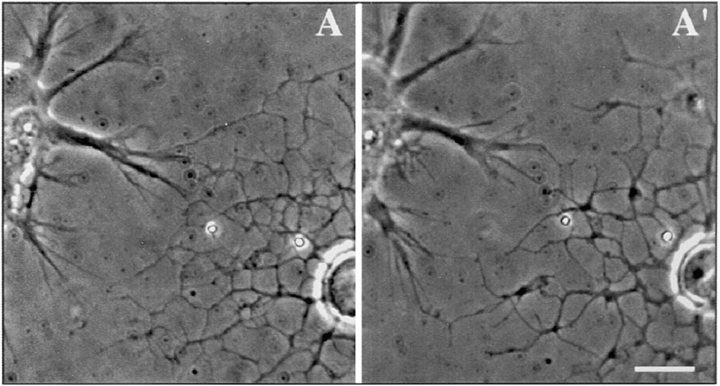
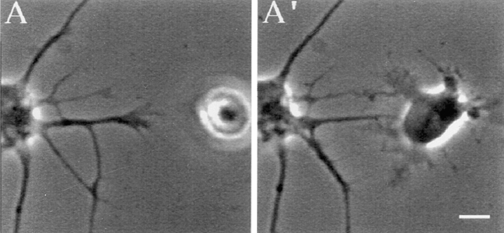
During central nervous system development, neurons differentiate distinct axonal and dendritic processes whose outgrowth is influenced by environmental cues. Given the known intrinsic differences between axons and dendrites and that little is known about the response of dendrites to inhibitory cues, we tested the hypothesis that outgrowth of differentiating axons and dendrites of hippocampal neurons is differentially influenced by inhibitory environmental cues. A sensitive growth cone behavior assay was used to assess responses of differentiating axonal and dendritic growth cones to oligodendrocytes and oligodendrocyte- derived, myelin-associated glycoprotein (MAG). We report that >90% of axonal growth cones collapsed after contact with oligodendrocytes. None of the encounters between differentiating, MAP-2 positive dendritic growth cones and oligodendrocytes resulted in growth cone collapse. The insensitivity of differentiating dendritic growth cones appears to be acquired since they develop from minor processes whose growth cones are inhibited (nearly 70% collapse) by contact with oligodendrocytes. Recombinant MAG(rMAG)-coated beads caused collapse of 72% of axonal growth cones but only 29% of differentiating dendritic growth cones. Unlike their response to contact with oligodendrocytes, few growth cones of minor processes were inhibited by rMAG-coated beads (20% collapsed). These results reveal the capability of differentiating growth cones of the same neuron to partition the complex molecular terrain they navigate by generating unique responses to particular inhibitory environmental cues.


Figures

Figure 1
Figure 1
Postnatal hippocampal pyramidal-like neurons differentiate mature axons and dendrites. (A) Hippocampal pyramidal-like neuron cultured for 1.5 DIV. One neurite, the presumptive axon, has extended at a greater rate than the other minor processes. (B) MAP-2 stains all neurites (the presumptive axon [arrow] and all minor processes) at this early developmental stage. (C) After 5–6 DIV, postnatal hippocampal neurons extend a long, thin axon and several shorter tapering presumptive dendrites. (D) The differentiation of mature dendrites was exhibited by the specific localization of MAP-2 to the dendro-somatic region of all cultured hippocampal pyramidal-like neurons observed after 5–6 DIV. The mature axon, indicated by the arrow, does not express MAP-2. Bar, 25 μm.
Figure 2
Figure 2
Primary cultured oligodendrocytes express MAG in culture. A dropped, differentiated oligodendrocyte used for coculture experiments was stained with anti-MAG antibody. Phase images of the cocultured hippocampal neuron and oligodendrocyte are shown in A. The corresponding fluorescent image is shown in B. The spread oligodendrocyte shows positive immunoreactivity for MAG, as do 94.6% of the cultured oligodendrocytes used in our experiments. Neuronal processes were not immunopositive for MAG. Bar, 10 μm.
Figure 3
Figure 3
Phase images of hippocampal axonal growth cones encountering a primary cultured oligodendrocyte and 3T3 fibroblast. An axonal growth cone (arrowheads) contacted the extended membrane of an oligodendrocyte (A, upper right corner, arrows) and sampled the membrane with motile filopodia and lamellipodium (3.2 min after initial contact, A′). After 16.6 min of contact the growth cone collapsed. The lamellipodium folded in on itself becoming club shaped and phase dark and the growth cone retracted except for the thin filopodial thread that remained (A′′). The collapse was manifested as the loss of 60% of the original growth cone surface area. A 3T3 fibroblast was dropped into the path of an axonal growth cone (B). Filopodia of an axonal growth cone contacted the spread fibroblast membrane (B′). After 10.4 min the axonal growth cone had traversed over the 3T3 fibroblast as well as a distal neurite (B′′) and had increased its growth cone surface area by 35%. The axonal growth cone continued to elongate for the duration of the experiment (∼80 min). Fibroblasts were often stimulated to move away from the site of contact with neuronal growth cones (B′′). Bar, 10 μm.
Figure 4
Figure 4
Hippocampal dendritic growth cone encountering a primary cultured oligodendrocyte. (A) Dendritic growth cone encounters the web-like membrane of an oligodendrocyte with its extended filopodia. (A′) After 17.2 min of contact and throughout the experiment (120 min) the dendritic growth cone retains active filopodia and has increased its growth cone lamellipodium surface area by 24%. Bar, 10 μm.
Figure 5
Figure 5
Growth cone of a hippocampal minor process encounters a primary cultured oligodendrocyte. (A) The growth cone of a minor process extended as a dropped oligodendrocyte settled to the dish and began to spread its processes. (A′) After 18.9 min of contact the growth cone has collapsed. The encounter results in a 98% loss of the original growth cone surface area. Bar, 10 μm.
Figure 6
Figure 6
Axonal growth cones of rat hippocampal neuron encountering rMAG-coated and control beads. (A) Several filopodia of the large active axonal growth cone contact rMAG-coated beads. At contact the growth cone exhibits a spread lamellipodial veil. (A′) After 17.3 min of contact the growth cone lamellipodium has collapsed leaving only 22% behind as a club-shaped, phase-dark growth cone with one or more thin threads remaining in contact with the bead as the neurite retracts. Similar to oligodendrocyte-induced collapse of axonal growth cones, this growth cone does not recover from rMAG-induced collapse during the period of observation (85 min). (B) An axonal growth cone elongated toward a control, denatured rMAG-coated bead. (B′) The growth cone contacted the beads and elongated underneath them without effecting the overall activity or morphology of the growth cone. The growth cone did not show significant changes in growth cone surface area. Bar, 10 μm.
Figure 7
Figure 7
Growth cones of dendritic and minor processes encountering rMAG-coated and control beads. (A) A dendritic growth cone extends thin filopodia to contact a dropped rMAG-coated bead. (A′) After 17.9 min of contact the dendritic growth cone remains active with motile filopodia and a spread lamellipodium. (B) Growth cone of a minor process encounters a rMAG-coated bead and is unaffected by the encounter. The growth cone contacts the bead with filopodia and the distal edge of its lamellipodium. (B′) 19.2 min after initial contact the growth cone has nearly doubled its original growth cone surface (increase of 82%). For the remainder of the experiment (∼87 min) the growth cone continued motile activity but did not elongate. Bar, 10 μm.

Similar articles

Cited by

References

    1. Angevine JB., Jr Time of neural origins in the hippocampal region. An autoradiographic study in the mouse. Expl Neurol Suppl. 1965;2:1–70. - PubMed
    1. Angevine, J.B., Jr. 1975. Development of the hippocampal region. In The Hippocampus Vol. 1: Structure and Development. R.L. Isaacson, K.H. Pribram, editors. Plenum Press, New York. 61–94.
    1. Autillo-Touati A, Chamak B, Araud D, Vuillet J, Seite R, Prochiantz A. Region-specific neuro-astroglial interactions: ultrastructural study of the in vitroexpression of neuronal polarity. J Neurosci Res. 1988;19:326–342. - PubMed
    1. Bandtlow CE, Zachleder T, Schwab ME. Oligodendrocytes arrest neurite growth by contact inhibition. J Neurosci. 1990;10:3837–3848. - PMC - PubMed
    1. Banker GA, Cowan WM. Rat hippocampal neurons in dispersed cell culture. Brain Res. 1977;126:397–425. - PubMed

Publication types

Substances