. 1998 Jul 13;142(1):229-40.
doi: 10.1083/jcb.142.1.229.

Dual actions of sphingosine-1-phosphate: extracellular through the Gi-coupled receptor Edg-1 and intracellular to regulate proliferation and survival

Affiliations

Dual actions of sphingosine-1-phosphate: extracellular through the Gi-coupled receptor Edg-1 and intracellular to regulate proliferation and survival

J R Van Brocklyn et al. J Cell Biol. .

Abstract

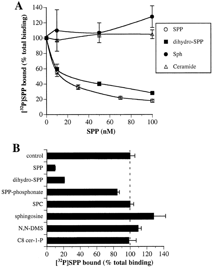
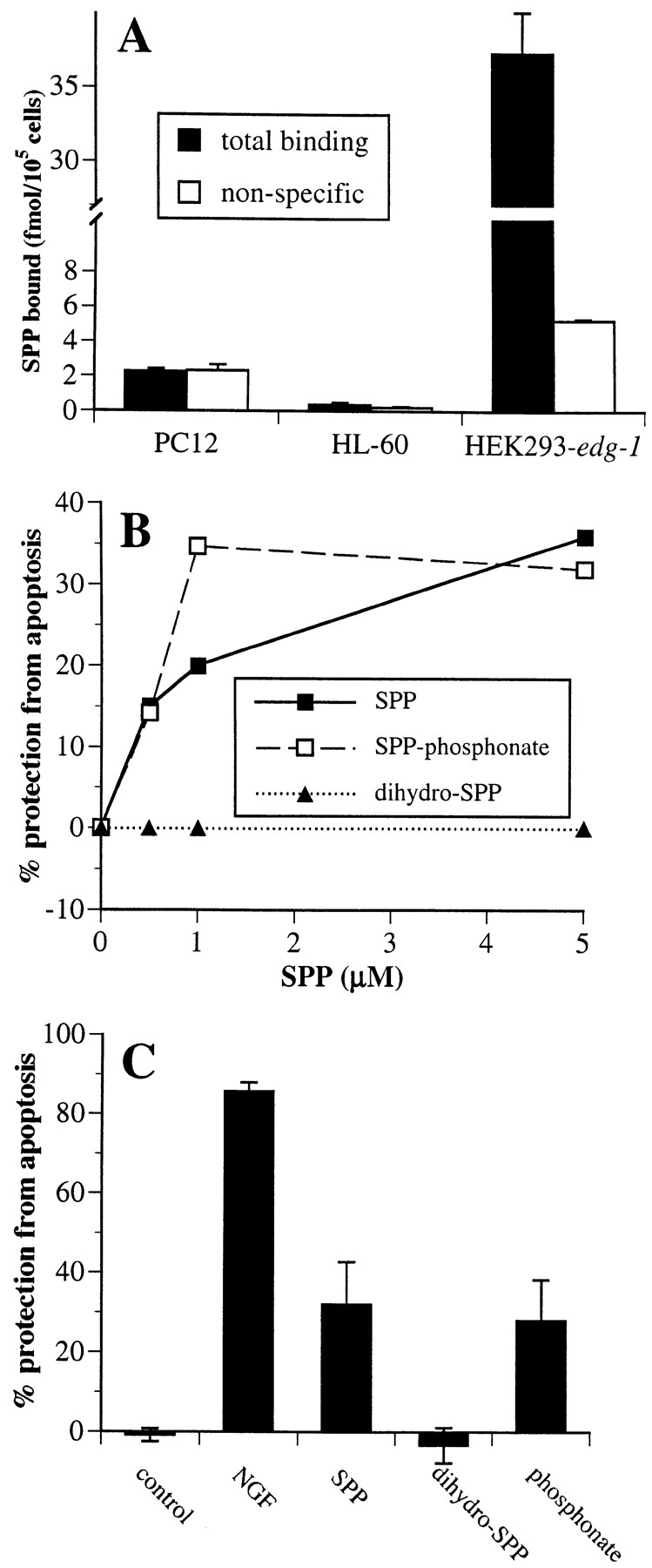
Sphingosine-1-phosphate (SPP), a bioactive lipid, acts both intracellularly and extracellularly to cause pleiotropic biological responses. Recently, we identified SPP as a ligand for the G protein-coupled receptor Edg-1 (Lee, M.-J., J.R. Van Brocklyn, S. Thangada, C.H. Liu, A.R. Hand, R. Menzeleev, S. Spiegel, and T. Hla. 1998. Science. 279:1552-1555). Edg-1 binds SPP with remarkable specificity as only sphinganine-1-phosphate displaced radiolabeled SPP, while other sphingolipids did not. Binding of SPP to Edg-1 resulted in inhibition of forskolin-stimulated cAMP accumulation, in a pertussis toxin-sensitive manner. In contrast, two well-characterized biological responses of SPP, mitogenesis and prevention of apoptosis, were clearly unrelated to binding to Edg-1 and correlated with intracellular uptake. SPP also stimulated signal transduction pathways, including calcium mobilization, activation of phospholipase D, and tyrosine phosphorylation of p125(FAK), independently of edg-1 expression. Moreover, DNA synthesis in Swiss 3T3 fibroblasts was significantly and specifically increased by microinjection of SPP. Finally, SPP suppresses apoptosis of HL-60 and pheochromocytoma PC12 cells, which do not have specific SPP binding or expression of Edg-1 mRNA. Conversely, sphinganine-1-phosphate, which binds to and signals via Edg-1, does not have any significant cytoprotective effect. Thus, SPP is a prototype for a novel class of lipid mediators that act both extracellularly as ligands for cell surface receptors and intracellularly as second messengers.


Figures

Figure 1
Figure 1
Effects of SPP analogues and other lipids on specific binding of [32P]SPP to Edg-1. Competition of SPP binding by related lipids. HEK293–edg-1 cells were incubated in the presence of 1 nM [32P]SPP with increasing concentrations of unlabeled SPP, dihydro-SPP, sphingosine, or C2-ceramide (A) or in the presence of 100 nM of the indicated lipids (B), and binding was measured as described in Materials and Methods. Specific binding of SPP to HEK293–edg-1 (total binding minus binding in the presence of 100-fold excess unlabeled SPP) was 32 fmol/105 cells. Results are means ± standard deviations of triplicate determinations.
Figure 2
Figure 2
SPP decreases cAMP levels through the Edg-1 Gi-coupled receptor. (A) HEK293-vector (open bars) or HEK293–edg-1 cells (filled bars) were stimulated with 10 μM forskolin in the presence of 0.5 mM IBMX and the indicated concentrations of SPP for 15 min, and levels of cAMP were then measured. The levels of cAMP in untreated and forskolin-treated cells were 20 ± 1.7 and 420 ± 30 pmol/106 cells and 10 ± 1.5 and 820 ± 60 pmol/ 106 cells for vector and edg-1–overexpressing cells, respectively. Results are means ± SD of triplicate determinations. (B) Effects of SPP analogues on cAMP accumulation. HEK293–edg-1 cells were treated with lipids or SPP analogues (100 nM), and forskolin-stimulated cAMP accumulation was determined as described in A. (C) Pertussis toxin inhibits the effect of SPP on cAMP accumulation. Cells were pretreated in the absence (filled bars) or presence (open bars) of 200 ng/ml pertussis toxin (PTX) for 3 h and then were treated for 15 min with forskolin (10 μM) and/or SPP (100 nM), or combinations, as indicated, and cAMP accumulation was measured.
Figure 3
Figure 3
SPP stimulates non–Edg-1 receptor-mediated signal transduction pathways. (A) SPP stimulates tyrosine phosphorylation of p125FAK independently of Edg-1 expression. HEK293– edg-1 and vector-transfected HEK293 cells were treated with the indicated concentrations of SPP for various times, and cell lysates were immunoprecipitated with anti-p125FAK mAb and analyzed by Western blotting with anti-Tyr(P) antibody. The arrow indicates the migration p125FAK. (B) SPP-induced changes in intracellular free calcium. Vector and edg-1 stably transfected HEK293 cells were loaded with fura-2/AM, washed, and incubated at 37°C in Locke's buffer. At the indicated times, SPP (100 nM or 10 μM) was added, and [Ca2+]i was determined by fura-2 imaging (Mattie et al., 1994). Subsequent addition of ionomycin always caused increased [Ca2+]i. (C) SPP-induced phosphatidic acid accumulation. Vector and edg-1 stably transfected HEK293 cells and NIH 3T3 fibroblasts were prelabeled with [32P]i for 24 h and then stimulated with vehicle (control) or the indicated concentrations of SPP for 1 h. Lipids were extracted and separated by thin-layer chromatography, and [32P]phosphatidic acid was measured (Zhang et al., 1990). Data are expressed as fold- increases of [32P]phosphatidic acid. The incorporations of [32P] into phosphatidic acid in untreated cells were 1,500 ± 50 and 1,000 ± 100 cpm, and 2,500 ± 600 and 3,200 ± 200 cpm for vector and edg-1 stably transfected HEK293 cells and NIH 3T3 fibroblasts, respectively, determined from an aliquot of the phospholipid extract containing 150,000 cpm.
Figure 4
Figure 4
SPP-stimulated DNA synthesis does not correlate with Edg-1 expression. (A) Swiss 3T3 fibroblasts (filled bars), vector-transfected NIH 3T3 cells (hatched bars), NIH 3T3 cells stably transfected with edg-1 (open bars), HEK293-vector (stippled bars), and HEK293–edg-1 cells (gray bars) were grown to confluence in 24-well tissue culture plates, washed with serum-free DME containing 5 μg/ml transferrin and 20 μg/ml BSA, and then stimulated with the indicated concentration of SPP. After 16 h, cells were pulsed with 1.0 μCi of [3H]thymidine for 8 h, and incorporation of [3H]thymidine into trichloroacetic acid–insoluble material was measured. Values are the means of triplicate determinations, and standard deviations were routinely less than 10% of the mean. (B) Northern analysis of Edg-1 expression was performed on total RNA from the indicated cells as described in Materials and Methods. A probe corresponding to the mouse edg-1 gene (Liu and Hla, 1997) was used to detect the low level of edg-1 mRNA present in Swiss 3T3 cells. For analysis of HEK293–edg-1 and HEK293-vector cells, a probe corresponding to the human edg-1 gene was used (Hla and Maciag, 1990). (C) Specific [32P]SPP binding to various cell types. Nonspecific (open bars) and total binding (filled bars) of 1 nM [32P]SPP to Swiss 3T3 fibroblasts, vector-transfected NIH 3T3 cells, NIH 3T3 cells stably transfected with edg-1, vector-transfected HEK293 cells, and edg-1-transfected HEK293 were measured as described in Materials and Methods.
Figure 5
Figure 5
SPP-stimulated DNA synthesis correlates with uptake of SPP. (A) Swiss 3T3 fibroblasts, cultured in serum-free DME containing 4 μg/ml insulin, 4 μg/ml transferrin, and 20 μg/ml BSA, were treated with the indicated concentrations of SPP at 4 (filled bars) or 37°C (open bars) for 30 min, followed by washing and replacement in the same medium without SPP. Cells were then incubated for 16 h at 37°C, and [3H]thymidine incorporation was measured. (B) In duplicate cultures, uptake was measured after 30 min incubation with 10 μM [32P]SPP. (C) Swiss 3T3 fibroblasts cultured in serum-free DME were treated with the indicated concentrations of SPP added as a complex with 4 mg/ml BSA (open symbols) or with 5 μl/ml lipofectamine (filled symbols) for 30 min followed by washing and replacement with DME containing 4 μg/ml insulin, 4 μg/ml transferrin, and 20 μg/ml BSA, and DNA synthesis was measured 16 h later. (D) In duplicate cultures, uptake of [32P]SPP was measured in the absence (open bars) or presence (filled bars) of lipofectamine.
Figure 6
Figure 6
Microinjected SPP stimulates DNA synthesis. Serum-starved Swiss 3T3 cells were microinjected with rabbit IgG together with vehicle (A–C) or SPP (D–F). Cells were visualized by Texas red fluorescence (red), demonstrating immunoglobulin G localization after microinjection (A and D). BrdU incorporation into injected and uninjected cells on the same coverslips was visualized by green FITC fluorescence (B and E). C and F are superimposed images visualized using a triple band pass filter. Yellow-orange color indicates colocalization of microinjected SPP with BrdU staining. In G, stimulation of DNA synthesis in cells injected (open bars) with IgG in the absence or presence of SPP or C8-cer-1-P, as well as in adjacent uninjected cells (filled bars), was assessed by BrdU incorporation. Values (means ± SD) are the percentage of cells positive for BrdU staining and correspond to the average of three measurements in which 100 cells were scored. At least 400 cells were microinjected and scored in each experiment. For comparison, BrdU staining of Swiss 3T3 fibroblasts treated with vehicle (cross-hatched bar) or with 10 μM exogenous SPP (dotted bar) was also determined. (H) Swiss 3T3 cells were serum-starved 24 h in the presence of 5 μg/ml insulin and then treated with or without 200 ng/ml pertussis toxin for 3 h. Cells were then microinjected with rabbit IgG together with vehicle or SPP, or treated with 10 μM exogenous SPP and BrdU incorporation was assessed. In the experiment where SPP was microinjected, 14.5% of adjacent, uninjected cells were positive for BrdU incorporation.
Figure 6
Figure 6
Microinjected SPP stimulates DNA synthesis. Serum-starved Swiss 3T3 cells were microinjected with rabbit IgG together with vehicle (A–C) or SPP (D–F). Cells were visualized by Texas red fluorescence (red), demonstrating immunoglobulin G localization after microinjection (A and D). BrdU incorporation into injected and uninjected cells on the same coverslips was visualized by green FITC fluorescence (B and E). C and F are superimposed images visualized using a triple band pass filter. Yellow-orange color indicates colocalization of microinjected SPP with BrdU staining. In G, stimulation of DNA synthesis in cells injected (open bars) with IgG in the absence or presence of SPP or C8-cer-1-P, as well as in adjacent uninjected cells (filled bars), was assessed by BrdU incorporation. Values (means ± SD) are the percentage of cells positive for BrdU staining and correspond to the average of three measurements in which 100 cells were scored. At least 400 cells were microinjected and scored in each experiment. For comparison, BrdU staining of Swiss 3T3 fibroblasts treated with vehicle (cross-hatched bar) or with 10 μM exogenous SPP (dotted bar) was also determined. (H) Swiss 3T3 cells were serum-starved 24 h in the presence of 5 μg/ml insulin and then treated with or without 200 ng/ml pertussis toxin for 3 h. Cells were then microinjected with rabbit IgG together with vehicle or SPP, or treated with 10 μM exogenous SPP and BrdU incorporation was assessed. In the experiment where SPP was microinjected, 14.5% of adjacent, uninjected cells were positive for BrdU incorporation.
Figure 7
Figure 7
Effects of SPP and analogues on suppression of apoptosis. (A) Lack of specific [32P]SPP binding to HL-60 and PC12 cells. Nonspecific (open bars) and total binding (filled bars) of 1 nM [32P]SPP to HL-60 and PC12 cells was measured. (B) Inhibition of sphingomyelinase-induced DNA fragmentation by SPP in HL-60 cells. HL-60 cells were incubated with [3H]thymidine (1 μCi/ml) for 24 h to label DNA, washed, and then treated without or with 100 mU/ml Staphylococcus aureus sphingomyelinase in the presence of vehicle or with the indicated concentrations of SPP (filled squares), SPP-phosphonate (open squares), or dihydro-SPP (filled triangles), added as BSA complexes. After 5 h, DNA fragmentation was determined from the ratio of unfragmented/fragmented DNA (Cuvillier et al., 1996). Data are expressed as percent inhibition of DNA fragmentation. Percent protection from apoptosis = 100 × [(fragmentation induced by SMase) − (fragmentation induced by SMase in the presence of cytoprotective agents)] / [(fragmentation induced by SMase) − (fragmentation of untreated controls)]. (C) SPP and its hydrolysis-resistant analogue suppress apoptosis in PC12 cells induced by trophic factor withdrawal. PC12 cells were incubated in serum-free medium in the absence or presence of 5 μM SPP, dihydro-SPP, SPP-phosphonate, or NGF (100 ng/ml) for 15 h, fixed in 3.7% formaldehyde, and then stained with bisbenzimide trihydrochloride (24 μg/ml in 50% glycerol/PBS; Hoechst #33258, Calbiochem) (Edsall et al., 1997). Cells with chromatin condensation or segmentation of nuclei into three or more fragments were considered to be apoptotic. A minimum of 2,000 cells in each field was scored. Values are means ± SD of triplicate determinations. Percent protection from apoptosis = 100 × [(number of cells with fragmented nuclei) / (number of cells with fragmented nuclei + number of intact cells)].

Similar articles

Cited by

References

    1. An S, Bleu T, Huang W, Hallmark OG, Coughling SR, Goetzel EJ. Identification of cDNAs encoding two G protein-coupled receptors for lysosphingolipids. FEBS Lett. 1997;417:279–282. - PubMed
    1. Blakesly VA, Beitner-Johnson D, Van Brocklyn JR, Rani S, Shen-Orr Z, Stannard BS, Spiegel S, LeRoith D. Sphingosine 1-phosphate stimulates tyrosine phosphorylation of Crk. J Biol Chem. 1997;272:16211–16215. - PubMed
    1. Bornfeldt KE, Graves LM, Raines EW, Igarashi Y, Wayman G, Yamamura S, Yatomi Y, Sidhu JS, Krebs EG, Hakomori S, Ross R. Sphingosine-1-phosphate inhibits PDGF-induced chemotaxis of human arterial smooth muscle cells: spatial and temporal modulation of PDGF chemotactic signal transduction. J Cell Biol. 1995;130:193–206. - PMC - PubMed
    1. Bouaboula M, Poinot-Chazel C, Bourrié B, Canat X, Calandra B, Rinaldi-Carmona M, Le Fur G, Casellas P. Activation of mitogen-activated protein kinases by stimulation of the central cannabinoid receptor CB1. Biochem J. 1995;312:637–641. - PMC - PubMed
    1. Buehrer BM, Bardes ES, Bell RM. Protein kinase C-dependent regulation of human erythroleukemia (HEL) cell sphingosine kinase activity. Biochim Biophys Acta. 1996;1303:233–242. - PubMed

Publication types

MeSH terms