. 1998 Jul 13;142(1):251-61.
doi: 10.1083/jcb.142.1.251.

Structural requirements for outside-in and inside-out signaling by Drosophila neuroglian, a member of the L1 family of cell adhesion molecules

Affiliations

Structural requirements for outside-in and inside-out signaling by Drosophila neuroglian, a member of the L1 family of cell adhesion molecules

M Hortsch et al. J Cell Biol. .

Abstract

Expression of the Drosophila cell adhesion molecule neuroglian in S2 cells leads to cell aggregation and the intracellular recruitment of ankyrin to cell contact sites. We localized the region of neuroglian that interacts with ankyrin and investigated the mechanism that limits this interaction to cell contact sites. Yeast two-hybrid analysis and expression of neuroglian deletion constructs in S2 cells identified a conserved 36-amino acid sequence that is required for ankyrin binding. Mutation of a conserved tyrosine residue within this region reduced ankyrin binding and extracellular adhesion. However, residual recruitment of ankyrin by this mutant neuroglian molecule was still limited to cell contacts, indicating that the lack of ankyrin binding at noncontact sites is not caused by tyrosine phosphorylation. A chimeric molecule, in which the extracellular domain of neuroglian was replaced with the corresponding domain from the adhesion molecule fasciclin II, also selectively recruited ankyrin to cell contacts. Thus, outside-in signaling by neuroglian in S2 cells depends on extracellular adhesion, but does not depend on any unique property of its extracellular domain. We propose that the recruitment of ankyrin to cell contact sites depends on a physical rearrangement of neuroglian in response to cell adhesion, and that ankyrin binding plays a reciprocal role in stabilizing the adhesive interaction.


Figures

Figure 1
Figure 1
cDNA constructs introducing various deletions and point mutations into the cytoplasmic domain of Drosophila neuroglian. Truncations at the amino- and carboxyl-terminal end of the neuroglian167 cytoplasmic domain, an internal deletion, and tyrosine to phenylalanine point mutations were generated by PCR as described in the Materials and Methods section. Lines represent the wild-type or mutated neuroglian cytoplasmic domains, starting at the first basic amino acid residue after the membrane-spanning segment and ending at the carboxy-terminus of the protein. The results of the yeast two-hybrid interaction between Drosophila ankyrin and the neuroglian cytoplasmic deletion proteins, as well as the ability of these deleted neuroglian molecules to induce S2 cell aggregation and to recruit ankyrin to S2 cell contacts are summarized on the righ.Wild-type activity levels are indicated with ++. At the bottom of the figure, cytoplasmic amino acid residues which are conserved in more than 90% of all known L1 family members are shown.
Figure 2
Figure 2
A qualitative and quantitative yeast two-hybrid analysis maps the minimal ankyrin binding site to a 17-amino acid segment within the Drosophila neuroglian cytoplasmic domain with the next 19 carboxy-terminal amino acid residues being required for high efficiency binding. (A) Qualitative yeast two-hybrid analysis of the interaction between neuroglian cytoplasmic domain fragments and Drosophila ankyrin. Blue colonies result from the induction of β-galactosidase activity and indicate an interaction between the two GAL4 fusion proteins. Top row, yeast colonies containing a pACTII–Drosophila ankyrin construct (expressing a GAL4 activation domain–Drosophila ankyrin fusion protein) as well as a pAS1–CYH2 construct (expressing a fusion protein consisting of the GAL4 DNA-binding domain and a wild-type or deleted neuroglian cytoplasmic domain); bottom row, yeast colonies containing the same pAS1–CYH2 constructs and a pACTII vector control that did not result in an activation of β-galactosidase expression. (B) Quantitative analysis of the effects of neuroglian cytoplasmic deletions on the yeast two-hybrid neuroglian–ankyrin interaction. Data bars represent the mean ± SD from three different yeast colonies performed in triplicate determinations.
Figure 2
Figure 2
A qualitative and quantitative yeast two-hybrid analysis maps the minimal ankyrin binding site to a 17-amino acid segment within the Drosophila neuroglian cytoplasmic domain with the next 19 carboxy-terminal amino acid residues being required for high efficiency binding. (A) Qualitative yeast two-hybrid analysis of the interaction between neuroglian cytoplasmic domain fragments and Drosophila ankyrin. Blue colonies result from the induction of β-galactosidase activity and indicate an interaction between the two GAL4 fusion proteins. Top row, yeast colonies containing a pACTII–Drosophila ankyrin construct (expressing a GAL4 activation domain–Drosophila ankyrin fusion protein) as well as a pAS1–CYH2 construct (expressing a fusion protein consisting of the GAL4 DNA-binding domain and a wild-type or deleted neuroglian cytoplasmic domain); bottom row, yeast colonies containing the same pAS1–CYH2 constructs and a pACTII vector control that did not result in an activation of β-galactosidase expression. (B) Quantitative analysis of the effects of neuroglian cytoplasmic deletions on the yeast two-hybrid neuroglian–ankyrin interaction. Data bars represent the mean ± SD from three different yeast colonies performed in triplicate determinations.
Figure 3
Figure 3
Quantitative evaluation of S2 cell aggregation induced by the expression of mutated neuroglian. For a quantification of S2 cell aggregation, duplicate cultures were prepared, induced for 15 h at low cell density, and then processed as described in the Materials and Methods section. Cells that had not aggregated were counted (minimal aggregate size >5 cells) and the number of aggregated cells was calculated as the difference to the total number of cells. Shown is the percentage of total cells which had aggregated after 2 h of shaking. Data bars represent the mean ± SD from two independent experiments.
Figure 4
Figure 4
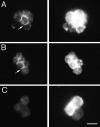
A conserved membrane-proximal segment of the neuroglian cytoplasmic domain is dispensable for recruitment of ankyrin to cell contacts in S2 cells. S2 cells expressing wild-type or cytoplasmic deletion Drosophila neuroglian were allowed to aggregate and were double stained with a polyclonal rabbit anti- Drosophila ankyrin (left column) and a mouse anti-Drosophila neuroglian antibody (right column) followed by fluorescently- labeled secondary antibodies. Cells were permeabilized before staining by treatment with 0.1% Triton X-100. Drosophila ankyrin was recruited to cell contact sites (arrows) in cell aggregates expressing either wild-type neuroglian (A) or neuroglian in which the first 18 amino acid residues of the cytoplasmic domain had been deleted (construct no. 2 shown in row B). All other deletions in the cytoplasmic neuroglian domain abolished ankyrin recruitment (as shown for construct no. 9 in panels C). Bar, 10 μm.
Figure 5
Figure 5
Qualitative and quantitative yeast two-hybrid analysis of the interaction between Drosophila ankyrin and neuroglian167 cytoplasmic domains with tyrosine to phenylalanine amino acid substitutions. (A) Qualitative yeast two-hybrid analysis of the interaction between Y to F neuroglian cytoplasmic domain mutations and Drosophila ankyrin. Blue colonies result from the induction of β-galactosidase activity and indicate an interaction between the two GAL4 fusion proteins. Top row, yeast colonies containing a pACTII–Drosophila ankyrin construct (expressing a GAL4 activation domain–Drosophila ankyrin fusion protein) as well as a pAS1-CYH2 construct (expressing a fusion protein consisting of the GAL4 DNA-binding domain and a neuroglian cytoplasmic domain, which was either wild-type or contained one or two point mutations resulting in Y to F amino acid substitutions); bottom row, yeast colonies containing the same pAS1–CYH2 constructs and a pACTII vector control. (B) Quantitative evaluation of β-galactosidase expression in the yeast cells shown in A. Data bars represent the mean ± SD from two different experiments performed in triplicate determinations.
Figure 5
Figure 5
Qualitative and quantitative yeast two-hybrid analysis of the interaction between Drosophila ankyrin and neuroglian167 cytoplasmic domains with tyrosine to phenylalanine amino acid substitutions. (A) Qualitative yeast two-hybrid analysis of the interaction between Y to F neuroglian cytoplasmic domain mutations and Drosophila ankyrin. Blue colonies result from the induction of β-galactosidase activity and indicate an interaction between the two GAL4 fusion proteins. Top row, yeast colonies containing a pACTII–Drosophila ankyrin construct (expressing a GAL4 activation domain–Drosophila ankyrin fusion protein) as well as a pAS1-CYH2 construct (expressing a fusion protein consisting of the GAL4 DNA-binding domain and a neuroglian cytoplasmic domain, which was either wild-type or contained one or two point mutations resulting in Y to F amino acid substitutions); bottom row, yeast colonies containing the same pAS1–CYH2 constructs and a pACTII vector control. (B) Quantitative evaluation of β-galactosidase expression in the yeast cells shown in A. Data bars represent the mean ± SD from two different experiments performed in triplicate determinations.
Figure 6
Figure 6
A cytoplasmic tyrosine residue is required for efficient recruitment of ankyrin to cell contacts in neuroglian-expressing S2 cells. S2 cells were induced to express wild-type neuroglian (A), nrgY1217F (B), nrgY1234F (C and D), or double mutant neuroglian (E–G) for 24 h before processing for antibody labeling of neuroglian (left panels) and ankyrin (right panels) as in Fig. 4. Arrows, abundance of neuroglian at noncontact regions of the plasma membrane; arrowheads, cell contact sites in mutant cells with little or no recruitment of ankyrin. Bar, 10 μM.
Figure 7
Figure 7
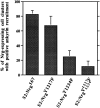
Quantification of ankyrin recruitment in S2 cells expressing neuroglian proteins mutated at cytoplasmic tyrosines. Populations of transfected S2 cells expressing wild-type neuroglian (Nrg167) or the indicated tyrosine mutations of neuroglian were double labeled with mouse anti-neuroglian and rabbit anti-ankyrin antibodies as in Fig. 3. In each experiment, 100 S2 cell clusters were identified by virtue of their neuroglian expression and then scored for the presence or absence of detectable ankyrin staining at cell contacts. Data bars represent the mean ± SD from two independent experiments.
Figure 8
Figure 8
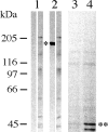
Neuroglian is not phosphorylated on tyrosine residues in S2 cells. Western blots of total S2 cell proteins from uninduced cells (lane 1) or cells induced to express neuroglian (lane 2) were stained with mouse anti-neuroglian (lanes 1 and 2, left columns) or rabbit anti-phosphotyrosine antibodies (right columns). Antibody binding was detected with alkaline phosphatase-conjugated secondary antibodies. Total proteins from control PC12 cells (lane 3) and nerve growth factor-stimulated PC12 cells (lane 4) were also stained with anti-phosphotyrosine antibody. The positions of neuroglian (*), MAP kinase (**) and molecular weight standards (left) are indicated.
Figure 9
Figure 9
S2 cells expressing neuroglian with a cytoplasmic Y1234F mutation exhibit a reduced level of cell aggregation. S2 cell lines that had been transfected with either wild-type (B) or mutated neuroglian cDNA constructs (C, NrgY1217F; D, NrgY1234F; and E, NrgnrgY1217/1234F) were induced for 15 h with Cu2+ ions and subsequently incubated at room temperature on a shaking platform at 200 rpm for 4 h. (A) Control cells not expressing neuroglian. S2 cells cultures were processed in parallel for Western blot analysis and probed with the neuroglian-specific 3F4 mAb (F). The equivalent of 5 × 10⁵ cells was loaded in each lane. Bar, 200 μm.
Figure 9
Figure 9
S2 cells expressing neuroglian with a cytoplasmic Y1234F mutation exhibit a reduced level of cell aggregation. S2 cell lines that had been transfected with either wild-type (B) or mutated neuroglian cDNA constructs (C, NrgY1217F; D, NrgY1234F; and E, NrgnrgY1217/1234F) were induced for 15 h with Cu2+ ions and subsequently incubated at room temperature on a shaking platform at 200 rpm for 4 h. (A) Control cells not expressing neuroglian. S2 cells cultures were processed in parallel for Western blot analysis and probed with the neuroglian-specific 3F4 mAb (F). The equivalent of 5 × 10⁵ cells was loaded in each lane. Bar, 200 μm.
Figure 10
Figure 10
Western blots of fasciclin II, neuroglian, and fasciclin IIEC-neuroglianTM+CP hybrid proteins expressed in S2 cells. S2 cells, which had been transfected with a fasciclin II (PEST form) (lanes 1), a neuroglian167 (lanes 2), or a fasciclin IIEC-neuroglianTM+CP (lanes 3) cDNA construct, were induced overnight and processed for Western blot analysis. Blot A was incubated with the neuroglian-specific mAb 3F4 which recognizes an epitope on the neuroglian extracellular domain, blot B with a rat polyclonal antiserum which was raised against the entire fasciclin II protein, blot C with the mAb 1D4 which binds to a cytoplasmic epitope of the fasciclin II (PEST) protein form, and blot D with a polyclonal rat antiserum which was raised against a small peptide representing the eight carboxy-terminal amino acid residues of the neuroglian167 form.
Figure 11
Figure 11
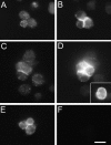
Selective recruitment of ankyrin to cell contacts in S2 cells expressing a chimeric neuroglian molecule. S2 cells were induced to express the adhesion molecule fasciclin II (A and B) or a chimeric molecule (fasciclin IIEC-neuroglianTM+CP) in which the extracellular neuroglian domain was replaced with the corresponding domain from fasciclin II (C–F). Cells were induced for 48 h and then processed for immunofluorescent staining with antibodies against ankyrin (A, C, and E) and against the extracellular domain of either fasciclin II (B and D) or neuroglian (F). Control experiments (D, inset) revealed that fasciclin II was abundantly detected at all regions of the plasma membrane when the detergent permeabilization step was omitted. Bar, 10 μM.

Similar articles

Cited by

References

    1. Ausubel, F.M., R. Brent, R.E. Kingston, D.D. Moore, J.G. Seidman, J.A. Smith, and K. Struhl. 1988. Current Protocols in Molecular Biology. J. Wiley & Sons, Inc., New York.
    1. Bartel, P.L., C.-t. Chien, R. Sternglanz, and S. Fields. 1993. Using the two- hybrid system to detect protein-protein interactions. In Cellular Interactions in Development: A Practical Approach. D.A. Hartley, editor. IRL Press, Oxford, UK. 153–179.
    1. Bieber, A.J. 1994. Analysis of cellular adhesion in cultured cells. In Drosophila melanogaster: Practical Uses in Cell Biology. L. Goldstein and E. Fyrberg, editors. 44:683–696. Academic Press, San Diego, CA. - PubMed
    1. Bieber AJ, Snow PM, Hortsch M, Patel NH, Jacobs JR, Traquina ZR, Schilling J, Goodman CS. Drosophilaneuroglian: a member of the immunoglobulin superfamily with extensive homology to the vertebrate neural adhesion molecule L1. Cell. 1989;59:447–460. - PubMed
    1. Boulton TG, Nye SH, Robbins DJ, Ip NY, Radziejewska E, Morgenbesser SD, DePinho RA, Panayotatos N, Cobb MH, Yancopoulos GD. ERKs: a family of protein-serine/threonine kinases that are activated and tyrosine phosphorylated in response to insulin and NGF. Cell. 1991;65:663–675. - PubMed

Publication types

MeSH terms