. 1998 Jul 13;142(1):285-94.
doi: 10.1083/jcb.142.1.285.

Cartilage fibrils of mammals are biochemically heterogeneous: differential distribution of decorin and collagen IX

Affiliations

Cartilage fibrils of mammals are biochemically heterogeneous: differential distribution of decorin and collagen IX

R Hagg et al. J Cell Biol. .

Abstract

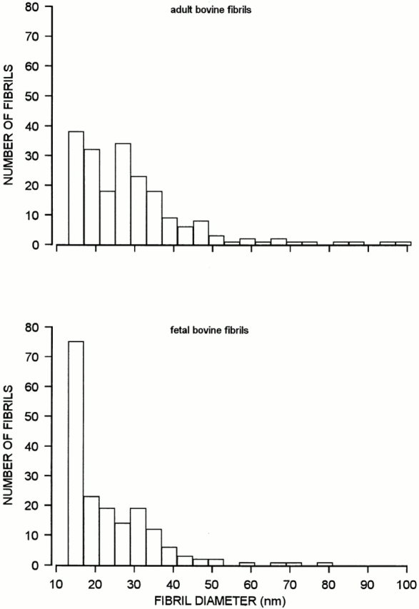
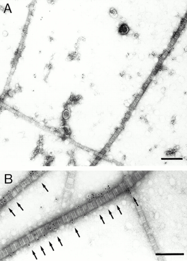
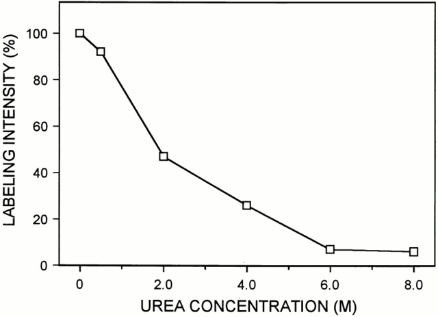
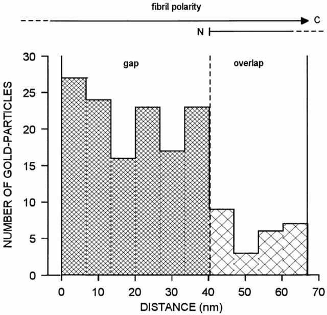
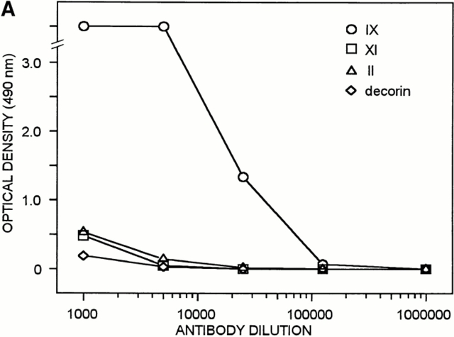
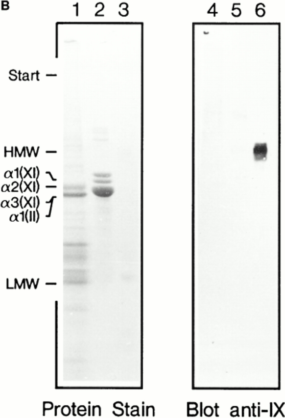
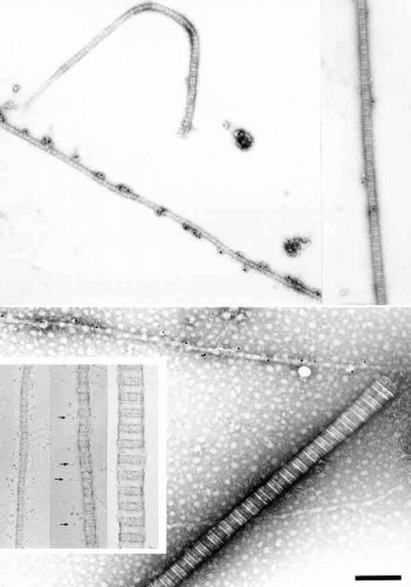
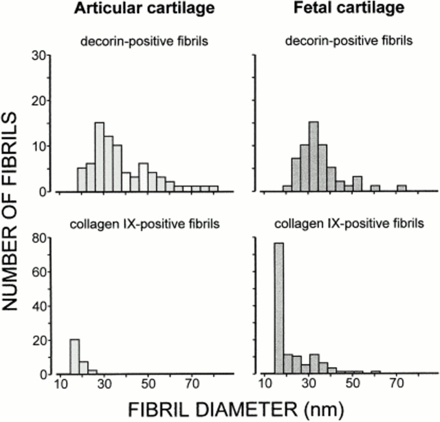
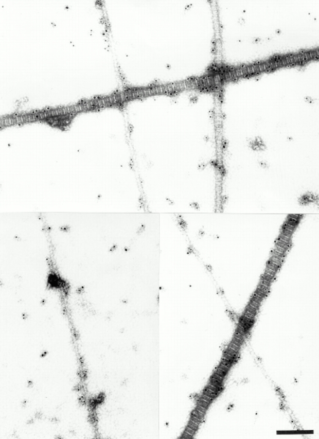
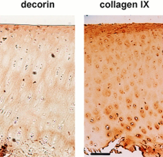
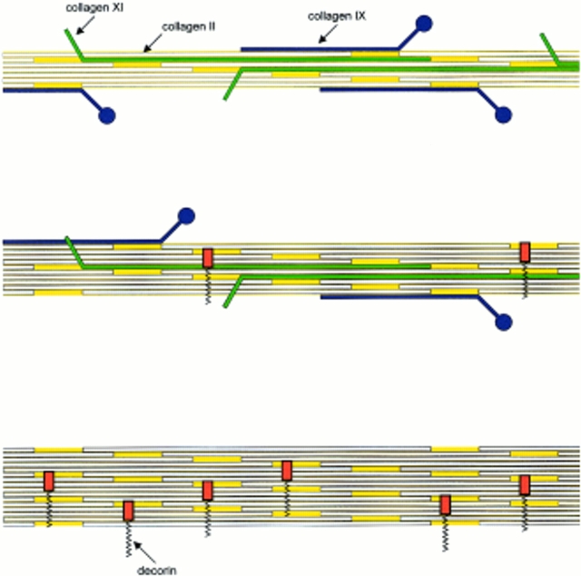
Cartilage fibrils contain collagen II as the major constituent, but the presence of additional components, minor collagens, and noncollagenous glycoproteins is thought to be crucial for modulating several fibril properties. We have examined the distribution of two fibril constituents-decorin and collagen IX-in samples of fibril fragments obtained after bovine cartilage homogenization. Decorin was preferentially associated with a population of thicker fibril fragments from adult articular cartilage, but was not present on the thinnest fibrils. The binding was specific for the gap regions of the fibrils, and depended on the decorin core protein. Collagen IX, by contrast, predominated in the population with the thinnest fibrils, and was scarce on wider fibrils. Double-labeling experiments demonstrated the coexistence of decorin and collagen IX in some fibrils of intermediate diameter, although most fibril fragments from adult cartilage were strongly positive for one component and lacked the other. Fibril fragments from fetal epiphyseal cartilage showed a different pattern, with decorin and collagen IX frequently colocalized on fragments of intermediate and large diameters. Hence, the presence of collagen IX was not exclusive for fibrils of small diameter. These results establish that articular cartilage fibrils are biochemically heterogeneous. Different populations of fibrils share collagen II, but have distinct compositions with respect to macromolecules defining their surface properties.


Figures

Figure 1
Figure 1
Histograms showing the diameter distribution of fibril fragments from bovine adult articular cartilage (top) and fetal cartilage (bottom), as determined by electron microscopy after negative staining. The fibril extracts were those described in the footnotes to Tables I and II. The overall number of measured fibrils were 202 and 181, respectively.
Figure 2
Figure 2
Ultrastructural immunolocalization of decorin in adult bovine articular cartilage extracts. (A) Fibril fragments having similar morphological appearance are either unlabeled or strongly labeled with gold probes (12 nm) for decorin. Note also the specific decorin labeling in structures apart from fibrils. (B) Electron micrographs at higher magnification indicate the preferential localization of colloidal gold to the gap regions (arrows). Bars, 200 nm.
Figure 3
Figure 3
Effect of urea treatment on the association of decorin within fibril fragments extracted from adult bovine articular cartilage. For each urea concentration, several electron micrographs were taken, gold particles associated with fibrils were counted, and the average number of probes per D-period was calculated. The results are shown as relative labeling intensities, with the density of markers in the sample not treated with urea set to 100%.
Figure 4
Figure 4
Frequency distribution of decorin-directed gold particles along fibril fragments. Distances were measured between the NH2-terminal starting point of the gap region and the center of each gold particle within the respective D-period. For the evaluation, D-periods were considered only if the polarity of the collagen molecules within the fibril could be clearly identified.
Figure 5
Figure 5
Characterization of polyclonal mouse antiserum against bovine collagen IX by ELISA and immunoblotting. (A) The antigens tested by ELISA were collagen IX (○), collagen XI (□), collagen II (▵), and decorin (⋄). (B) Noncollagenous proteins were extracted from chondroitinase ABC–digested pieces of bovine articular cartilage with boiling SDS-PAGE sample buffer containing 2% β-mercaptoethanol. Aliquots were electrophoresed on 4.5–15% polyacrylamide gels (lanes 1 and 4). Portions of a sample containing purified collagens II and XI were loaded in lanes 2 and 5. Pepsin-resistant collagen IX, i.e., the antigen, was applied in lanes 3 and 6. Proteins in the gel were stained with Coomassie Blue (lanes 1–3) or transferred to nitrocellulose and subjected to immunoblotting (lanes 4–6).
Figure 5
Figure 5
Characterization of polyclonal mouse antiserum against bovine collagen IX by ELISA and immunoblotting. (A) The antigens tested by ELISA were collagen IX (○), collagen XI (□), collagen II (▵), and decorin (⋄). (B) Noncollagenous proteins were extracted from chondroitinase ABC–digested pieces of bovine articular cartilage with boiling SDS-PAGE sample buffer containing 2% β-mercaptoethanol. Aliquots were electrophoresed on 4.5–15% polyacrylamide gels (lanes 1 and 4). Portions of a sample containing purified collagens II and XI were loaded in lanes 2 and 5. Pepsin-resistant collagen IX, i.e., the antigen, was applied in lanes 3 and 6. Proteins in the gel were stained with Coomassie Blue (lanes 1–3) or transferred to nitrocellulose and subjected to immunoblotting (lanes 4–6).
Figure 6
Figure 6
Ultrastructural localization of collagen IX in adult bovine articular cartilage extracts. Typical immunolabeling patterns are shown. A strongly labeled 20-nm fibril is seen together with a unlabeled 30-nm-fibril (top left). A 40-nm fibril demonstrates extremely weak labeling (top right). A thick 80-nm fibril is completely unlabeled, whereas the adjoining thin fibril is densely populated by gold particles (bottom). Rotary shadowing images of three selected fibril fragments are shown in the inset panel. Collagen IX projections (arrows) are seen with a rigid stalk (COL3) and a terminal globule (NC4). A typical thin fibril (∼20 nm) displays abundant collagen IX projections (left of inset), a fibril of intermediate thickness shows fewer of these projections (middle of inset), and the thick fibril fragment appears to be devoid of collagen IX (right of inset). Bar, 200 nm.
Figure 7
Figure 7
Distribution of diameters of extracted fibril fragments from adult articular cartilage and fetal bovine cartilage stained for decorin or collagen IX. Only fibrils with an average density of ≥5 gold particles per 10 D-periods were considered in this analysis. Thereby, labeling above background was ensured in all cases.
Figure 8
Figure 8
Combined immunolocalization of decorin and collagen IX on fibril fragments from adult bovine articular cartilage. Rabbit anti-decorin antibodies were detected by 18-nm gold probes and mouse anti-collagen IX antibodies by 12-nm probes. Two representative examples of fibrils with diameters between 30 and 40 nm are shown (top). These fibrils, although weakly labeled (<5 gold particles per 10 D-periods), demonstrate colocalization of the two fibril constituents. A typical thin fibril carries dense label with collagen IX-probes only, whereas the thick 60-nm fibril is labeled exclusively for decorin (bottom). Bar, 200 nm.
Figure 9
Figure 9
Combined immunolocalization of decorin and collagen IX in fetal bovine cartilage extracts. Rabbit anti-decorin antibodies were detected by 18-nm gold probes and mouse anti-collagen IX antibodies by 12-nm probes. Note that fibrils of all diameters are labeled with collagen IX-probes. The coexistence of decorin and collagen IX is obvious for fibrils thicker than 20 nm. Bar, 200 nm.
Figure 10
Figure 10
Localization of decorin and collagen IX in adult bovine articular cartilage. Sections of tarsometatarsal joint cartilage were incubated with antiserum against decorin (left) and collagen IX (right). Bound antibodies were detected by indirect immunoperoxidase staining. Decorin and collagen IX show differential distribution with partial overlapping. Both appear enriched in the most superficial cartilage. In the zone representing the deeper two-thirds of the tissue, strong staining for collagen IX is restricted to the territorial matrix surrounding the chondrocytes. Staining for decorin in the deep zone, by contrast, is strongest in the interterritorial matrix. Bar, 70 μm.
Figure 11
Figure 11
Schematic representation of the biochemical heterogeneity of D-periodic fibrils in mature articular cartilage. The thinnest fibrils (17–20 nm) are enriched in collagens IX and XI. Fibrils of intermediate size have lower contents of collagen IX, and the small proteoglycan decorin can occur as an additional component. The coexistence of collagen IX and decorin on the fibril surface is a notable feature. The thickest fibrils lack collagens IX and XI, but they frequently contain decorin. The suggested distribution of collagen IX and decorin is based on our data, whereas the distribution of collagen XI is drawn by analogy to that observed in human rib and fetal calf cartilage (Keene et al., 1995).

Similar articles

Cited by

References

    1. Birk DE, Fitch JM, Babiarz JP, Linsenmayer TF. Collagen type I and type V are present in the same fibrils in the avian corneal stroma. J Cell Biol. 1988;106:999–1008. - PMC - PubMed
    1. Birk DE, Nurminskaya MV, Zycband EI. Collagen fibrillogenesis in situ: fibril segments undergo post-depositional modifications resulting in linear and lateral growth during matrix development. Dev Dyn. 1995;202:229–243. - PubMed
    1. Brown DC, Vogel KG. Characteristics of the in vitro interaction of a small proteoglycan (PGII) of bovine tendon with type I collagen. Matrix. 1989;9:468–478. - PubMed
    1. Bruckner P, Mendler M, Steinmann B, Huber S, Winterhalter KH. The structure of human collagen type IX and its organization in fetal and infant cartilage fibrils. J Biol Chem. 1988;263:16911–16917. - PubMed
    1. Danielson KG, Baribault H, Holmes DF, Graham H, Kadler KE, Iozzo RV. Targeted disruption of decorin leads to abnormal collagen fibril morphology and skin fragility. J Cell Biol. 1997;136:729–743. - PMC - PubMed

Publication types