. 1998 Aug 1;510 ( Pt 3)(Pt 3):773-81.
doi: 10.1111/j.1469-7793.1998.773bj.x.

Angiotensin AT1 receptor-mediated excitation of rat carotid body chemoreceptor afferent activity

Affiliations

Angiotensin AT1 receptor-mediated excitation of rat carotid body chemoreceptor afferent activity

A M Allen. J Physiol. .

Abstract

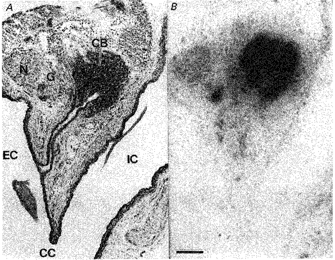
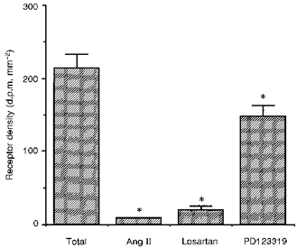
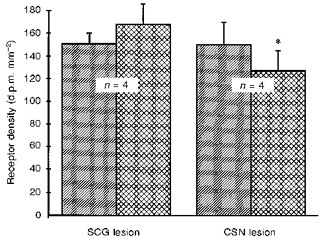
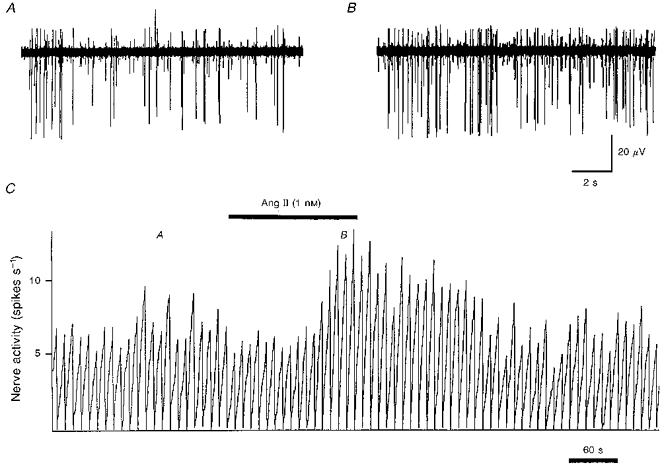
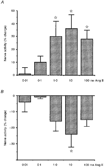
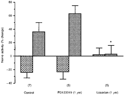
1. A high density of angiotensin II receptors was observed in the rat carotid body by in vitro autoradiography employing 125I-[Sar1, Ile8]-angiotensin II as radioligand. Displacement studies demonstrated that the receptors were of the AT1 subtype. 2. The binding pattern indicated that the AT1 receptors occurred over clumps of glomus cells, the principal chemoreceptor cell of the carotid body. Selective lesions of the sympathetic or afferent innervation of the carotid body had little effect on the density of receptor binding, demonstrating that the majority of AT1 receptors were intrinsic to the glomus cells. 3. To determine the direct effect of angiotensin II on chemoreceptor function, without the confounding effects of the vasoconstrictor action of angiotensin II, carotid sinus nerve activity was recorded from the isolated carotid body in vitro. The carotid body was superfused with Tyrode solution saturated with carbogen (95 % O2, 5 % CO2), maintained at 36 C, and multi-unit nerve activity recorded with a suction electrode. 4. Angiotensin II elicited a dose-dependent excitation of carotid sinus nerve activity (maximum increase of 36 +/- 11 % with 10 nM angiotensin II) with a threshold concentration of 1 nM. The response was blocked by the addition of an AT1 receptor antagonist, losartan (1 microM), but not by the addition of an AT2 receptor antagonist, PD123319 (1 microM). 5. In approximately 50 % of experiments the excitation was preceded by an inhibition of activity (maximum decrease of 24 +/- 8 % with 10 nM angiotensin II). This inhibitory response was markedly attenuated by losartan but not affected by PD123319. 6. These observations demonstrate that angiotensin II, acting through AT1 receptors located on glomus cells in the carotid body, can directly alter carotid chemoreceptor afferent activity. This provides a means whereby humoral information about fluid and electrolyte homeostasis might influence control of cardiorespiratory function.


Figures

Figure 1
Figure 1. Angiotensin AT1 receptor distribution in the rat carotid body and carotid bifurcation
A, a haematoxylin- and eosin-stained 20 μm section of the carotid bifurcation region. B, an X-ray film depicting binding of 125I-[Sar1,Ile8]-Ang II to the section adjacent to that in A. Black silver deposits in B indicate the presence of Ang II AT1 receptors. The binding is totally displaced by 10 μm losartan or 1 μm Ang II amide. The calibration bar represents 250 μm. CB, carotid body; CC, common carotid artery; EC, external carotid artery; IC, internal carotid artery; G, isolated clump of ganglion cells; N, sympathetic nerve bundle arising from the superior cervical ganglion.
Figure 2
Figure 2. Density of Ang II AT1 and AT2 receptor subtypes in the rat carotid body
Bar graph demonstrating the density of 125I-[Sar1,Ile8]-Ang II binding to the rat carotid body alone and in the presence of 1 μm Ang II amide, 10 μm losartan or 10 μm PD123319. The asterisk denotes a significant difference in receptor density compared to total binding (P < 0.05).
Figure 3
Figure 3. Effect of selective nerve lesions on Ang II receptor density in the rat carotid body
Bar graph showing the effect of superior cervical ganglionectomy (SCG lesion) or carotid sinus nerve section (CSN lesion) on AT1 receptor binding in the rat carotid body. Hatched bars represent the sham-lesioned contralateral carotid body which served as control. Cross-hatched bars are the lesioned carotid bodies. The asterisk denotes a significant difference from control (P < 0.05).
Figure 4
Figure 4. Carotid sinus nerve activity during application of Ang II (1 nm) to the carotid body in vitro
An example of the change in carotid sinus nerve activity in response to superfusion of the carotid body in vitro with 1 nm Ang II in Tyrode solution. Raw multi-unit activity is shown before (A) and during (B) superfusion with Ang II. The integrated spike activity (10 s bins) is shown in C. The times of the recordings shown in A and B are denoted by the respective letters in C. It can be observed, particularly with the largest spike, that the increase in multi-unit activity is due to an increase in the rate of firing, and not just recruitment of new neurons.
Figure 5
Figure 5. Change in carotid sinus nerve activity in response to carotid body superfusion with Ang II
Histogram demonstrating mean ±s.e.m. changes in multi-unit carotid sinus nerve activity in response to various doses of Ang II applied to the carotid body in vitro. A, mean increases in activity which occurred in the majority of experiments 123 ± 3 s after exposure to Ang II. B, mean decreases in activity which preceded the excitation in approximately 50% of experiments at a time of 50 ± 5 s after exposure to Ang II. Activity is expressed as a percentage change from the preceding control period in each case. The stars denote that the mean is significantly different from control (P < 0.05, ANOVA followed by Fisher's LSD test).
Figure 6
Figure 6. The effect of AT1 and AT2 receptor antagonists on the changes in chemoreceptor activity in response to superfusion with 10 nm Ang II
Bar graph indicating changes in carotid sinus nerve activity (means ±s.e.m.) in response to superfusion with 10 nm Ang II alone (control), or in the presence of 1 μm PD123319 or 1 μm losartan. The values depicted by the cross-hatched bars are for the initial inhibitory period whilst the hatched bars are for the later excitatory response. The numbers in parentheses indicate the number of experiments at each point. The asterisk denotes that the excitatory response to Ang II is significantly decreased in the presence of losartan (P < 0.05).

Similar articles

Cited by

References

    1. Allen AM, Chai SY, Clevers J, McKinley MJ, Paxinos G, Mendelsohn FAO. Localization and characterization of angiotensin II receptor binding and angiotensin converting enzyme in the human medulla oblongata. Journal of Comparative Neurology. 1988;269:249–264. - PubMed
    1. Allen AM, Paxinos G, McKinley MJ, Chai SY, Mendelsohn FAO. Localization and characterization of angiotensin II receptor binding sites in the human basal ganglia, thalamus, midbrain, pons and cerebellum. Journal of Comparative Neurology. 1991;312:291–298. - PubMed
    1. Assmussen E, Nielsen M. Studies on the initial increase in O2-capacity of the blood at low oxygen pressure. Acta Physiologica Scandinavica. 1945;9:75–87.
    1. Castren E, Kurihara M, Gutkind JS, Saavedra JM. Specific angiotensin binding sites in the rat stellate and superior cervical ganglia. Brain Research. 1987;422:347–351. - PubMed
    1. Donnelly DF. Does catecholamine secretion mediate the hypoxia-induced increase in nerve activity? Biological Signals. 1995;4:304–309. - PubMed

Publication types

MeSH terms

LinkOut - more resources