

Sympathetic attenuation of parasympathetic vasodilatation in oro-facial areas in the cat
- PMID: 9660902
- PMCID: PMC2231085
- DOI: 10.1111/j.1469-7793.1998.915bj.x
Sympathetic attenuation of parasympathetic vasodilatation in oro-facial areas in the cat
Abstract
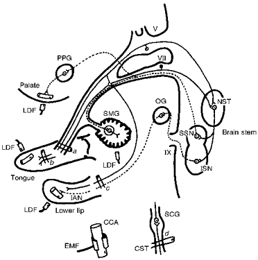
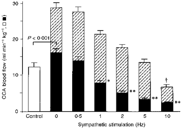
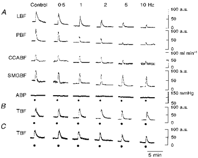
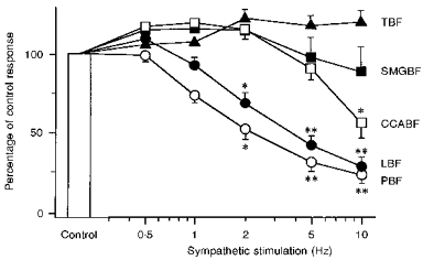
1. The present study was designed to examine the interaction between sympathetic and parasympathetic influences on blood flow in oro-facial areas such as lower lip, palate and submandibular gland (SMG) and in the common carotid artery (CCA) in anaesthetized cats. 2. Section of the ipsilateral superior cervical sympathetic trunk (CST) increased the basal CCA blood flow significantly. The control level with the nerve intact was comparable with that seen at 0.5-1 Hz CST stimulation, suggesting a spontaneous discharge of around 0. 5-1 Hz in the CST fibres innervating the beds supplied by the CCA. The basal blood flow at all sites examined was reduced by CST stimulation in a frequency-dependent manner. 3. Electrical stimulation of the central end of the lingual nerve (LN) evoked blood flow increases in the lower lip and palate. These blood flow increases were markedly reduced by concurrent CST stimulation in a manner that was frequency dependent, but not simply related to the vasoconstrictor effect of CST stimulation. This effect of CST stimulation was not observed in tongue or SMG, even though CST stimulation evoked vasoconstriction in these tissues. A significant reduction in the level of CCA blood flow attained during LN stimulation was observed on repetitive CST stimulation only at 10 Hz, indicating that this response behaved in a fashion different from that seen in the lower lip, palate, tongue and SMG. 4. The present study suggests that concurrent repetitive CST stimulation reduces parasympathetically mediated blood flow increases in certain oro-facial areas (such as the lower lip and palate), but not in the tongue and SMG. This inhibitory action was not a simple additive effect (between vasoconstriction and vasodilatation) and it disappeared rapidly after the cessation of CST stimulation.
Figures
Similar articles
-
Escape of parasympathetic vasodilatation from sympathetic attenuation in oro-facial areas in the cat.Tohoku J Exp Med. 1999 Jun;188(2):153-60. doi: 10.1620/tjem.188.153. Tohoku J Exp Med. 1999. PMID: 10526877
-
Functional roles played by the sympathetic supply to lip blood vessels in the cat.Am J Physiol. 1999 Sep;277(3):R682-9. doi: 10.1152/ajpregu.1999.277.3.R682. Am J Physiol. 1999. PMID: 10484484
-
Nifedipine-induced inhibition of parasympathetic-mediated vasodilation in the orofacial areas of the cat.Am J Physiol Regul Integr Comp Physiol. 2000 Jul;279(1):R332-9. doi: 10.1152/ajpregu.2000.279.1.R332. Am J Physiol Regul Integr Comp Physiol. 2000. PMID: 10896897
-
Reflex parasympathetic vasodilatation in facial skin.Gen Pharmacol. 1995 Mar;26(2):237-44. doi: 10.1016/0306-3623(94)00155-g. Gen Pharmacol. 1995. PMID: 7590072 Review.
-
Nervous control of blood flow in the orofacial region.Pharmacol Ther. 1999 Feb;81(2):141-61. doi: 10.1016/s0163-7258(98)00040-0. Pharmacol Ther. 1999. PMID: 10190584 Review.
Cited by
-
Occurrence of parasympathetic vasodilator fibers in the lower lip of the guinea-pig.J Comp Physiol B. 2008 Mar;178(3):297-305. doi: 10.1007/s00360-007-0222-z. Epub 2007 Nov 21. J Comp Physiol B. 2008. PMID: 18030480
-
Effect of propofol on salivary secretion from the submandibular, sublingual, and labial glands during intravenous sedation.J Dent Anesth Pain Med. 2023 Jun;23(3):153-162. doi: 10.17245/jdapm.2023.23.3.153. Epub 2023 May 26. J Dent Anesth Pain Med. 2023. PMID: 37313266 Free PMC article.
-
Differences in the autonomic regulation of temperature in the lower lip and tongue during activation of the lingual nerve.J Physiol Sci. 2025 Jul;75(2):100028. doi: 10.1016/j.jphyss.2025.100028. Epub 2025 Jun 4. J Physiol Sci. 2025. PMID: 40483984 Free PMC article.
-
Effect of the parasympathetic vasodilation on temperature regulation via trigeminal afferents in the orofacial area.J Physiol Sci. 2020 Mar 31;70(1):22. doi: 10.1186/s12576-020-00749-y. J Physiol Sci. 2020. PMID: 32234014 Free PMC article.
-
Species differences in the reflex effects of lingual afferent nerve stimulation on lip blood flow and arterial pressure.J Comp Physiol B. 2003 Nov;173(8):629-36. doi: 10.1007/s00360-003-0363-7. Epub 2003 Aug 15. J Comp Physiol B. 2003. PMID: 12920546
References
-
- Bartsch T, Habler H-J, Jänig W. Functional properties of postganglionic sympathetic neurones supplying the submandibular gland in the anaesthetized rat. Neuroscience Letters. 1996;214:143–146. - PubMed
-
- Habler H-J, Jänig W, Krummel M, Peters OA. Reflex patterns in postganglionic neurons supplying skin and skeletal muscle of the rat hindlimb. Journal of Neurophysiology. 1994;72:2222–2236. - PubMed
-
- Habler H-J, Wasner G, Jänig W. Interaction of sympathetic vasoconstriction and antidromic vasodilatation in the control of skin blood flow. Experimental Brain Research. 1997;113:402–410. - PubMed
-
- Hall GT, Gardner TD, Potter EK. Attenuation of long-lasting effects of sympathetic stimulation after repeated stimulation. Circulation Research. 1990;67:193–198. - PubMed
Publication types
MeSH terms
LinkOut - more resources
Full Text Sources
Miscellaneous