

Signalling of static and dynamic features of muscle spindle input by cuneate neurones in the cat
- PMID: 9660903
- PMCID: PMC2231082
- DOI: 10.1111/j.1469-7793.1998.923bj.x
Signalling of static and dynamic features of muscle spindle input by cuneate neurones in the cat
Abstract
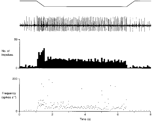
1. The capacity of cuneate neurones to signal information derived from muscle spindle afferent fibres about static stretch or vibration of forearm extensor muscles was examined electrophysiologically in anaesthetized cats. 2. Static stretch (>= 2 mm in amplitude) and sinusoidal vibration (at frequencies of 50-800 Hz) were applied longitudinally to individual muscle tendons by means of a feedback controlled mechanical stimulator, and responses were recorded from individual cuneate neurones and from individual spindle afferent fibres. 3. Cuneate neurones sampled were located caudal to the obex and displayed a sensitivity to both vibration and static stretch of forearm muscles that was consistent with their input arising from primary spindle endings. In response to static muscle stretch, they displayed graded and approximately linear stimulus-response relations, and a stability of response level at fixed lengths that was consistent with these neurones contributing discriminative information about static muscle stretch. 4. In response to sinusoidal muscle vibration the cuneate neurones also showed graded stimulus-response relations (in contrast to spindle afferents which at low vibration amplitudes attain a plateau response level corresponding to a discharge of 1 impulse on each vibration cycle). Lowest thresholds were at 100-300 Hz and bandwidths of vibration sensitivity extended up to approximately 800 Hz. 5. Temporal precision in cuneate responses to muscle vibration was assessed by constructing phase scatter and cycle histograms from which measures of vector strength could be calculated. Cuneate responses displayed somewhat poorer phase locking (and lower vector strengths) than spindle afferent responses to vibration (a reflection of uncertainties associated with synaptic transmission). Nevertheless, the remarkable feature of cuneate responses to muscle vibration is the preservation of tight phase locking at frequencies up to 400-500 Hz, which presumably enables these central neurones to contribute accurate temporal information for the kinaesthetic sense in a variety of circumstances involving dynamic perturbations to skeletal muscle.
Figures


Similar articles
-
Signalling of static and dynamic features of muscle spindle input by external cuneate neurones in the cat.J Physiol. 1999 Sep 1;519 Pt 2(Pt 2):559-69. doi: 10.1111/j.1469-7793.1999.0559m.x. J Physiol. 1999. PMID: 10457071 Free PMC article.
-
Coding of information about tactile stimuli by neurones of the cuneate nucleus.J Physiol. 1978 Dec;285:493-513. doi: 10.1113/jphysiol.1978.sp012585. J Physiol. 1978. PMID: 745115 Free PMC article.
-
Inhibition of cuneate neurones: its afferent source and influence on dynamically sensitive "tactile" neurones.J Physiol. 1977 Jun;268(1):251-70. doi: 10.1113/jphysiol.1977.sp011856. J Physiol. 1977. PMID: 874897 Free PMC article.
-
Information transmission by isolated frog muscle spindle.Biol Cybern. 1985;52(3):165-76. doi: 10.1007/BF00339945. Biol Cybern. 1985. PMID: 2992613 Review.
-
Synaptic transmission between single tactile and kinaesthetic sensory nerve fibers and their central target neurones.Behav Brain Res. 2002 Sep 20;135(1-2):197-212. doi: 10.1016/s0166-4328(02)00166-3. Behav Brain Res. 2002. PMID: 12356451 Review.
Cited by
-
Somatosensory properties of cuneocerebellar neurones in the main cuneate nucleus of the rat.Cerebellum. 2003;2(2):131-45. doi: 10.1080/14734220309406. Cerebellum. 2003. PMID: 12880181
-
Processing afferent proprioceptive information at the main cuneate nucleus of anesthetized cats.J Neurosci. 2010 Nov 17;30(46):15383-99. doi: 10.1523/JNEUROSCI.2193-10.2010. J Neurosci. 2010. PMID: 21084595 Free PMC article.
-
Signalling of static and dynamic features of muscle spindle input by external cuneate neurones in the cat.J Physiol. 1999 Sep 1;519 Pt 2(Pt 2):559-69. doi: 10.1111/j.1469-7793.1999.0559m.x. J Physiol. 1999. PMID: 10457071 Free PMC article.
References
-
- Amassian VE, Macy J, Jr, Waller HJ, Leader HS, Swift M. Transformation of afferent activity at the cuneate nucleus. In: Gerard RW, Duyff JW, editors. Information Processing in the Nervous System. Amsterdam: Excerpta Medica Foundation; 1962. pp. 235–254.
-
- Andersen P, Eccles JC, Oshima T, Schmidt RF. Mechanisms of synaptic transmission in the cuneate nucleus. Journal of Neurophysiology. 1964;27:1096–1116. - PubMed
-
- Barker D. The innervation of mammalian skeletal muscle. In: de Reuck AVS, Knight J, editors. Myotatic, Kinesthetic and Vestibular Mechanisms. London: Churchill; 1967. pp. 3–15. Ciba Foundation Symposium.
-
- Bessou P, Laporte Y. Responses from primary and secondary endings of the same neuromuscular spindle of the tenuissimus muscle of the cat. In: Barker D, editor. Symposium on Muscle Receptors. Hong Kong: Hong Kong University Press; 1962. pp. 105–119.
-
- Bianconi R, Van der Meulen JP. The response to vibration of the end organs of the mammalian muscle spindles. Journal of Neurophysiology. 1963;26:177–190. - PubMed
Publication types
MeSH terms
LinkOut - more resources
Full Text Sources
Miscellaneous