. 1998 Jul;117(3):1015-22.
doi: 10.1104/pp.117.3.1015.

Multiple genes encoding the conserved CCAAT-box transcription factor complex are expressed in Arabidopsis

Affiliations

Multiple genes encoding the conserved CCAAT-box transcription factor complex are expressed in Arabidopsis

D Edwards et al. Plant Physiol. 1998 Jul.

Abstract

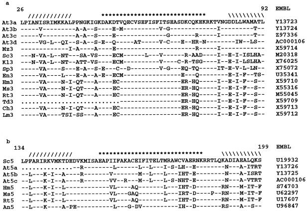
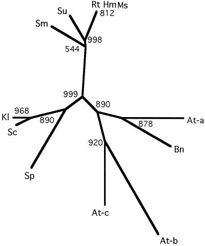
The CCAAT motif is found in the promoters of many eukaryotic genes. In yeast a single complex of three proteins, termed HAP2, HAP3, and HAP5, binds to this sequence, and in mammals the three components of the equivalent complex (called variously NF-Y, CBF, or CP1) are also represented by single genes. Here we report the presence of multiple genes for each of the components of the CCAAT-binding complex, HAP2, 3,5, from Arabidopsis. Three independent Arabidopsis HAP subunit 2 (AtHAP2) cDNAs were cloned by functional complementation of a yeast hap2 mutant, and two independent forms each of AtHAP3 and AtHAP5 cDNAs were detected in the expressed sequence tag database. Additional homologs (two of AtHAP3 and one of AtHAP5) have been identified from available Arabidopsis genomic sequences. Northern-blot analysis indicated ubiquitous expression for each AtHAP2 and AtHAP5 cDNA in a range of tissues, whereas expression of each AtHAP3 cDNA was under developmental and/or environmental regulation. The unexpected presence of multiple forms of each HAP homolog in Arabidopsis, compared with the single genes in yeast and vertebrates, suggests that the HAP2,3,5 complex may play diverse roles in gene transcription in higher plants.


Figures

Figure 1
Figure 1
Complementation of yeast hap2 with AtHAP2 cDNAs. S. cerevisiae strain JO1-1a (MY69) was transformed with three independent Arabidopsis HAP2 cDNAs (P1, P3, and P5 for AtHAP2a, AtHAP2b, and AtHAP2c, respectively) in the yeast expression vector pFL61. After primary selection on medium without uracil, two different dilutions of transformants were plated onto lactate medium along with Δhap2 itself (MY69) and the parental strain with a wild-type HAP2 gene (MY68). A reduced level of complementation was observed for AtHAP2b (P3).
Figure 2
Figure 2
Protein sequence alignment of the subunit association and DNA-binding domains of HAP2 from a range of organisms. Numbering relates to AtHAP2b. Sequences were identified in the EMBL database (accession numbers are given in the right column) and were aligned using the Lineup program (Genetics Computer Group). Dots indicate gaps in the sequence; dashes indicate identity with AtHAP2a. At, Arabidopsis; Bn, B. napus; Sc, S. cerevisiae; Kl, Kluyveromyces lactis; Sp, S. pombe; Hm, human; Ms, mouse; Rt, rat; Sm, Schistosoma mansoni; and Su, sea urchin.
Figure 3
Figure 3
Alignment of HAP3 and HAP5 core sequences from different organisms. Sequences were identified in the EMBL database (accession numbers are given in the right column). The symbols in the top line (/ / / / / / /********///////) denote the position of the predicted histone fold triple helix. Numbering refers to the position within AtHAP3a or S. cerevisiae HAP5. a, Alignment of HAP3 sequences. b, Alignment of HAP5 sequences. It is clear that AtHAP5a is incomplete because it is missing the first of the conserved helices. Dots indicate gaps in the sequence; dashes indicate sequence identity. At, Arabidopsis; Mz, maize; Sc, S. cerevisiae; Kl, K. lactis; Sp, S. pombe; En, Emericella nidulans; Hm, human; Ms, mouse; Rt, rat; Td, toad; Ch, chicken; Lm, lamprey; and An, Aspergillus nidulans.
Figure 4
Figure 4
Expression of AtHAP cDNAs in various tissues from Arabidopsis. RNA was extracted from leaves (lane 1), flowers and siliques (lane 2), roots (lane 3), whole seedlings grown under a 16-h day/8-h night (lane 4), and whole seedlings grown in the dark for 48 h (lane 5). Ten micrograms of total RNA was loaded onto each lane, separated by electrophoresis, and blotted onto a nylon membrane. Membranes were probed with each radiolabeled AtHAP cDNA, followed by autoradiography. The bands were quantified by densitometry and normalized against the hybridization of an rDNA probe to the same filter. The relative values are shown in the histograms to the right of the autoradiographs. The sizes of the transcripts in kilobase pairs are indicated on the left.
Figure 5
Figure 5
Southern-blot analysis of Arabidopsis genomic DNA. Ten micrograms of DNA was digested with EcoRI (lane 1), BamHI (lane 2), or HindIII (lane 3), separated by gel electrophoresis, and blotted onto a nylon membrane. This was probed with radiolabeled AtHAP5a or AtHAP5b cDNAs, followed by autoradiography. The sizes of the bands in kilobase pairs are indicated to the right of each blot.
Figure 6
Figure 6
Unrooted phylogenetic tree relating HAP2 homologs from different organisms. Multiple sequence alignment and generation of the tree was the program ClustalW (Genetics Computer Group), excluding gap positions and with correction for multiple substitutions. Bootstrap values are for 1000 replicates. Abbreviations are as in Figure 2.

References

    1. Albani D, Robert LS. Cloning and characterization of a Brassica napus gene encoding a homologue of the B-subunit of a heteromeric CCAAT-binding factor. Gene. 1995;167:209–213. - PubMed
    1. Arents G, Moudrianakis EN. The histone fold: a ubiquitous architectural motif utilized in DNA compaction and protein dimerization. Proc Natl Acad Sci USA. 1995;92:11170–11174. - PMC - PubMed
    1. Baxevanis AD, Arents G, Moudrianakis EN, Landsman D. A variety of DNA-binding and multimeric proteins contain the histone fold motif. Nucleic Acids Res. 1995;23:2685–2691. - PMC - PubMed
    1. Becker DM, Fikes JD, Guarente L. A cDNA encoding a human CCAAT-binding protein cloned by functional complementation in yeast. Proc Natl Acad Sci USA. 1991;88:1968–1972. - PMC - PubMed
    1. Bellorini M, Kun Lee D, Dantonel JC, Zemozoumi K, Roeder RG, Tora L, Mantovani R. CCAAT binding NF-Y-TBP interactions: NF-YB and NF-YC require short domains adjacent to their histone fold motifs for association with TBP basic residues. Nucleic Acids Res. 1997;25:2174–2181. - PMC - PubMed