. 1998 Jul;153(1):109-19.
doi: 10.1016/s0002-9440(10)65551-4.

Expression and localization of matrix metalloproteinase-12 in the aorta of cholesterol-fed rabbits: relationship to lesion development

Affiliations

Expression and localization of matrix metalloproteinase-12 in the aorta of cholesterol-fed rabbits: relationship to lesion development

S Matsumoto et al. Am J Pathol. 1998 Jul.

Abstract

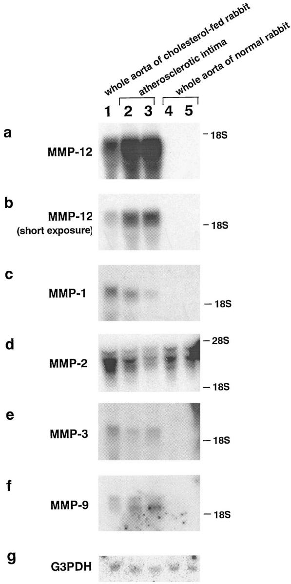
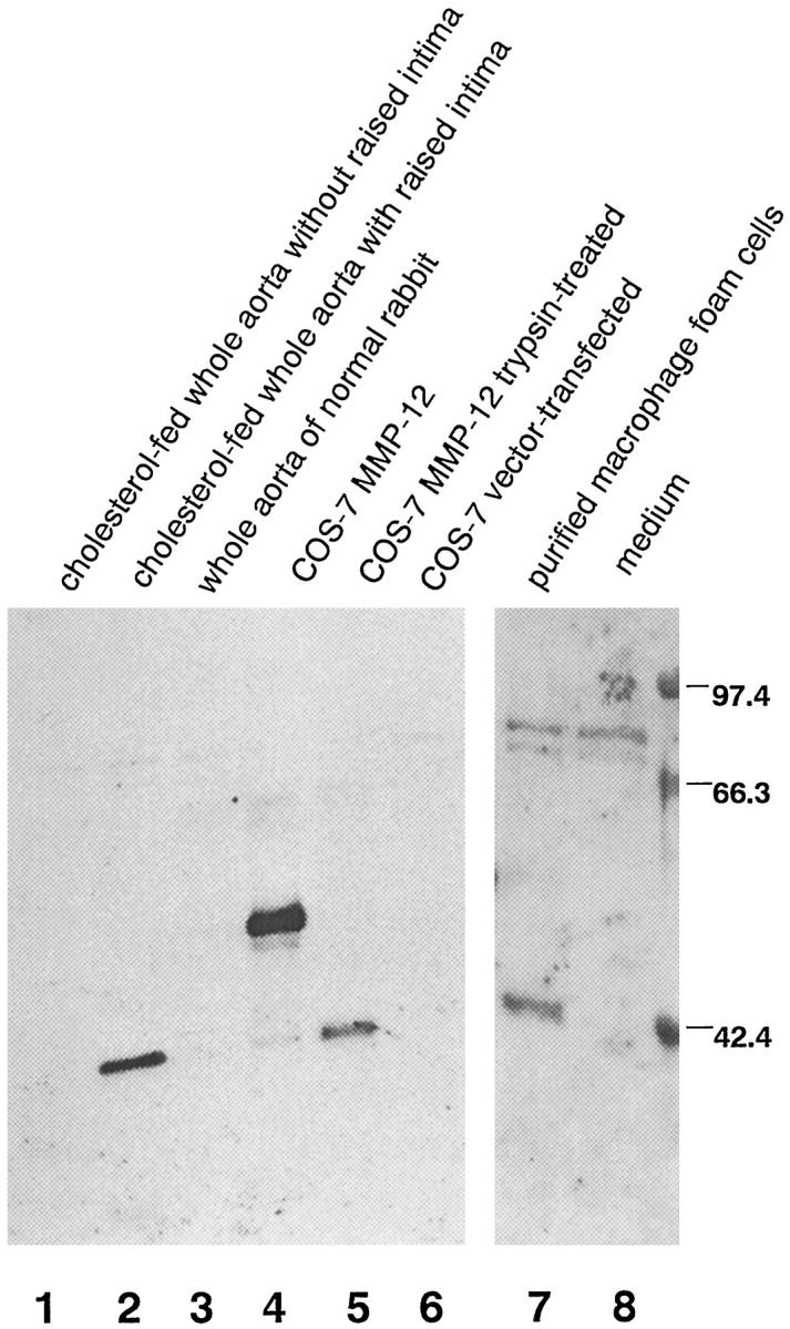
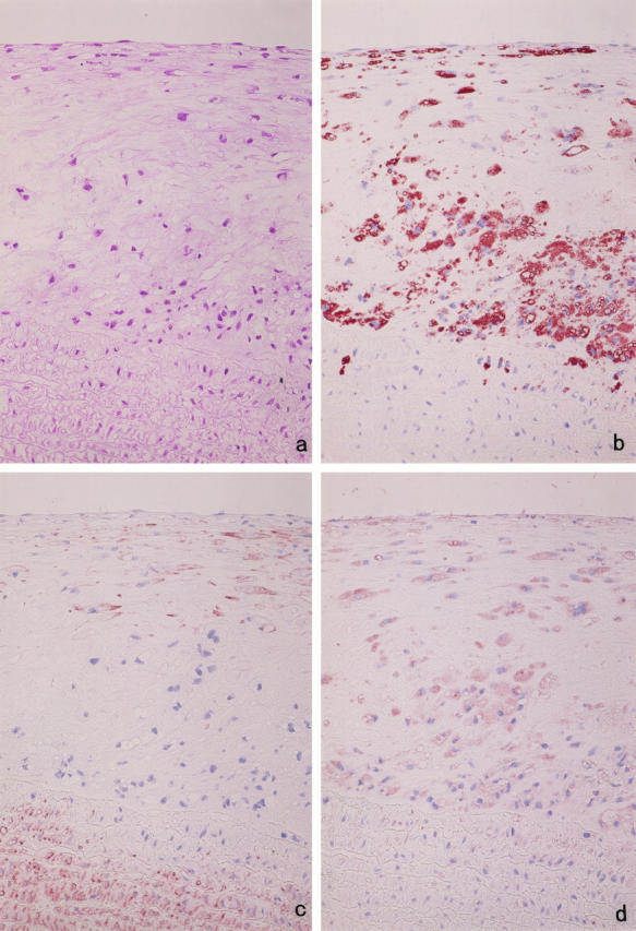
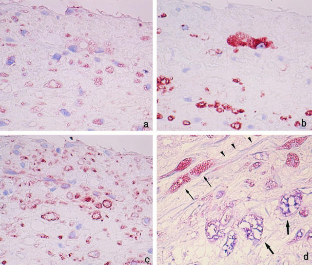
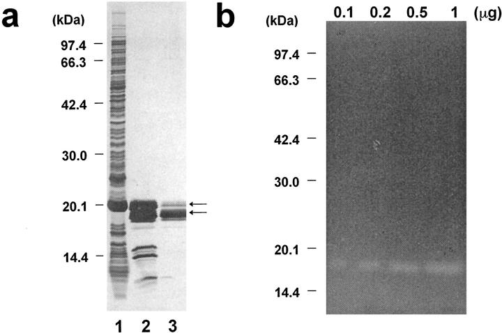
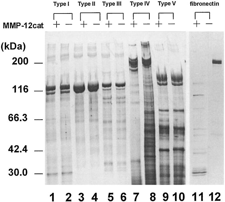
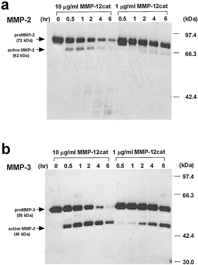
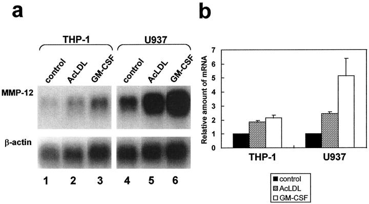
Degradation of extracellular matrix (ECM) proteins in the aorta is a critical step for the development of atherosclerosis. Expression of matrix metalloproteinase (MMP)-12 (macrophage elastase), an elastin-degrading proteinase in the MMP family, was investigated in the thoracic aorta of rabbits fed a 1% cholesterol-containing diet for 16 weeks. In the atherosclerotic lesions, MMP-12 was produced abundantly at both the mRNA and protein levels, whereas no expression was observed in the normal rabbit aortas. The principal source of MMP-12 was macrophage foam cells (MFCs) that had infiltrated the atherosclerotic intima; this was demonstrated in both in vitro culture studies of MFCs purified from atherosclerotic lesions and immunohistochemical studies of aortic lesions. Additional biochemical studies using recombinant rabbit MMP-12 revealed that MMP-12 digested elastin, type IV collagen, and fibronectin and also activated MMP-2 and MMP-3. Expression of MMP-12 by human macrophage cell lines was increased by stimulation with acetylated low-density lipoprotein, implying augmentation of MMP-12 production during foam cell formation. Increased expression of MMP-12 in atherosclerotic lesions, concomitant with foam cell generation, which triggers the acceleration of ECM breakdown, is likely to be a critical step in the initiation and progression of the atherosclerotic cascade.


Figures

Figure 1.
Figure 1.
Northern blot analysis of MMPs in aortas of normal and cholesterol-fed rabbits. Total RNA was isolated from the whole aorta of a cholesterol-fed rabbit (lane 1), atherosclerotic intima of a cholesterol-fed rabbit (lanes 2 and 3), and the whole aorta of normal rabbits (lanes 4 and 5). Total RNA (30 μg each) was blotted on a nylon membrane, and full-length cDNA fragments of rabbit MMP-1, -2, -3, -9, and -12 and human glyceraldehyde 3-phosphate dehydrogenase (G3PDH) were used as probes. The hybridized membranes were exposed to x-ray films for 1 day (a and d), 3 days (c, e, and f), or 5 hours (b). This is a representative result of three independent experiments.
Figure 2.
Figure 2.
A schematic illustration of rabbit MMP-12. The latent form of rabbit MMP-12 (top), the active form of MMP-12 (middle), and the E. coli-expressed catalytic domain of MMP-12 (bottom) are illustrated. The meshed box in pro-MMP-12 indicates the signal peptides. In the structure of MMP-12cat, MAS represents starting amino acids Met, Ala, and Ser added at the amino terminus to be expressed in E. coli. The arrow shows the 19-kd MMP-12cat, which was obtained by purification. In the amino-terminal amino acid sequence of the 19-kd MMP-12cat, Met, Ala, Ser, and Phe were cleaved and the enzymes were started with Lys.
Figure 3.
Figure 3.
MMP-12 expression by cells from aorta with atherosclerotic intima. The conditioned medium of dissociated cells from a cholesterol-fed rabbit’s whole aorta without raised intima (lane 1) and with raised intima (lane 2) and cells from the whole aorta of a normal rabbit (lane 3) were subjected to Western blot analysis with an anti-MMP-12 polyclonal Ab. Also analyzed was the conditioned medium of purified MFCs from the intima of an atherosclerotic aorta (lane 7). Rabbit pro-MMP-12 expressed by COS-7 cells (lane 4), active MMP-12 (lane 5), which was trypsin-treated conditioned medium of MMP-12-expressed COS-7 cells, conditioned medium of vector-transfected COS-7 cells (lane 6), and medium incubated without cells (lane 8) were used as control samples. This is a representative result of three independent experiments.
Figure 4.
Figure 4.
Immunohistochemical localization of MMP-12. Serial sections of cholesterol-fed rabbit aorta were stained with hematoxylin and eosin (a) and immunostained with anti-rabbit macrophage MAb RAM-11 (b), anti-muscle actin MAb HHF-35 (c), and anti-rabbit MMP-12 polyclonal Ab (d). The sections indicate that most of the RAM-11-positive MFCs are stained with anti-MMP-12 Ab. Counterstaining with hematoxylin (b to d); original magnification, ×200.
Figure 5.
Figure 5.
Cellular localization of MMP-12 in superficial region of the intima. Serial sections were immunostained by anti-MMP-12 Ab (a), anti-macrophage Ab RAM-11 (b), and anti-muscle actin Ab HHF-35 (c). In addition to RAM-11-positive MFCs, HHF-35-positive SMCs are variably stained with anti-MMP-12 Ab. Counterstaining was with hematoxylin. Double immunostaining was performed for MMP-12 and the RAM-11 (d). MMP-12-positive macrophages are stained red-blue (thick arrows), whereas MMP-12-positive SMCs are stained red only (thin arrows). Note also double-negative cells (MMP-12-negative SMCs, arrowheads). Original magnification, ×400 (a to c) and ×1000 (d).
Figure 6.
Figure 6.
E. coli expression of catalytic domain of MMP-12. a: Purification of MMP-12cat, analyzed by SDS-PAGE and silver staining. Lane 1, whole E. coli extract; lane 2, MMP-12cat refolded from the inclusion body; lane 3, MMP-12cat purified on a heparin-Sepharose column. The arrows indicate two sizes, 20.5 and 19 kd, of MMP-12cat. b: Elastin zymography of MMP-12cat, with 0.1, 0.2, 0.5, and 1 μg of purified MMP-12cat applied to κ-elastin containing 10% SDS-PAGE gel. Elastin zymography was carried out by incubating the gel at 37°C for 72 hours.
Figure 7.
Figure 7.
Degradation of ECM proteins by MMP-12cat. Type I (lanes 1 and 2), type II (lanes 3 and 4), type III (lanes 5 and 6), type IV (lanes 7 and 8), and type V (lanes 9 and 10) collagens and fibronectin (lanes 11 and 12) were incubated with MMP-12cat (lanes 1, 3, 5, 7, 9, and 11) or alone (lanes 2 , 4, 6, 8, 10, and 12). The reaction products were analyzed by SDS-PAGE under reducing conditions and silver stained.
Figure 8.
Figure 8.
Time- and concentration-dependent pro-MMP-2 and -3 activation by MMP-12cat. a: Activation of pro-MMP-2 by MMP-12cat. Five microliters of conditioned medium of rabbit MMP-2-expressing COS-7 cells was reacted with 10 or 1 μg/ml MMP-12cat in reaction buffer (2 μmol/L pepstatin A, 2 mg/ml Pefabloc, and 1 μg/ml leupeptin in buffer A). The reactions were quenched at 0, 0.5, 1, 2, 4, or 6 hours with EDTA at a final concentration of 50 mmol/L. The reaction products were analyzed by Western blot using an anti-human MMP-2 MAb. The pro-MMP-2 (72 kd) and active MMP-2 (62 kd) are indicated by arrows. b: Activation of pro-MMP-3 by MMP-12cat. Conditioned medium of COS-7 cells containing rabbit MMP-3 was reacted with MMP-12cat, and an anti-rabbit MMP-3 MAb, MP1807, was used for Western blot analysis. The pro-MMP-3 (58 kd) and active MMP-3 (46 kd) are indicated. This is a representative experiment of two performed.
Figure 9.
Figure 9.
MMP-12 expression is enhanced by stimulation with AcLDL. a: Northern blot analysis of MMP-12 expression by macrophage-differentiated THP-1 (lanes 1 to 3) and U937 (lanes 4 to 6) cells under stimulation with AcLDL (lanes 2 and 5) or GM-CSF (lanes 3 and 6). Radiolabeled human MMP-12 cDNA (top) and human β-actin cDNA (bottom) were used as probe DNAs. This is a representative experiment of three performed. b: The relative amounts of mRNA for MMP-12 were standardized with the amount of mRNA for β-actin and are indicated as 1 for the amount of MMP-12 in each cell line under normal conditions. The values are the mean ± SD of triplicate cultures.

References

    1. Ross R: The pathogenesis of atherosclerosis: a perspective for the 1990s. Nature 1993, 362:801-809 - PubMed
    1. Watanabe T, Tokunaga O, Fan J, Shimokama T: Atherosclerosis and macrophages. Acta Pathol Jpn 1989, 39:473-486 - PubMed
    1. Dollery CM, McEwan JR, Henney AM: Matrix metalloproteinases and cardiovascular disease. Circ Res 1995, 77:863-868 - PubMed
    1. Holvoet P, Collen D: Oxidized lipoproteins in atherosclerosis and thrombosis. FASEB J 1994, 8:1279-1284 - PubMed
    1. Steinberg D, Parthasarathy S, Carew TE, Khoo JC, Witztum JL: Beyond cholesterol: modifications of low-density lipoprotein that increase its atherogenicity. N Engl J Med 1989, 320:915-924 - PubMed

Publication types

MeSH terms