

Mantle cell lymphomas lack expression of p27Kip1, a cyclin-dependent kinase inhibitor
- PMID: 9665478
- PMCID: PMC1852938
- DOI: 10.1016/S0002-9440(10)65558-7
Mantle cell lymphomas lack expression of p27Kip1, a cyclin-dependent kinase inhibitor
Abstract
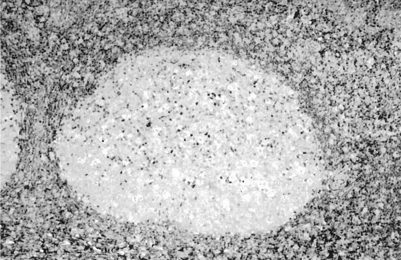
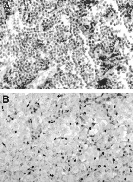
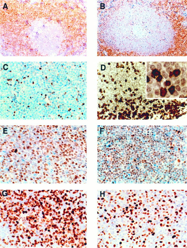
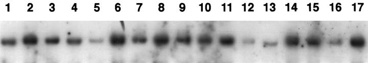
p27Kip1 is a cyclin-dependent kinase inhibitor that regulates the decision to enter S phase or withdraw from the cell cycle. In resting cells, the level of p27Kip1 provides an inhibitory threshold above which G1 cyclin D/E/cyclin-dependent kinases accumulate before activation; however, in cycling cells, p27Kip1 protein is sequestered by high levels of active cyclin D/cyclin-dependent kinase 4 complexes. As a group, the cyclin-dependent kinase inhibitors have been proposed to act as tumor suppressor genes, and several members have been implicated in the pathogenesis of a variety of human cancers. We examined p27Kip1 expression in 116 non-Hodgkin's lymphomas including 50 cases of MCL (40 typical and 10 blastic variants), 21 follicular lymphomas, 20 diffuse large B-cell lymphomas, 16 chronic lymphocytic leukemias, 8 marginal zone B-cell lymphomas, and 1 splenic marginal zone lymphoma, and correlated its expression with that of the proliferation marker Ki67 (MiB1) and with p53. p27Kip1 gene structure was analyzed by Southern blot in the group of MCLs. In all cases of non-Hodgkin's lymphoma other than MCL, p27Kip1 expression was inversely related to the proliferation index as measured by Ki67. In contrast, in typical MCL, p27Kip1 expression was negative in 35 of 40 (88%) cases, irrespective of the proliferative rate (median 15%; range 2 to 90%). Paradoxically, in the blastic variant of MCL, 8 of 10 (80%) cases showed expression of p27Kip1, despite a high proliferation rate (median 60%; range 32 to 100%). However, the staining in most of the cases was less intense than in the reactive T lymphocytes. Deletions of p27Kip1 gene were not found in any of the 25 cases examined. p53 expression was found in 15 of 50 cases of MCL: 7 of 10 (70%) in the blastic variant and 8 of 40 (20%) in the typical MCL (70% vs. 20%, P < 0.0045). These results demonstrate that MCLs, in contrast to other non-Hodgkin's lymphomas and normal lymphoid tissue, fail to correlate p27Kip1 expression with the proliferation rate. This peculiar uncoupling of p27Kip1 protein expression from the proliferation rate may be related to the high levels of cyclin D1 expressed in MCL and is likely to have profound effects on cell cycle regulation and contribute to the pathogenesis of MCL.
Figures
References
-
- Sherr CJ: Cancer cell cycles. Science 1996, 274:1672-1677 - PubMed
-
- Sherr CJ, Roberts JM: Inhibitors of mammalian G1 cyclin-dependent kinases. Genes Dev 1995, 9:1149-1163 - PubMed
-
- Polyak K, Lee M-H, Erdjument-Bromage H, Koff A, Roberts JM, Tempst P, Massague J: Cloning of p27KIP-1, a cyclin-dependent kinase inhibitor and a potential mediator of extracellular antimitogenic signals. Cell 1994, 78:59-66 - PubMed
-
- Kato J, Matsuoka M, Polyak K, Massague J, Sherr CJ: Cyclic AMP-induced G1 phase arrest mediated by an inhibitor (p27KIP1) of cyclin-dependent kinase 4 activation. Cell 1994, 79:487-496 - PubMed
-
- Nourse J, Firpo E, Flanagan WM, Coats S, Polyak K, Lee M-H, Massague J, Crabtree GR, Roberts JM: Interleukin-2-mediated elimination of the p27KIP-1 cyclin-dependent kinase inhibitor prevented by rapamycin. Nature 1994, 372:570-573 - PubMed
Publication types
MeSH terms
Substances
LinkOut - more resources
Full Text Sources
Other Literature Sources
Research Materials
Miscellaneous
