Comparative Study
. 1998 Jul;153(1):233-45.
doi: 10.1016/S0002-9440(10)65564-2.

Immunohistochemical localization of somatostatin receptors sst2A in human tumors

Affiliations
Comparative Study

Immunohistochemical localization of somatostatin receptors sst2A in human tumors

J C Reubi et al. Am J Pathol. 1998 Jul.

Abstract

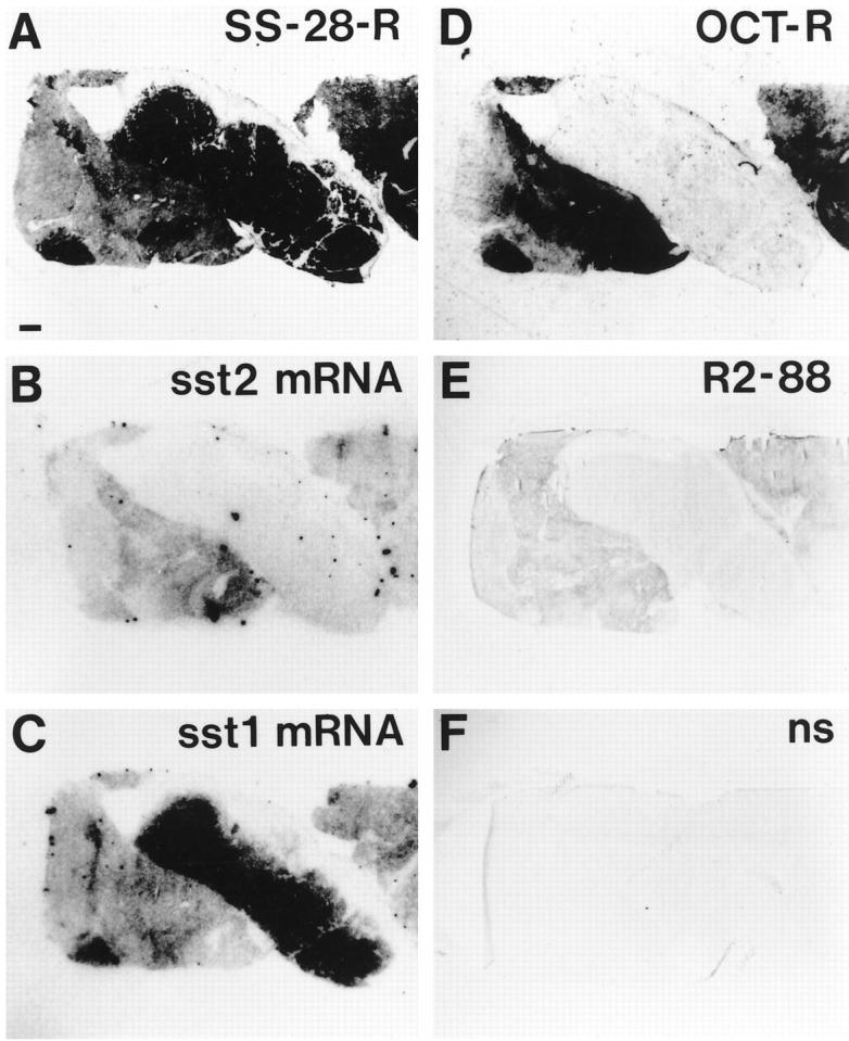
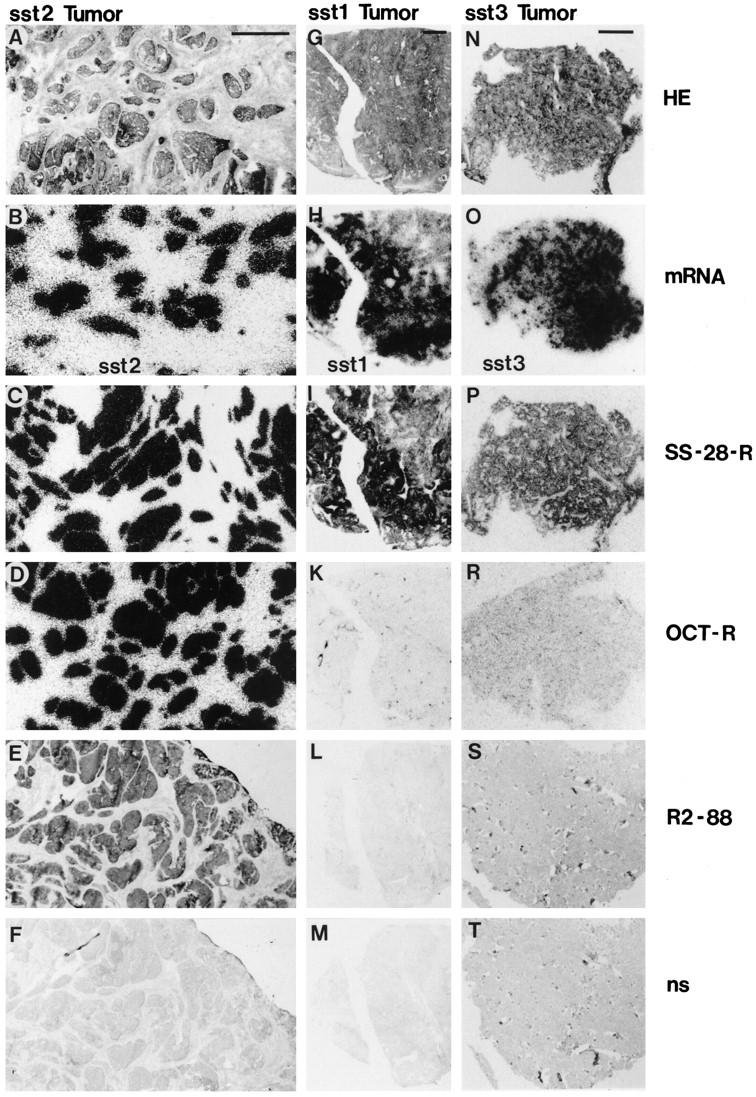
Human tumors frequently express somatostatin receptors. However, none of the receptor subtype proteins have been individually visualized in normal or neoplastic human tissues. Here, the distribution of the sst2A receptor was investigated using immunohistochemistry with the specific anti-peptide antibody R2-88 in 47 human tumors. All tumors selected for their abundance of sst2 mRNA and/or strong binding of the sst2-preferring ligand 125I-labeled Tyr3-octreotide were specifically immunostained with R2-88. Conversely, all tumors without somatostatin binding or expressing predominantly other somatostatin receptor subtype mRNAs (sst1 or sst3) were not specifically immunostained by R2-88. Specificity was shown in immunoblots, demonstrating receptor migration as a 70-kd broad band. In immunohistochemical and immunoblotting experiments, the abolition of staining after antibody blockade with antigen peptide was demonstrated. Immunostaining was identified in cryostat and in formalin-fixed, paraffin-embedded sections. Heat-induced epitope retrieval was necessary to visualize sst2A receptors in formalin-fixed sections. Moreover, because of occasional high nonspecific staining, the demonstration of complete abolition of immunostaining by treatment with antigen peptide was a prerequisite for the correct identification of sst2A-positive tumors. The sst2A receptors were clearly located at the membrane of the tumor cells. These results provide the first localization of a somatostatin receptor subtype in human tissues at the cellular level. The sst2A receptor identification and visualization in tumors with simple immunohistochemical methods in formalin-fixed, paraffin-embedded material will open new diagnostic opportunities for pathologists.


Figures

Figure 1.
Figure 1.
Western blot analysis of sst2A receptor immunoreactivity in tumor tissue. Membrane proteins from sst2A-transfected GH-R2 cells (GH; 2.5 μg), a GH-producing pituitary adenoma (tumor 5 in Table 2 ▶ ) (Pit; 60 μg), and a meningioma (tumor 9 in Table 2 ▶ ) (Men; 90 μg) were separated by PAGE and electrophoretically transferred to PVDF membrane. The membrane was incubated with sst2A receptor antibody in the absence (left panel) or presence (right panel) of 100 nmol/L antigen peptide. Molecular size markers are shown on the left.
Figure 2.
Figure 2.
Immunohistochemical staining with R2–88 of a bronchial carcinoid tumor (frozen sample). Comparison with receptor autoradiography and in situ hybridization. A: Autoradiogram showing total binding of 125I-labeled LTT-SS-28 in the whole tumor, with particularly strong labeling of the central part. Bar, 1 mm. B: Autoradiogram showing sst2 mRNA located in the lateral parts of the tumor but not in the central part. C: Autoradiogram showing sst1 mRNA. It is abundant in the central part and detected in moderate amounts in the lateral parts. D: Autoradiogram showing total binding of 125I-labeled Tyr3-octreotide located in the lateral parts but not in the central part. E: Immunohistochemical staining with R2–88 located in the lateral parts but not in the central part. This cryostat section was not hematoxylin counterstained. F: Control, showing lack of R2–88 staining after absorption with 100 nmol/L peptide antigen (nonspecific staining); section not counterstained.
Figure 3.
Figure 3.
Effects of various pretreatments on the immunohistochemical staining with R2–88 of a paraffin-embedded duodenal neuroendocrine carcinoma. A and B: Identification of 125I-labeled Tyr3-octreotide binding in cryostat sections of a frozen sample of the tumor. A: H&E-stained section. T, tumor. Bar, 1 mm. B: Autoradiogram of the total binding of 125I-labeled Tyr3-octreotide showing strong labeling of the tumor. C to H: Immunohistochemical R2–88 staining of paraffin-embedded samples adjacent to the above-mentioned frozen sample. T, tumor; m, mucosa. C: Pretreatment with boiling in a pressure cooker results in marked immunoreactivity of the tumor. D: Absorption with 100 nmol/L peptide antigen (nonspecific staining) of a section pretreated with boiling in a pressure cooker abolishes the immunoreactivity of the tumor. E: Pretreatment with boiling in a microwave oven results in a positive immunoreactivity comparable to that seen in C. F: Absorption with 100 nmol/L peptide antigen (nonspecific staining) of a section pretreated with boiling in a microwave oven abolishes the immunoreactivity. G: Pretreatment with Pronase does not yield a satisfactory immunoreactivity. H: Pretreatment with trypsin yields a negative result as in G. Only pretreatments with boiling in a pressure cooker or microwave oven give a strong and specific immunostaining.
Figure 4.
Figure 4.
R2–88 immunohistochemistry of a gastrinoma: effect of tyramide amplification compared with boiling (microwave oven) pretreatment (paraffin sections). A: Boiling pretreatment showing the tumor R2–88 immunostaining. Bar, 1 mm. B: Boiling pretreatment including absorption with 100 nmol/L peptide antigen abolishes the reactivity of the tumor (nonspecific staining). C: Tyramide amplification of a non-pretreated, adjacent section of the tumor results in a moderate R2–88 reactivity of the tumor. D: Tyramide amplification, including absorption with 100 nmol/L peptide antigen, shows a marked nonspecific staining of the tumor. The tumor is less intensely immunostained with tyramide amplification than with boiling pretreatment. Moreover, the nonspecific staining is more intense with tyramide amplification.
Figure 5.
Figure 5.
R2–88 immunohistochemistry in two somatostatin receptor-negative pancreatic tumors: a poorly differentiated pancreatic carcinoma (A to C, left vertical column) and a well differentiated ductal pancreatic adenocarcinoma (D to F, right vertical column). Paraffin sections. A and D: H&E-stained sections. Bar, 1 mm. Arrowheads indicate ductal carcinoma, and arrows indicate pancreatic islets. B and E: Immunohistochemistry with R2–88. No immunostaining is seen in B. E, however, shows stained islets (arrows) and a stained ductal carcinoma with tubulopapillary structure (arrowheads). C and F: Immunohistochemistry with R2–88 after absorption with 100 nmol/L peptide antigen. In F, the staining of the islets is completely abolished whereas the staining of the neoplastic ducts remains visible. The ductal staining is therefore not recognized by the antipeptide antibody R2–88 (nonspecific staining). There is no counterstain in E and F. Whereas both tumors are sst2A receptor negative, the staining of the islets is inhibited by the peptide, demonstrating that it is specific and representing an internal control.
Figure 6.
Figure 6.
R2–88 immunohistochemical staining of tumors in relation to their respective somatostatin receptor subtype expression. The sst2A receptor immunostaining is compared with the receptor content measured by receptor autoradiography and to the mRNA content measured by in situ hybridization in an sst2-expressing gastrointestinal carcinoid (A to F, first vertical column), an sst1-expressing leiomyosarcoma (G to M, middle vertical column), and an sst3-expressing insulinoma (N to T, last vertical column). A, G, and N: H&E-stained sections. Bar, 1 mm. B, H, and O: Autoradiograms showing in situ hybridization for sst2 mRNA (B), for sst1 mRNA (H), and for sst3 mRNA (O). The sst1- and the sst3-expressing tumors do not express measurable amounts of sst2 receptors. C, I, and P: Autoradiograms showing total binding of the universal ligand 125I-labeled LTT-SS-28. All three tumors were labeled. D, K, and R: Autoradiograms showing total binding of the sst2-preferring 125I-labeled Tyr3-octreotide. Only the sst2-expressing tumor (D) was labeled. E, L, and S: Immunohistochemical R2–88 staining. Only the sst2-expressing tumor was stained (E). E and L were cut from frozen samples; S was from formalin-fixed, paraffin-embedded tissue. F, M, and T: Controls, showing lack of R2–88 immunohistochemical staining after absorption with 100 nmol/L peptide antigen (nonspecific staining). The tumor strands stained in E are no longer immunoreactive in F; only the weak hematoxylin counterstain remains visible.
Figure 7.
Figure 7.
Comparison of the R2–88 immunohistochemistry in paraffin-embedded and in frozen samples of the same sst2-expressing glucagonoma. A: Autoradiogram showing total binding of 125I-labeled Tyr3-octreotide in this tumor. B: R2–88 immunohistochemistry in a frozen sample of the tumor described above. Section is adjacent to that used in A. The tumor is immunoreactive. C: R2–88 immunohistochemistry in a paraffin-embedded sample fixed for 12 hours in formalin. The tumor is immunoreactive. D: R2–88 immunohistochemistry in another paraffin-embedded sample of the same tumor fixed for 14 days in formalin. The tumor is also immunoreactive. In B to D, absorption with 100 nmol/L peptide antigen on adjacent sections prevented completely the immunostaining (not shown).
Figure 8.
Figure 8.
R2–88 immunohistochemistry showing the membrane-bound localization of the sst2A receptors and its tissue selectivity in a gastrinoma at high magnification (paraffin sections). A: The brown immunoreactivity is predominantly located on the cell membrane of the tumor cells. Bar, 25 μm. B: The tumor cells but not the three pancreatic ducts (middle) are sst2A receptor positive. Bar, 50 μm. C: Membrane-bound sst2A receptors at high magnification. Bar, 16 μm. D: Adjacent section showing that absorption with 100 nmol/L peptide antigen abolishes the staining of the membrane. Weak residual, nonspecific brown staining is seen in the connective tissue. Bar, 16 μm. In C and D, cell bodies are stained in blue (hematoxylin).

References

    1. Reubi JC: Relevance of somatostatin receptors and other peptide receptors in pathology. Endocr Pathol 1997, 8:11-20 - PubMed
    1. Krenning EP, Bakker WH, Breeman WAP, Koper JW, Kooij PPM, Ausema L, Lameris JS, Reubi JC, Lamberts SWJ: Localisation of endocrine-related tumours with radioiodinated analogue of somatostatin. Lancet 1989, I:242-244 - PubMed
    1. Lamberts SWJ, Bakker WH, Reubi JC, Krenning EP: Somatostatin-receptor imaging in the localization of endocrine tumors. N Engl J Med 1990, 323:1246-1249 - PubMed
    1. Reubi JC: Neuropeptide receptors in health and disease: the molecular basis for in vivo imaging. J Nucl Med 1995, 36:1825-1835 - PubMed
    1. Lamberts SWJ, Krenning EP, Reubi JC: The role of somatostatin and its analogs in the diagnosis and treatment of tumors. Endocr Rev 1991, 12:450-482 - PubMed

Publication types

Substances

LinkOut - more resources