

Comparative phenotypic studies of duct epithelial cell lines derived from normal human pancreas and pancreatic carcinoma
- PMID: 9665487
- PMCID: PMC1852927
- DOI: 10.1016/S0002-9440(10)65567-8
Comparative phenotypic studies of duct epithelial cell lines derived from normal human pancreas and pancreatic carcinoma
Abstract
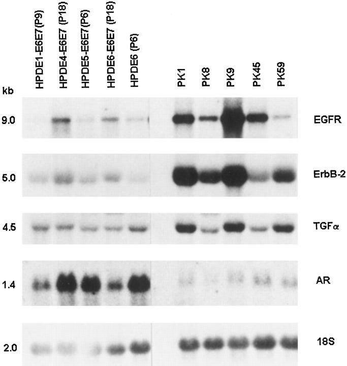
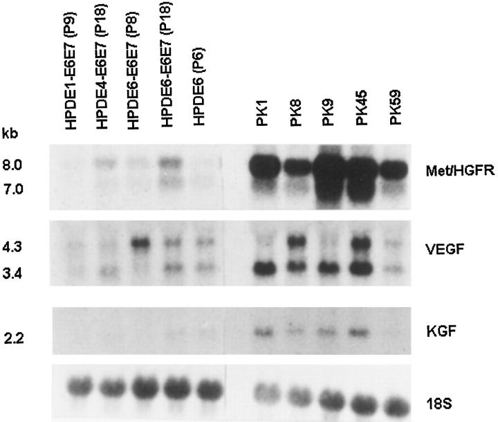
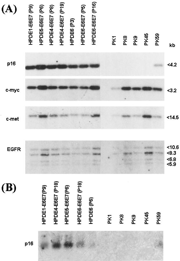
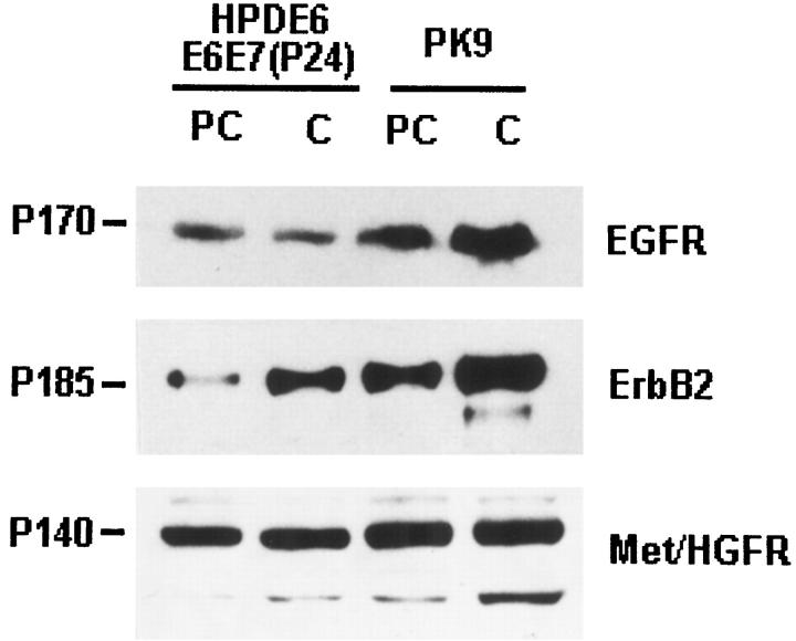
We have investigated the mRNA/protein expression of several tyrosine kinase receptors, growth factors, and p16INK4A cyclin inhibitor in cell lines derived from normal human pancreatic duct epithelium (HPDE) and compared them with those of five pancreatic ductal carcinoma cell lines. Cultured HPDE cells express low levels of epidermal growth factor receptor (EGFR), erbB2, transforming growth factor (TGF)-alpha, Met/hepatocyte growth factor receptor (HGFR), vascular endothelial growth factor (VEGF), and keratinocyte growth factor (KGF). They also expressed high levels of amphiregulin but did not express EGF and cripto. The expression levels were similar in primary normal HPDE cells and those expressing transfected E6E7 genes of human papilloma virus-16, but their immortalization appeared to enhance the expression of EGFR and Met/HGFR. In comparison, pancreatic carcinoma cell lines commonly demonstrated overexpression of EGFR, erbB2, TGF-alpha, Met/HGFR, VEGF, and KGF, but they consistently showed marked down-regulation of amphiregulin mRNA expression. In contrast to all carcinoma cell lines that showed deletions of the p16 gene, HPDE cells consistently demonstrated normal p16 genotype and its mRNA expression. This is the first report that compares the phenotypic expression of cultured pancreatic ductal carcinoma cells with epithelial cell lines derived from normal human pancreatic ducts. The findings confirm that malignant transformation of human pancreatic duct cells commonly results in a deregulation of expression of various growth factors and receptors.
Figures
References
-
- Parker SL, Tong T, Bolden S, Wingo PA: Cancer statistics, 1997. CA Cancer J Clin 1997, 47:5-27 - PubMed
-
- Kloppel G: Pathology of nonendocrine pancreatic tumours. Go VLW Dimagno EP Gardner JD Lebenthal E Reber HA Scheele GA eds. The Pancreas: Biology, Pathobiology, and Disease. 1993, :pp 871-897 Raven Press, New York
-
- Baumel H, Huguier M, Manderscheid JC, Fabre JM, Houry S, Fagot H: Results of resection for cancer of exocrine pancreas: a study from the French Association of Surgery. Br J Surg 1994, 81:102-107 - PubMed
-
- Murr MM, Sarr MG, Oishi AJ, van Heerden JA: Pancreatic cancer. CA Cancer J Clin 1994, 44:304-318 - PubMed
-
- DiGiuseppe JA, Hruban RH: Pathobiology of cancer of the pancreas. Semin Surg Oncol 1995, 11:87-96
Publication types
MeSH terms
Substances
LinkOut - more resources
Full Text Sources
Other Literature Sources
Medical
Research Materials
Miscellaneous
