Comparative Study
. 1998 Jul;5(4):438-45.
doi: 10.1128/CDLI.5.4.438-445.1998.

Inducible nitric oxide synthase and nitrotyrosine are found in monocytes/macrophages and/or astrocytes in acute, but not in chronic, multiple sclerosis

Affiliations
Comparative Study

Inducible nitric oxide synthase and nitrotyrosine are found in monocytes/macrophages and/or astrocytes in acute, but not in chronic, multiple sclerosis

E L Oleszak et al. Clin Diagn Lab Immunol. 1998 Jul.

Abstract

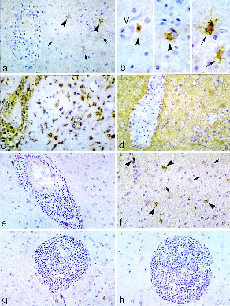
We have examined the localization of inducible nitric oxide synthase (iNOS) and nitrotyrosine (the product of nitration of tyrosine by peroxynitrite, a highly reactive derivative of nitric oxide [NO]) in demyelinating lesions from (i) two young adult patients with acute multiple sclerosis (MS), (ii) a child with MS (consistent with diffuse sclerosis), and (iii) five adult patients with chronic MS. Previous reports have suggested a possible correlation between iNOS, peroxynitrite, related nitrogen-derived oxidants, and the demyelinating processes in MS. We have demonstrated iNOS-immunoreactive cells in both acute-MS and diffuse-sclerosis-type lesions. In acute-MS lesions, iNOS was localized in both monocytes/macrophages and reactive astrocytes. However, foamy (myelin-laden) macrophages and the majority of reactive astrocytes were iNOS negative. In specimens from the childhood MS patient, iNOS protein was present only in a subpopulation of reactive or hypertrophic astrocytes. In contrast, no iNOS staining was detected in chronic-MS lesions. Immunohistochemical staining of acute-MS lesions with an antibody to nitrotyrosine revealed codistribution of iNOS- and nitrotyrosine-positive cells, although nitrotyrosine staining was more widespread in cells of the monocyte/macrophage lineage. In diffuse-sclerosis-type lesions, nitrotyrosine staining was present in hypertrophic astrocytes, whereas it was absent in chronic-MS lesions. These results suggest that NO and nitrogen-derived oxidants may play a role in the initiation of demyelination in acute-MS lesions but not in the later phase of the disease.


Figures

FIG. 1
FIG. 1
Immunocytochemical detection of iNOS protein (a and b), HAM-56 (c), GFAP (d), and nitrotyrosine (e, f, and g) in active, monophasic, demyelinating lesions consistent with acute MS. (a) Evidence of florid demyelinating process characterized by perivascular inflammatory infiltrates (lymphocytes and monocytes/macrophages), myelin pallor, and a mixture of mononuclear cells and reactive astrocytes in the white matter. iNOS localization is present in scattered cells of the monocyte/macrophage lineage (large arrowheads). In this field, there is lack of iNOS staining in reactive astrocytes (small arrows) and in perivascular inflammatory cell cuffs. Original magnification, ×400. (b) Higher magnification (under oil-immersion lens) of a field adjoining that of panel a. iNOS-positive cells of the monocyte/macrophage series (large arrowheads) and somewhat larger, multipolar cells consistent with reactive astrocytes (small arrows) are visible. It should be noted that only a minority (<10%) of mononuclear cells and reactive astrocytes combined are iNOS positive in these acute lesions. Original magnification, ×1,000. (c) Deeper, sequential tissue section relative to that of panel a. Widespread HAM-56 staining of monocytes/macrophages is visible in the perivascular space and in the white matter. Note morphologic variation among these cells, including round (monocyte-like), plump (macrophage-like), rod-shaped, and ameboid (microglia-like) forms. Original magnification, ×400. (d) Homologous microscopic field in a subserial section relative to sections shown in panels a and c. There is diffuse GFAP staining in reactive astrocytes and their fibrillary processes, surrounding a Virchow-Robin (perivascular) space packed with GFAP-negative foamy (myelin-laden) macrophages. Due to the abundance of macrophages, the vascular lumen is obliterated. Original magnification, ×400. (e) Perivascular mononuclear cell cuffing in a white matter blood vessel. Note scattered monocytes/macrophages within the inflammatory cuff, showing nitrotyrosine-like immunoreactivity. Original magnification, ×400. (f) Nearby field relative to panel e field. Nitrotyrosine-like staining is visible in cells of the monocyte/macrophage lineage (large arrowheads). In this field, reactive astrocytes (small arrows) are nitrotyrosine negative. During the acute phase of demyelination, the majority of nitrotyrosine-labeled cells are monocytes/macrophages; only rare reactive astrocytes are nitrotyrosine positive (not shown). Original magnification, ×400. (g) Dense perivascular mononuclear cell infiltrates. As in panel f, nitrotyrosine-like immunoreactivity is present in scattered monocytes/macrophages. Also, note nitrotyrosine-positive monocytic elements in the contiguous white matter (upper right corner). Original magnification, ×400. (h) Deeper tissue section relative to panel g section. A lack of staining is apparent in perivascular mononuclear cell infiltrates after the use of nonspecific IgG in lieu of the primary antibody. Original magnification, ×400.
FIG. 2
FIG. 2
Immunocytochemical detection of iNOS protein (a and c), GFAP (b), and nitrotyrosine (d) in active, confluent demyelinating lesions consistent with diffuse sclerosis (Schilder type). (a) Extensive, confluent demyelinating lesions involving cerebral white matter. Scattered iNOS-positive reactive or hypertrophic astrocytes are visible amidst heavy accumulations of iNOS-negative foamy macrophages. Original magnification, ×100. (b) Subserial section relative to that of panel a. Accumulations of GFAP-positive hypertrophic astrocytes intermingled with GFAP-negative foamy macrophages are visible. Original magnification, ×100. (c) Higher magnification of the microscopic field depicted in panel a. There is distinctive iNOS immunoreactivity in large astrocytic cell bodies; however, some degree of staining variability among morphologically identical hypertrophic astrocytes is visible. The inset shows discrete iNOS localization in hypertrophic astrocytes and its processes encroaching on the adventitia of a parenchymal blood vessel. Original magnification, ×400. (d) Robust nitrotyrosine-like staining in hypertrophic astrocytes surrounding a white matter blood vessel. Also, linear-type staining in the luminal endothelial surface, as well as lack of immunoreactivity in foamy macrophages (inset), is visible. Original magnification, ×400. (e to h) Immunocytochemical profile of HAM-56 (e), GFAP (f), iNOS (g), and nitrotyrosine (h) in subserial sections from a chronic-MS plaque. (e) Perivascular aggregates of HAM-56-positive monocytes/macrophages surrounding a sclerotic vessel within a chronic-MS plaque. Original magnification, ×400. (f) GFAP staining highlights fibrous gliosis surrounding an intralesional blood vessel. GFAP-negative mononuclear cells are visible in the perivascular space. Original magnification, ×400. (g) Lack of iNOS staining in perivascular mononuclear cells and in fibrous astrocytes. Original magnification, ×400. (h) Lack of nitrotyrosine-like staining in perivascular mononuclear cells and in fibrous astrocytes mirroring the profile of iNOS. Original magnification, ×400.

Similar articles

Cited by

References

    1. Adams C W, Poston R N. Macrophage histology in paraffin-embedded multiple sclerosis plaques is demonstrated by the monoclonal pan-macrophage marker HAM-56: correlation with chronicity of the lesion. Acta Neuropathol. 1990;80:208–211. - PubMed
    1. Adams C W M, Poston R N, Buk S J. Pathology, histochemistry and immunocytochemistry of lesions in acute multiple sclerosis. J Neurol Sci. 1989;92:291–306. - PubMed
    1. Bagasra O, Michaels F H, Zheng Y M, Bobroski L E, Spitsin S V, Fu Z F, Tawadros R, Koprowski H. Activation of the inducible form of nitric oxide synthase in the brains of patients with multiple sclerosis. Proc Natl Acad Sci USA. 1997;92:12041–12045. - PMC - PubMed
    1. Beckman J S, Crow J P. Pathological implications of nitric oxide, superoxide and peroxynitrite formation. Biochem Soc Trans. 1993;21:330–334. - PubMed
    1. Beckman J S, Ye Y Z, Anderson P G, Chen J, Accavitti M A, Tarpey M M, White C R. Extensive nitration of protein tyrosines in human atherosclerosis detected by immunohistochemistry. Biol Chem Hoppe-Seyler. 1994;375:81–88. - PubMed

Publication types

MeSH terms