

Detection of Mycobacterium avium subsp. paratuberculosis in infected tissues by new species-specific immunohistological procedures
- PMID: 9665946
- PMCID: PMC95597
- DOI: 10.1128/CDLI.5.4.446-451.1998
Detection of Mycobacterium avium subsp. paratuberculosis in infected tissues by new species-specific immunohistological procedures
Abstract
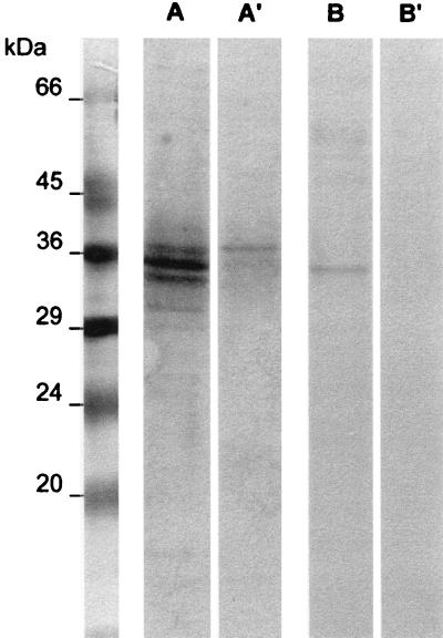
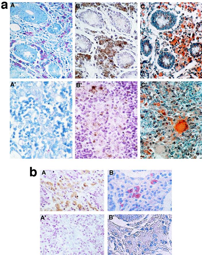
We have previously described the cloning and sequencing of a gene portion coding for the terminal part of a 34-kDa protein of Mycobacterium avium subsp. paratuberculosis, the etiological agent of Johne's disease (P. Gilot, M. De Kesel, L. Machtelinckx, M. Coene, and C. Cocito, J. Bacteriol. 175:4930-4935, 1993). The recombinant polypeptide (a362) carries species-specific B-cell epitopes which do not cross-react with other mycobacterial pathogens (M. De Kesel, P. Gilot, M.-C. Misonne, M. Coene, and C. Cocito, J. Clin. Microbiol. 31:947-954, 1993). The present work describes the preparation of polyclonal and monoclonal antibodies directed against a362 and the use of these immunoglobulins for histopathological diagnosis of Johne's disease. The new immunohistological procedures herewith detailed proved to be able to identify M. avium subsp. paratuberculosis antigens in the intestinal tissues and lymph nodes of cattle affected by either the paucibacillary or pluribacillary form of the disease. They yielded negative responses not only with healthy animals but also with those affected by tuberculosis (Mycobacterium bovis). Both immunohistological procedures proved to be as sensitive as or more sensitive than Ziehl-Neelsen staining and, in addition, to be endowed with species specificity.
Figures
Comment in
-
Specificity of the 34-kilodalton immunodominant protein of Mycobacterium avium sp. paratuberculosis.Clin Diagn Lab Immunol. 1999 Jan;6(1):146-8. doi: 10.1128/CDLI.6.1.146-148.1999. Clin Diagn Lab Immunol. 1999. PMID: 10068253 Free PMC article. No abstract available.
References
-
- Ackermans F, Nisol F, Bazin H. Immunization of rats. In: Bazin H, editor. Rat hybridomas and rat monoclonal antibodies. Boca Raton, Fla: CRC Press; 1990. pp. 75–85.
-
- Bazin H, Deckers C, Beckers A, Heremans J F. Transplantable immunoglobulin-secreting tumours in rats. I. General features of LOU-WS1 strain rat immunocytomas and their monoclonal proteins. Int J Cancer. 1972;10:568–580. - PubMed
-
- Bazin H, Beckers A, Querinjean P. Three classes and four (sub)classes of rat immunoglobulins: IgM, IgA, IgE and IgG1, IgG2a, IgG2b, IgG2c. Eur J Immunol. 1974;4:44–48. - PubMed
-
- Bazin H. Production of rat monoclonal antibodies with the LOU rat non-secreting IR983F myeloma cell line. In: Peeters H, editor. Protides of the biological fluids. New York, N.Y: Pergamon Press; 1982. pp. 615–618.
-
- Bazin H, Cormont F, De Clercq L. Rat monoclonal antibodies. II. A rapid and efficient method for purification from ascitic or serum. J Immunol Methods. 1984;71:9–16. - PubMed
Publication types
MeSH terms
Substances
LinkOut - more resources
Full Text Sources
