

Relevant criteria for detecting microsporidia in stool specimens
- PMID: 9666005
- PMCID: PMC105031
- DOI: 10.1128/JCM.36.8.2279-2283.1998
Relevant criteria for detecting microsporidia in stool specimens
Abstract
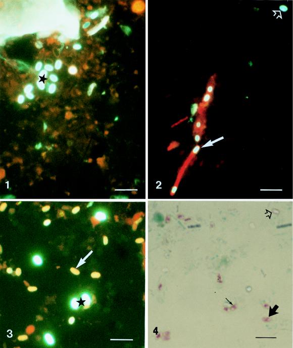
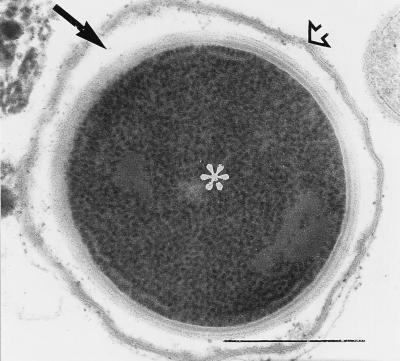
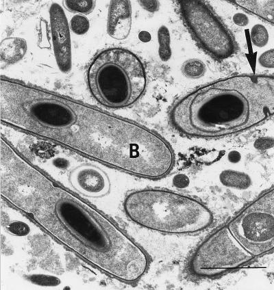
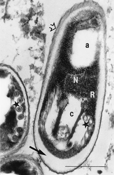
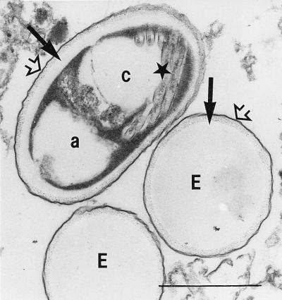
By using different staining techniques, 479 stool specimens from 212 diarrheic patients with AIDS were examined for microsporidian spores. Calcofluor fluorescence staining of 119 specimens revealed fluorescent ovoid structures of microsporidian size. Staining of these samples according to the method of Weber et al. (R. Weber, R. T. Bryan, R. L. Owen, C. M. Wilcox, L. Gorelkin, and G. S. Visvesvara, N. Engl. J. Med. 326:161-166, 1992) with trichrome produced six specimens with pinkish spores containing the characteristic microsporidian belt-like structure. The 6 specimens were processed for transmission electron microscopy, as were another 21 specimens which did not present the belt-like structure after trichrome staining but which looked highly suspicious after fluorescence staining. In these 21 samples, only fungal spores and, particularly, bacterial Clostridium spores were demonstrated, whereas in the 6 samples diagnosed positive after trichrome staining, the existence of microsporidia could be verified by electron microscopy. Based on our observations, we propose that the belt-like structure seen with the Weber stains in microsporidian spores corresponds to structures existing in priming-stage spores. The results suggest that routine microscopical fecal diagnosis for microsporidian infection should include a screening by fluorescence staining and, subsequently, a confirmatory viewing of fluorescence-positive samples after trichrome staining.
Figures
Similar articles
-
Comparison of three staining methods for detecting microsporidia in fluids.J Clin Microbiol. 1995 Dec;33(12):3138-45. doi: 10.1128/jcm.33.12.3138-3145.1995. J Clin Microbiol. 1995. PMID: 8586689 Free PMC article.
-
Modified technique to recover microsporidian spores in sodium acetate-acetic acid-formalin-fixed fecal samples by light microscopy and correlation with transmission electron microscopy.J Clin Microbiol. 1996 Nov;34(11):2670-3. doi: 10.1128/jcm.34.11.2670-2673.1996. J Clin Microbiol. 1996. PMID: 8897162 Free PMC article.
-
Fluorescence techniques for diagnosing intestinal microsporidiosis in stool, enteric fluid, and biopsy specimens from acquired immunodeficiency syndrome patients with chronic diarrhea.Arch Pathol Lab Med. 1996 Sep;120(9):847-53. Arch Pathol Lab Med. 1996. PMID: 9140290
-
Intestinal coinfection with Enterocytozoon bieneusi and Cryptosporidium in a human immunodeficiency virus-infected child with chronic diarrhea.Clin Infect Dis. 1993 Sep;17(3):480-3. doi: 10.1093/clinids/17.3.480. Clin Infect Dis. 1993. PMID: 8218693 Review.
-
Clinical features of microsporidiosis in patients with AIDS.Clin Infect Dis. 1994 May;18(5):819-25. doi: 10.1093/clinids/18.5.819. Clin Infect Dis. 1994. PMID: 7915548 Review.
Cited by
-
The microsporidian polar tube: origin, structure, composition, function, and application.Parasit Vectors. 2023 Aug 30;16(1):305. doi: 10.1186/s13071-023-05908-9. Parasit Vectors. 2023. PMID: 37649053 Free PMC article. Review.
-
Encephalitozoon cuniculi: Grading the Histological Lesions in Brain, Kidney, and Liver during Primoinfection Outbreak in Rabbits.J Pathog. 2016;2016:5768428. doi: 10.1155/2016/5768428. Epub 2016 Feb 28. J Pathog. 2016. PMID: 27022485 Free PMC article.
-
Real-time PCR method for detection of Encephalitozoon intestinalis from stool specimens.J Clin Microbiol. 2002 Nov;40(11):3922-8. doi: 10.1128/JCM.40.11.3922-3928.2002. J Clin Microbiol. 2002. PMID: 12409353 Free PMC article.
-
Practical Guidance for Clinical Microbiology Laboratories: Laboratory Diagnosis of Parasites from the Gastrointestinal Tract.Clin Microbiol Rev. 2017 Nov 15;31(1):e00025-17. doi: 10.1128/CMR.00025-17. Print 2018 Jan. Clin Microbiol Rev. 2017. PMID: 29142079 Free PMC article. Review.
-
Mechanics of Microsporidian Polar Tube Firing.Exp Suppl. 2022;114:215-245. doi: 10.1007/978-3-030-93306-7_9. Exp Suppl. 2022. PMID: 35544005
References
-
- Cali A, Meisler D M, Lowder C Y, Lembach R, Ayers L, Takvorian P M, Rutherford I, Longworth D L, McMahon J, Bryan R T. Corneal microsporidiosis: characterization and identification. J Protozool. 1991;38:215S–217S. - PubMed
-
- Cali A, Takvorian P M, Lewin S, Rendel M, Sian C, Wittner M, Weiss L M. Identification of a new Nosema-like microsporidian associated with myositis in an AIDS patient. J Eukaryot Microbiol. 1996;43:108S. - PubMed
-
- Cali A, Takvorian P M, Keohane E, Weiss L M. Opportunistic microsporidian infections associated with myositis. J Eukaryot Microbiol. 1997;44:86S. - PubMed
-
- Chioralia G, Trammer T, Maier W A, Seitz H M. Morphologic changes in Nosema algerae (Microspora) during extrusion. Parasitol Res. 1998;84:123–131. - PubMed
-
- Conteas C, Donovan J, Berlin O G W, Sowerby T M, La Riviere M. Comparison of fluorescence and standard light microscopy for diagnosis of microsporidia in stools of patients with AIDS and chronic diarrhea. AIDS. 1997;11:386–387. - PubMed
MeSH terms
Substances
LinkOut - more resources
Full Text Sources
Medical
