. 1998 Jul 20;188(2):297-304.
doi: 10.1084/jem.188.2.297.

CD4 T cell cytokine differentiation: the B cell activation molecule, OX40 ligand, instructs CD4 T cells to express interleukin 4 and upregulates expression of the chemokine receptor, Blr-1

Affiliations

CD4 T cell cytokine differentiation: the B cell activation molecule, OX40 ligand, instructs CD4 T cells to express interleukin 4 and upregulates expression of the chemokine receptor, Blr-1

S Flynn et al. J Exp Med. .

Abstract

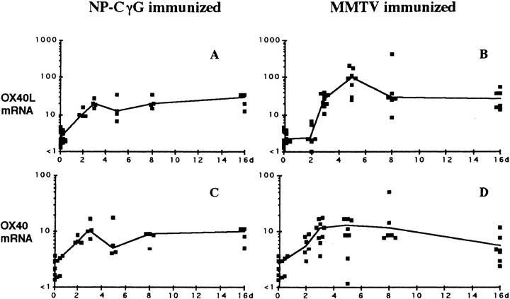
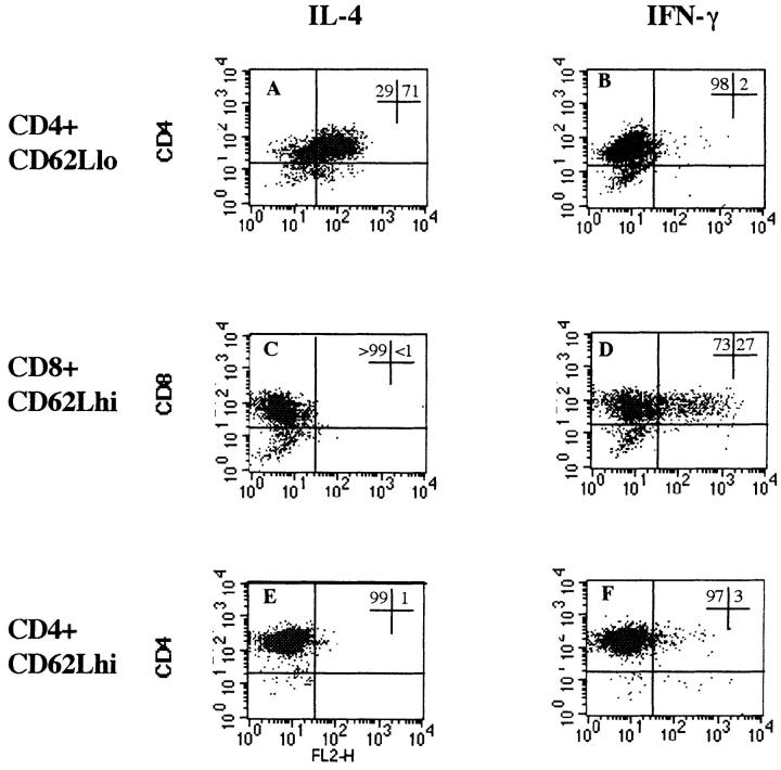
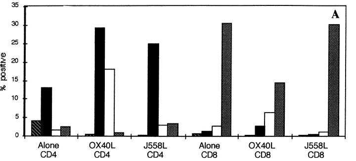
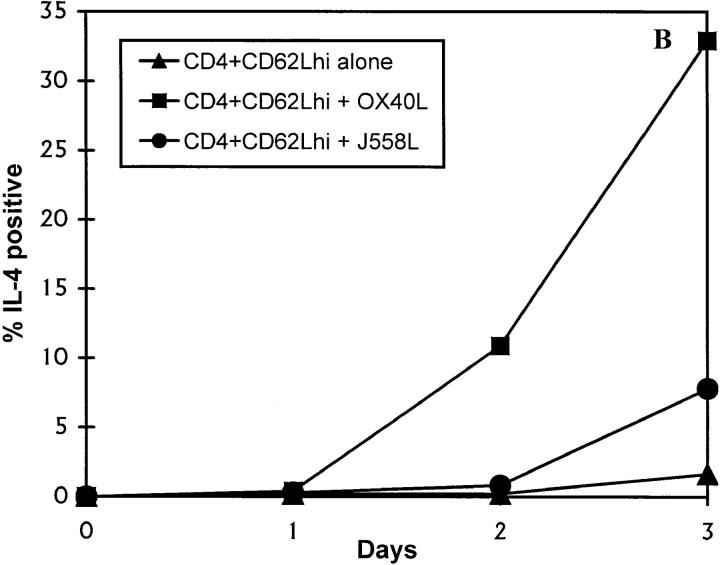
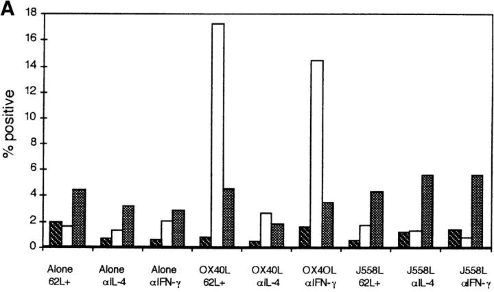
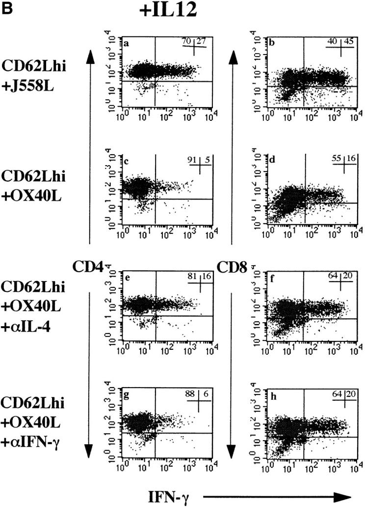
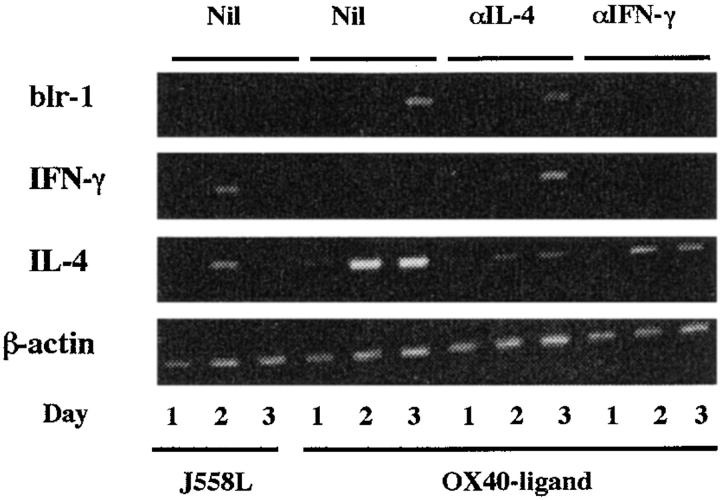
This report investigates the role of OX40 ligand (OX40L) and its receptor, OX40, expressed on activated B and T cells, respectively, in promoting the differentiation of T helper type 2 (Th2) CD4 T cells. These molecules are expressed in vivo by day 2 after priming with T cell- dependent antigens. Their expression coincides with the appearance of immunoglobulin (Ig)G switch transcripts and mRNA for interleukin (IL)-4 and interferon (IFN)-gamma, suggesting that this molecular interaction plays a role in early cognate interactions between B and T cells. In vitro, we report that costimulation of naive, CD62Lhigh CD4 T cells through OX40 promotes IL-4 expression and upregulates mRNA for the chemokine receptor, blr-1, whose ligand is expressed in B follicles and attracts lymphocytes to this location. Furthermore, T cell stimulation through OX40 inhibits IFN-gamma expression in both CD8 T cells and IL-12-stimulated CD4 T cells. Although this signal initiates IL-4 expression, IL-4 itself is strongly synergistic. Our data suggest that OX40L on antigen-activated B cells instructs naive T cells to differentiate into Th2 cells and migrate into B follicles, where T cell-dependent germinal centers develop.


Figures

Figure 1
Figure 1
Induction of OX40 and OX40L after immunization. Time course of induction of mRNA for OX40 and OX40L in popliteal LNs in mice after immunization with NP-CγG (plus killed Bordetella pertussis 5 × 10⁸) or MMTV. Symbols show the amount of mRNA for OX40L (A and B) and OX40 mRNA (C and D) per LN section by semiquantitative PCR. Symbols, Results for individual mice; solid lines, median values.
Figure 2
Figure 2
Intracellular staining for IL-4 and IFN-γ on activated subpopulations of T cells. Purified subpopulations of CD4 and CD8 T cells, which had been activated for 3 d with immobilized mAb to CD3 and soluble mAb to CD28, were restimulated through CD3 for 4 h in the presence of GolgiStop™ to cause accumulation of intracellular cytokines before staining on the FACs®. Histograms show the proportion of cells positive compared with a negative control mAb: CD4+CD62Llow T cells enriched for activated and memory T cells (A) IL-4 and (B) IFN-γ; CD8+ CD62Lhigh naive T cells (C) IL-4 and (D) IFN-γ; and CD4+CD62Lhi naive T cells (E) IL-4 and (F) IFN-γ. lo, Low; hi, high.
Figure 3
Figure 3
Induction of IL-4 expression by OX40L. CD62Lhigh CD8 and CD4 T cells were activated through CD3 and CD28 for 3 d in the presence of a fixed OX40L transfectant or the control parental cell line. Cells were restimulated through CD3 for 4 h in the presence of GolgiStop™ and stained for intracellular expression. (A) Staining with a control mAb (striped bars), anti-CD40L (black bars), anti–IL-4 (white bars), and anti–IFN-γ (stippled bars), on purified CD4 or CD8 CD62Lhigh T cells. The above data are representative of at least 10 different experiments. (B) Timing of induction of IL-4 expression in naive T cells by OX40L. Cells were stimulated as above. Data are representative of two experiments. hi, High.
Figure 3
Figure 3
Induction of IL-4 expression by OX40L. CD62Lhigh CD8 and CD4 T cells were activated through CD3 and CD28 for 3 d in the presence of a fixed OX40L transfectant or the control parental cell line. Cells were restimulated through CD3 for 4 h in the presence of GolgiStop™ and stained for intracellular expression. (A) Staining with a control mAb (striped bars), anti-CD40L (black bars), anti–IL-4 (white bars), and anti–IFN-γ (stippled bars), on purified CD4 or CD8 CD62Lhigh T cells. The above data are representative of at least 10 different experiments. (B) Timing of induction of IL-4 expression in naive T cells by OX40L. Cells were stimulated as above. Data are representative of two experiments. hi, High.
Figure 4
Figure 4
(A) Dependence of OX40L effect on IL-4 and IFN-γ secretion. Purified CD4+CD62Lhigh T cells were stimulated as described in the legend to Fig. 3 in the presence or absence of neutralizing mAbs to IL-4 and IFN-γ at a final concentration of 50 μg/ml. Before restimulation, cells were washed three times in medium to remove excess cytokine antibody. This was done to prevent interference with subsequent staining for intracellular cytokines. A shows the percentage of IL-4– or IFN-γ–positive CD4+ T cells when T cells were stimulated alone, with J558L, or with OX40L-fixed transfectants. Data shown are representative of six experiments in Balb/c mice. C57Bl/6 mice gave qualitatively similar results, but the degree of IL-4 expression was less (three experiments). Striped bars, Control; white bars, IL-4; stippled bars, IFN-γ. (B) Effect of OX40L on intracellular expression of IFN-γ in CD4 and CD8 T cells. CD4 and CD8 T cells were activated for 3 d as described in the legend to Fig. 2. To induce IFN-γ expression in CD4 T cells, IL-12 was added to cultures at a final concentration of 50 ng/ml. a, c, e, and g, Effects of OX40L and blocking cytokine mAbs on IFN-γ expression in CD4 T cells. b, d, f, and h, Effects on CD8 T cells. Results are representative of five separate experiments. hi, High.
Figure 4
Figure 4
(A) Dependence of OX40L effect on IL-4 and IFN-γ secretion. Purified CD4+CD62Lhigh T cells were stimulated as described in the legend to Fig. 3 in the presence or absence of neutralizing mAbs to IL-4 and IFN-γ at a final concentration of 50 μg/ml. Before restimulation, cells were washed three times in medium to remove excess cytokine antibody. This was done to prevent interference with subsequent staining for intracellular cytokines. A shows the percentage of IL-4– or IFN-γ–positive CD4+ T cells when T cells were stimulated alone, with J558L, or with OX40L-fixed transfectants. Data shown are representative of six experiments in Balb/c mice. C57Bl/6 mice gave qualitatively similar results, but the degree of IL-4 expression was less (three experiments). Striped bars, Control; white bars, IL-4; stippled bars, IFN-γ. (B) Effect of OX40L on intracellular expression of IFN-γ in CD4 and CD8 T cells. CD4 and CD8 T cells were activated for 3 d as described in the legend to Fig. 2. To induce IFN-γ expression in CD4 T cells, IL-12 was added to cultures at a final concentration of 50 ng/ml. a, c, e, and g, Effects of OX40L and blocking cytokine mAbs on IFN-γ expression in CD4 T cells. b, d, f, and h, Effects on CD8 T cells. Results are representative of five separate experiments. hi, High.
Figure 5
Figure 5
OX40L induces IL-4 and blr-1 mRNA in naive CD4 T cells. The ethidium bromide gel shows semiquantitative RT-PCR for IL-4, IFN-γ, and blr-1 in cell samples of activated CD4 CD62Lhigh T cells at the times indicated after culture. Amounts of mRNA were adjusted to give comparable β-actin signals. T cells had been stimulated in the presence of fixed parental cell line J558L or OX40L transfectant with and without mAb to IL-4 and IFN-γ. RT-PCR of the fixed cell lines did not yield cDNA that could be amplified. Results are representative of three experiments. Nil, no blocking mAb added.

References

    1. Kumararatne DS. Tuberculosis and immunodeficiency— of mice and men. Clin Exp Immunol. 1997;107:11–14. - PMC - PubMed
    1. Toellner K, Luther S, Sze DM-Y, Choy RK-W, Taylor DR, MacLennan ICM, Acha-Orbea H. Th1 and Th2 characteristics start to develop during T cell priming and are associated with an immediate ability to induce immunoglobulin class switching. J Exp Med. 1998;187:1193–1201. - PMC - PubMed
    1. Trinchieri G. Interleukin-12: a proinflammatory cytokine with immunoregulatory functions that bridge innate resistance and antigen-specific adaptive immunity. Annu Rev Immunol. 1995;13:251–276. - PubMed
    1. Cella M, Scheidegger D, Palmer-Lehman K, Lane P, Lanzavecchia A, Alber G. Ligation of CD40 on dendritic cells triggers production of high levels of interleukin-12 and enhances T cell costimulatory capacity: T–T help via APC activation. J Exp Med. 1996;184:747–752. - PMC - PubMed
    1. Sousa CR, Hieny S, Scharton-Kersten T, Jankovic D, Charest H, Germain RN, Sher A. In vivo microbial stimulation induces rapid CD40 ligand–independent production of interleukin 12 by dendritic cells and their redistribution to T cell areas. J Exp Med. 1997;186:1819–1829. - PMC - PubMed

Publication types

MeSH terms