. 1998 Jul 20;188(2):351-64.
doi: 10.1084/jem.188.2.351.

Host resistance to intracellular infection: mutation of natural resistance-associated macrophage protein 1 (Nramp1) impairs phagosomal acidification

Affiliations

Host resistance to intracellular infection: mutation of natural resistance-associated macrophage protein 1 (Nramp1) impairs phagosomal acidification

D J Hackam et al. J Exp Med. .

Abstract

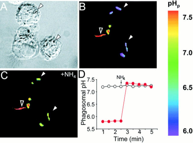
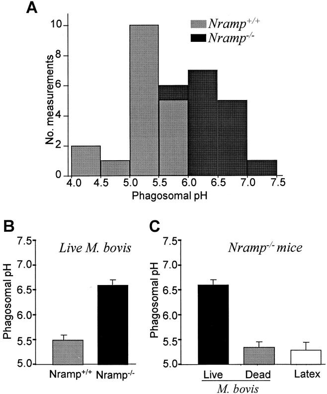
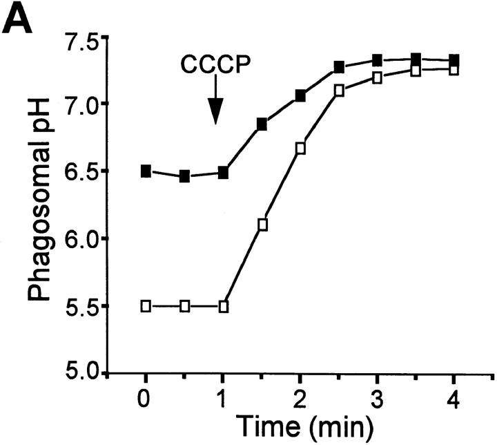
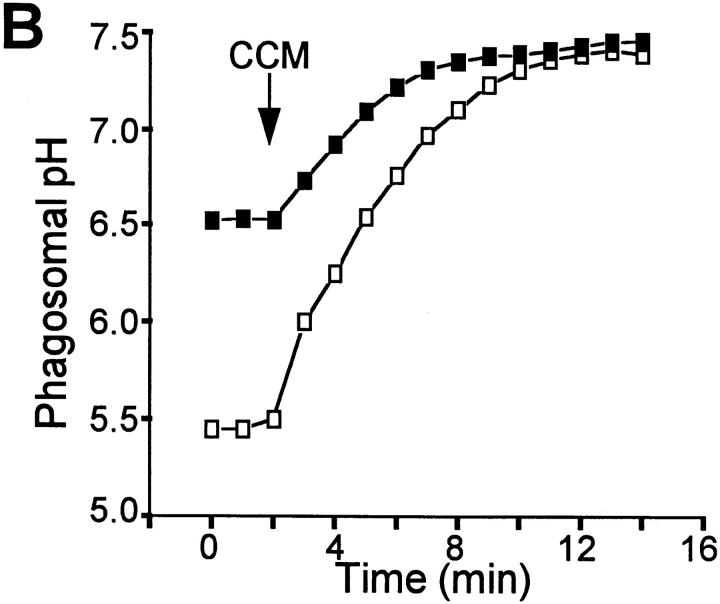
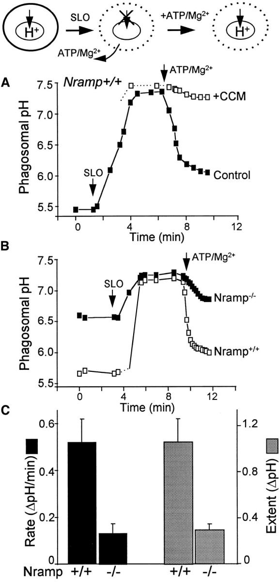
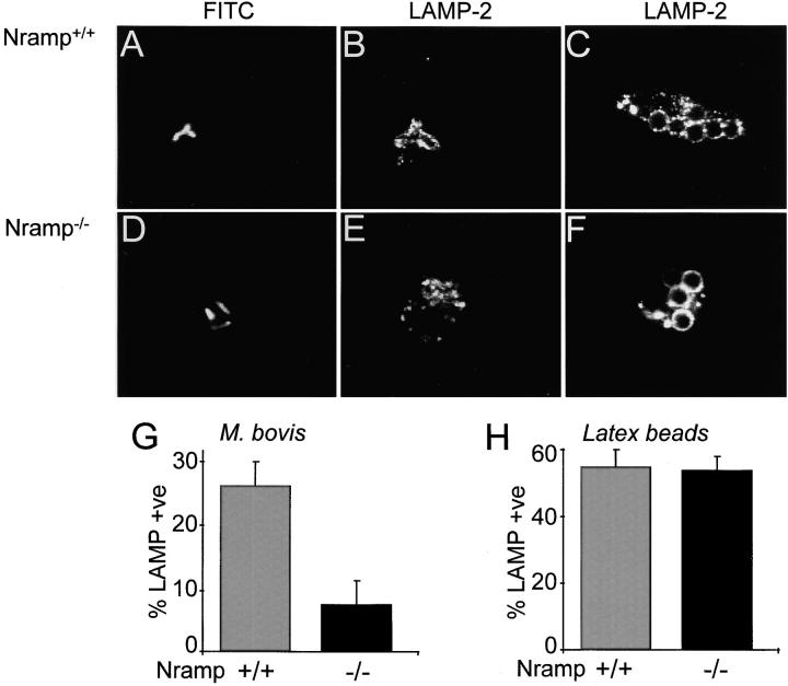
The mechanisms underlying the survival of intracellular parasites such as mycobacteria in host macrophages remain poorly understood. In mice, mutations at the Nramp1 gene (for natural resistance-associated macrophage protein), cause susceptibility to mycobacterial infections. Nramp1 encodes an integral membrane protein that is recruited to the phagosome membrane in infected macrophages. In this study, we used microfluorescence ratio imaging of macrophages from wild-type and Nramp1 mutant mice to analyze the effect of loss of Nramp1 function on the properties of phagosomes containing inert particles or live mycobacteria. The pH of phagosomes containing live Mycobacterium bovis was significantly more acidic in Nramp1- expressing macrophages than in mutant cells (pH 5.5 +/- 0.06 versus pH 6.6 +/- 0.05, respectively; P <0.005). The enhanced acidification could not be accounted for by differences in proton consumption during dismutation of superoxide, phagosomal buffering power, counterion conductance, or in the rate of proton "leak", as these were found to be comparable in wild-type and Nramp1-deficient macrophages. Rather, after ingestion of live mycobacteria, Nramp1-expressing cells exhibited increased concanamycin-sensitive H+ pumping across the phagosomal membrane. This was associated with an enhanced ability of phagosomes to fuse with vacuolar-type ATPase-containing late endosomes and/or lysosomes. This effect was restricted to live M. bovis and was not seen in phagosomes containing dead M. bovis or latex beads. These data support the notion that Nramp1 affects intracellular mycobacterial replication by modulating phagosomal pH, suggesting that Nramp1 plays a central role in this process.


Figures

Figure 1
Figure 1
Measurement of pHp. Peritoneal macrophages isolated from wild-type Nramp1-expressing mice (strain 129/sv) were allowed to interact with live, fluoresceinated M. bovis, some of which became internalized. The cells were then plated on glass coverslips, mounted in thermoregulated chambers, and visualized using differential interference contrast optics (A), while the fluorescence was measured with alternating excitation at 440 and 490 nm. The fluorescence ratio (B and C) was used for the measurement of the pH in the vicinity of the bacterial surface. After acquiring baseline pH measurements (B), the cells were exposed to 20 mM NH4 + and recording was resumed (C). A pseudocolor pH scale is shown to the right. Internalized bacteria, identified by their resting acidic pH and responsiveness to NH4 + (filled white arrowheads) and an adherent extracellular bacterium (open arrowhead) are indicated. A typical time course of pH determination in internalized (filled red circles) and adherent bacteria (open circles) are shown in D . The addition of 20 mM NH4 + is indicated. Representative of 37 individual experiments.
Figure 2
Figure 2
Determination of pHp in wild-type and Nramp1 −/− macrophages. Phagosomal pH was measured by ratio imaging as described for Fig. 1 (see Materials and Methods) in peritoneal macrophages obtained from wild-type (Nramp +/+) mice and Nramp1 mutant mice (Nramp1 −/−). Phagosome formation was induced using either live, fluoresceinated M. bovis (A, B, and leftmost bar in C), dead M. bovis (middle bar in C), or opsonized latex beads (rightmost bar in C). (A) frequency histogram comparing the pH in wild-type (Nramp +/+) mice and Nramp1 mutant mice (Nramp1 −/−); (B) summary of results comparing Nramp +/+ and Nramp1 −/− cells; data are means ± SE of 37 individual determinations; (C) summary of results comparing different phagocytic particles in Nramp1 −/− cells. Data are means ± SE of 24 determinations.
Figure 3
Figure 3
Effects of protonophores and V-ATPase inhibitors on pHp. pHp was measured by ratio imaging as described for Fig. 1 (see Materials and Methods) in peritoneal macrophages obtained from wild-type mice (open symbols) and Nramp1 −/− mice (filled symbols). Phagosome formation was induced using live, fluoresceinated M. bovis. After acquiring baseline measurements, 10 μM CCCP (A) or 100 nM concanamycin (B, CCM) was added to both samples. Data are representative of five determinations.
Figure 3
Figure 3
Effects of protonophores and V-ATPase inhibitors on pHp. pHp was measured by ratio imaging as described for Fig. 1 (see Materials and Methods) in peritoneal macrophages obtained from wild-type mice (open symbols) and Nramp1 −/− mice (filled symbols). Phagosome formation was induced using live, fluoresceinated M. bovis. After acquiring baseline measurements, 10 μM CCCP (A) or 100 nM concanamycin (B, CCM) was added to both samples. Data are representative of five determinations.
Figure 4
Figure 4
pHp determinations in SLO-permeabilized cells. The experimental protocol is diagramatically illustrated at the top. In brief, intact cells that normally have acidic phagosomes (left) were permeabilized using 0.1 μg/ml SLO in media devoid of ATP/Mg2+, resulting in inhibition of H+ pumping due to depletion of substrate (middle). Readdition of ATP/ Mg2+ induces resumption of pumping (right). Phagosomal pH was measured by ratio imaging as described for Fig. 1 (see Materials and Methods) in peritoneal macrophages obtained from wild-type (Nramp +/+) mice and from Nramp1 mutant mice (Nramp −/−). (A) Measurements in Nramp1 +/+ cells. Where indicated, cells were treated with SLO, and ATP and Mg were reintroduced in the absence (filled symbols) or presence of 100 nM concanamycin (CCM; open symbols). (B) Nramp1 +/+ cells (open symbols) or Nramp1 −/− cells (filled symbols) were treated with SLO. ATP and Mg were reintroduced where indicated. A and B are each representative of five determinations. (C) Summary of results from five experiments like that shown in B. The initial rate (black bars) and extent (stippled bars) of ATP/ Mg-induced acidification were measured. Data are means ± SE of five separate experiments.
Figure 5
Figure 5
Delivery of LAMP-2 to phagosomes in wild-type and Nramp1 −/− cells. Macrophages obtained from Nramp1 +/+ (A–C) or Nramp1 −/− (D–F) mice were allowed to internalize live, fluoresceinated M. bovis or opsonized latex beads as specified. The cells were then fixed, permeabilized, and incubated with antibodies to LAMP-2, followed by Cy3-labeled secondary antibodies. Representative confocal fluorescence images are shown in A–F. The fraction of phagosomes stained by anti–LAMP-2 antibodies in M. bovis phagosomes is summarized in G (means ± SE of seven determinations), and the fraction of phagosomes stained in latex bead phagosomes is shown in H (seven determinations).
Figure 6
Figure 6
Localization of V-ATPases in early endosomes and in phagosomes. (A–C) Macrophages obtained from Nramp1 −/− mice were allowed to internalize Texas red–labeled fixable (1 mg/ml) dextran for 15 min at 37°C. The cells were then fixed, permeabilized, and incubated with an affinity-purified polyclonal antibody against the 39-kD subunit of the V-ATPase, followed by an FITC-labeled secondary antibody. (A) Localization of Texas red–labeled dextran; (B) localization of the V-ATPase subunit; (C) areas of overlap between the dextran and the ATPase. (D–F) Macrophages obtained from Nramp1 −/− mice were allowed to internalize live, fluoresceinated M. bovis and were subsequently incubated with Texas red–labeled dextran for 15 min at 37°C. The cells were then fixed and visualized by Nomarski (D) and confocal immunofluorescence microscopy. (E) Localization of fluoresceinated bacteria; (F) distribution of Texas red–labeled dextran. Images are representative of at least five experiments of each kind. (G) Macrophages obtained from Nramp1 −/− mice were allowed to internalize FITC-labeled human holotransferrin (20 μg/ml), and pHe was measured using single-cell imaging. Where indicated, concanamycin (100 nM) was added. Data represent means ± SEM of three separate experiments.
Figure 6
Figure 6
Localization of V-ATPases in early endosomes and in phagosomes. (A–C) Macrophages obtained from Nramp1 −/− mice were allowed to internalize Texas red–labeled fixable (1 mg/ml) dextran for 15 min at 37°C. The cells were then fixed, permeabilized, and incubated with an affinity-purified polyclonal antibody against the 39-kD subunit of the V-ATPase, followed by an FITC-labeled secondary antibody. (A) Localization of Texas red–labeled dextran; (B) localization of the V-ATPase subunit; (C) areas of overlap between the dextran and the ATPase. (D–F) Macrophages obtained from Nramp1 −/− mice were allowed to internalize live, fluoresceinated M. bovis and were subsequently incubated with Texas red–labeled dextran for 15 min at 37°C. The cells were then fixed and visualized by Nomarski (D) and confocal immunofluorescence microscopy. (E) Localization of fluoresceinated bacteria; (F) distribution of Texas red–labeled dextran. Images are representative of at least five experiments of each kind. (G) Macrophages obtained from Nramp1 −/− mice were allowed to internalize FITC-labeled human holotransferrin (20 μg/ml), and pHe was measured using single-cell imaging. Where indicated, concanamycin (100 nM) was added. Data represent means ± SEM of three separate experiments.

References

    1. Young P. White House to expand response to infectious diseases. Am Soc Microbiol News. 1996;62:450–451.
    1. Berkelman RL, Bryan RT, Osterholm MT, Leduc JW, Hughes JM. Infectious disease surveillance: a crumbling foundation. Science. 1996;264:368–370. - PubMed
    1. Bloom BR. Tuberculosis: back to a frightening future. Nature. 1992;358:538–540. - PubMed
    1. Kaye K, Frieden TR. Tuberculosis control: the relevance of classic principles in an era of acquired immunodeficiency syndrome and multidrug resistance. Epidemiologic Rev. 1996;18:52–63. - PubMed
    1. Davies J. Inactivation of antibiotics and the dissemination of resistance genes. Science. 1994;264:375–382. - PubMed

Publication types

MeSH terms

Substances