

Nitric oxide donors induce late preconditioning against myocardial stunning and infarction in conscious rabbits via an antioxidant-sensitive mechanism
- PMID: 9670920
- PMCID: PMC3701311
- DOI: 10.1161/01.res.83.1.73
Nitric oxide donors induce late preconditioning against myocardial stunning and infarction in conscious rabbits via an antioxidant-sensitive mechanism
Abstract
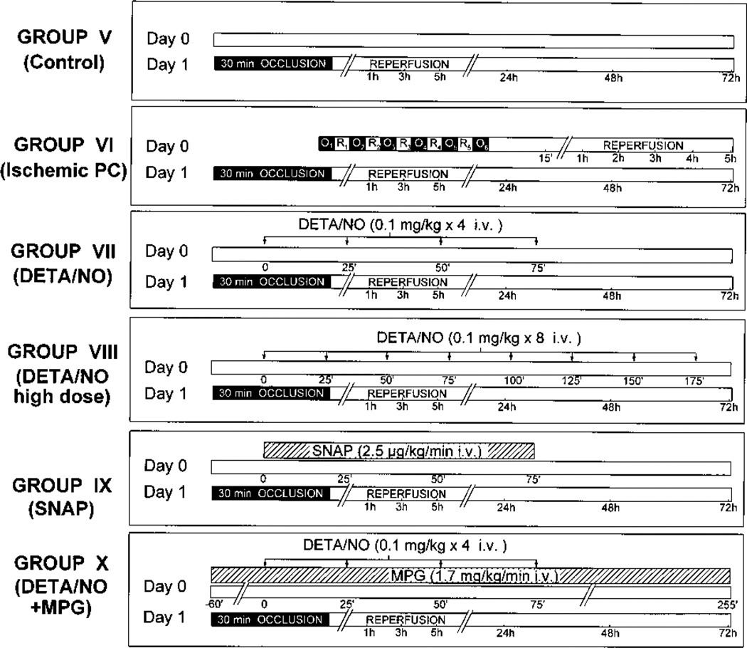
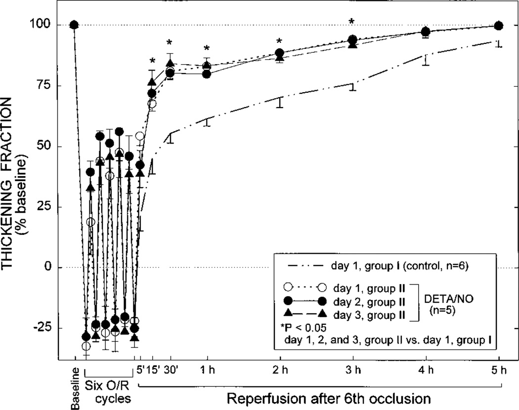
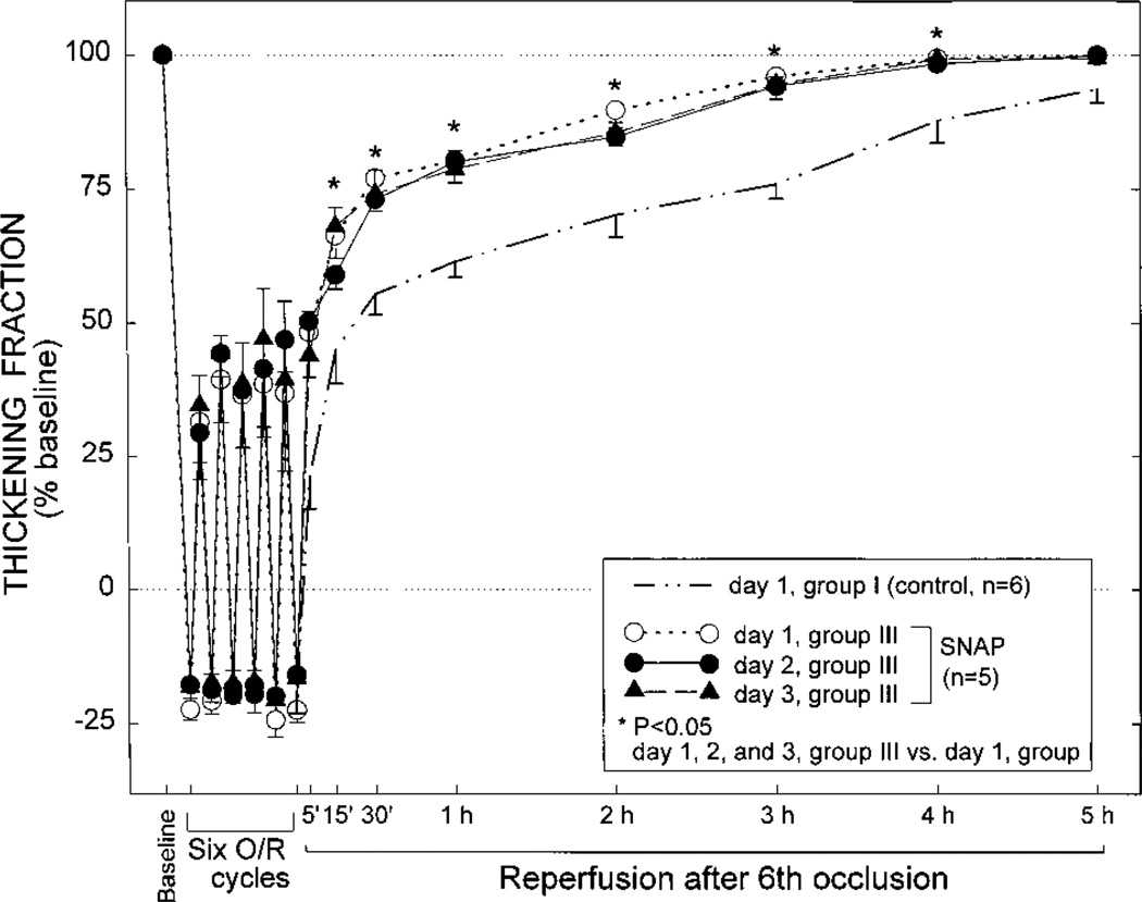
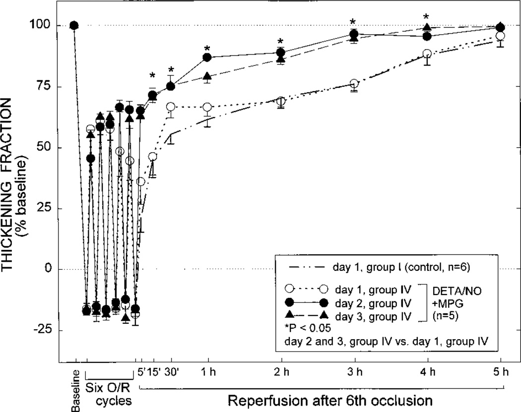
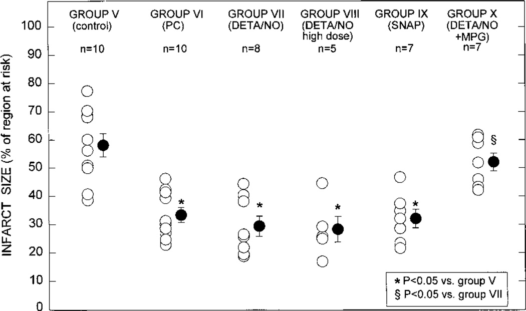
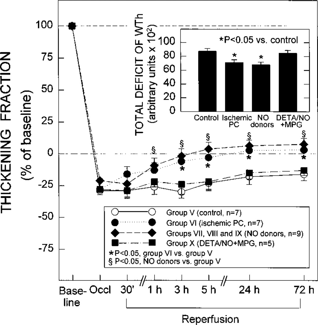
The goal of this study was to test the hypothesis that the cardioprotective effects of the late phase of ischemic preconditioning (PC) can be mimicked by treatment with NO donors. In phase I (studies of myocardial stunning), conscious rabbits underwent a sequence of six 4-minute coronary occlusion/4-minute reperfusion cycles for 3 consecutive days (days 1, 2, and 3). In group I (controls, n=6), the total deficit of systolic wall thickening (WTh) after the sixth reperfusion was reduced by 54% on days 2 and 3 compared with day 1 (P<0.05), indicating a late PC effect against myocardial stunning. When rabbits were given the NO donors diethylenetriamine/NO (DETA/NO, 0.1 mg/kg i.v., 4 times [group II, n=5]) or S-nitroso-N-acetylpenicillamine (SNAP, 2.5 microg x kg(-1) x min(-1) i.v. for 75 minutes [group III, n=51) 24 hours before the first sequence of occlusion/reperfusion cycles, the deficit of WTh on day 1 was 60% (group II) and 54% (group III) less than that observed in controls (P<0.05 for both). In both groups II and III, there was no further improvement in the deficit of WTh on days 2 and 3 compared with day 1. The protective effect of DETA/NO was completely abrogated when this agent was given in conjunction with the ONOO- and .OH scavenger mercaptopropionyl glycine (MPG) (group IV, n=5). In phase II (studies of myocardial infarction), conscious rabbits underwent a 30-minute coronary occlusion followed by 3 days of reperfusion. When rabbits were preconditioned 24 hours earlier with six 4-minute occlusion/4-minute reperfusion cycles, infarct size was reduced by 43% (33.2+/-2.7% versus 58.3+/-4.1% of the region at risk in controls, P<0.05), indicating a late PC effect against myocardial infarction. When rabbits were pretreated with DETA/NO (group VII, n=8) or SNAP (group IX, n=7) 24 hours before the 30-minute occlusion, infarct size was reduced by a similar degree (29.3+/-3.6% and 32.0+/-3.3% of the region at risk, respectively; P<0.05 versus controls). The degree of protection could not be increased by doubling the dose of DETA/NO (group VIII, n=5). Coadministration of MPG completely abrogated the infarct-sparing action of DETA/NO (group X, n=7). Taken together, these results demonstrate that in conscious rabbits the administration of 2 structurally unrelated NO donors induces protection 24 hours later against both reversible (stunning) and irreversible (infarction) ischemia/reperfusion injury and that the magnitude of this protection is indistinguishable from that observed during the late phase of ischemic PC. The fact that the late phase of ischemic PC can be mimicked by NO donors provides direct evidence that NO in itself is sufficient to elicit this cardioprotective mechanism. The fact that NO donor-induced late PC was abrogated by MPG indicates that the mechanism whereby NO induces this phenomenon involves the generation of oxidant species, possibly ONOO- and/or .OH. Since a relatively brief treatment with hemodynamically inactive doses of NO donors can induce long-lasting protective effects, these agents could be useful for preconditioning the heart in patients.
Figures


Similar articles
-
Isoform-selective activation of protein kinase C by nitric oxide in the heart of conscious rabbits: a signaling mechanism for both nitric oxide-induced and ischemia-induced preconditioning.Circ Res. 1999 Mar 19;84(5):587-604. doi: 10.1161/01.res.84.5.587. Circ Res. 1999. PMID: 10082480
-
Nitric oxide donors attenuate myocardial stunning in conscious rabbits.Am J Physiol. 1999 Dec;277(6):H2495-503. doi: 10.1152/ajpheart.1999.277.6.H2495. Am J Physiol. 1999. PMID: 10600874
-
The protective effect of late preconditioning against myocardial stunning in conscious rabbits is mediated by nitric oxide synthase. Evidence that nitric oxide acts both as a trigger and as a mediator of the late phase of ischemic preconditioning.Circ Res. 1997 Dec;81(6):1094-107. doi: 10.1161/01.res.81.6.1094. Circ Res. 1997. PMID: 9400391
-
Cardioprotective function of inducible nitric oxide synthase and role of nitric oxide in myocardial ischemia and preconditioning: an overview of a decade of research.J Mol Cell Cardiol. 2001 Nov;33(11):1897-918. doi: 10.1006/jmcc.2001.1462. J Mol Cell Cardiol. 2001. PMID: 11708836 Review.
-
Consequences of brief ischemia: stunning, preconditioning, and their clinical implications: part 1.Circulation. 2001 Dec 11;104(24):2981-9. doi: 10.1161/hc4801.100038. Circulation. 2001. PMID: 11739316 Review.
Cited by
-
Nitric oxide interacts with cholinoceptors to modulate insulin secretion by pancreatic β cells.Pflugers Arch. 2020 Oct;472(10):1469-1480. doi: 10.1007/s00424-020-02443-9. Epub 2020 Aug 16. Pflugers Arch. 2020. PMID: 32803305 Free PMC article.
-
Exogenous Nitric Oxide Protects Human Embryonic Stem Cell-Derived Cardiomyocytes against Ischemia/Reperfusion Injury.Oxid Med Cell Longev. 2016;2016:4298945. doi: 10.1155/2016/4298945. Epub 2016 Jun 15. Oxid Med Cell Longev. 2016. PMID: 27403231 Free PMC article.
-
Preconditioning in cardiac anesthesia…… where are we?Ann Card Anaesth. 2019 Oct-Dec;22(4):412-421. doi: 10.4103/aca.ACA_116_18. Ann Card Anaesth. 2019. PMID: 31621678 Free PMC article. Review.
-
Free-radical production triggered by hyperthermia contributes to heat stress-induced cardioprotection in isolated rat hearts.Br J Pharmacol. 2002 Apr;135(7):1776-82. doi: 10.1038/sj.bjp.0704619. Br J Pharmacol. 2002. PMID: 11934819 Free PMC article.
-
Delayed adaptation of the heart to stress: late preconditioning.Stroke. 2004 Nov;35(11 Suppl 1):2676-9. doi: 10.1161/01.STR.0000143220.21382.fd. Epub 2004 Sep 30. Stroke. 2004. PMID: 15459441 Free PMC article. Review.
References
-
- Marber MS, Latchman DS, Walker JM, Yellon DM. Cardiac stress protein elevation 24 hours after brief ischemia or heat stress is associated with resistance to myocardial infarction. Circulation. 1993;88:1264–1272. - PubMed
-
- Baxter GF, Marber MS, Patel VC, Yellon DM. Adenosine receptor involvement in a delayed phase of myocardial protection 24 hours after ischemic preconditioning. Circulation. 1994;90:2993–3000. - PubMed
-
- Bolli R, Bhatti ZA, Tang X-L, Qiu Y, Zhang Q, Guo Y, Jadoon AK. Evidence that late preconditioning against myocardial stunning in conscious rabbits is triggered by the generation of nitric oxide. Circ Res. 1997;81:42–52. - PubMed
-
- Qiu Y, Rizvi A, Tang X-L, Manchikalapudi S, Takano H, Jadoon AK, Wu W-J, Bolli R. Nitric oxide triggers late preconditioning against myocardial infarction in conscious rabbits. Am J Physiol. 1997;273:H2931–H2936. - PubMed
-
- Bolli R, Manchikalapudi S, Tang X-L, Takano H, Qiu Y, Guo Y, Zhang Q, Jadoon AK. The protective effect of late preconditioning against myocardial stunning in conscious rabbits is mediated by nitric oxide synthase: evidence that nitric oxide acts both as a trigger and as mediator of the late phase of preconditioning. Circ Res. 1997;81:1094–1107. - PubMed
Publication types
MeSH terms
Substances
Grants and funding
LinkOut - more resources
Full Text Sources
Medical
Miscellaneous
