. 1998 Aug;18(8):4471-87.
doi: 10.1128/MCB.18.8.4471.

High-mobility group chromatin proteins 1 and 2 functionally interact with steroid hormone receptors to enhance their DNA binding in vitro and transcriptional activity in mammalian cells

Affiliations

High-mobility group chromatin proteins 1 and 2 functionally interact with steroid hormone receptors to enhance their DNA binding in vitro and transcriptional activity in mammalian cells

V Boonyaratanakornkit et al. Mol Cell Biol. 1998 Aug.

Abstract

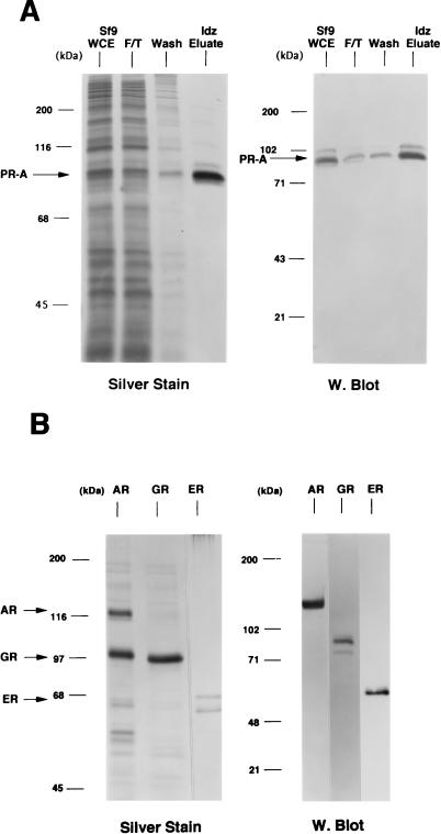
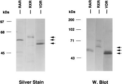
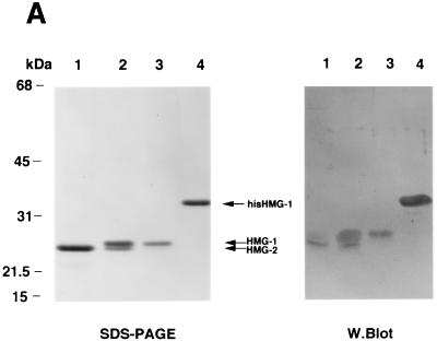
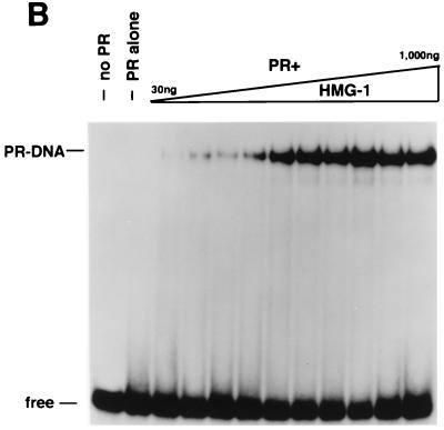
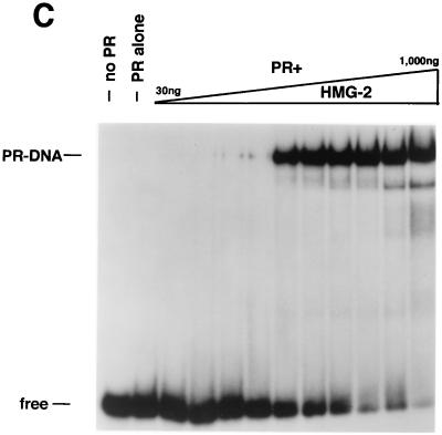
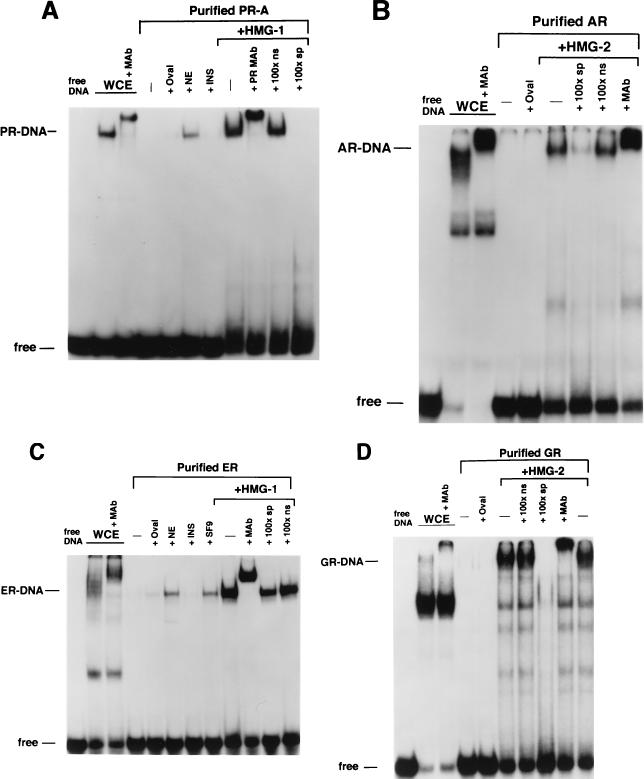
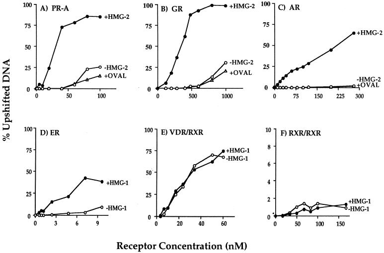
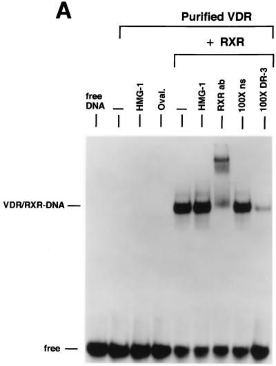
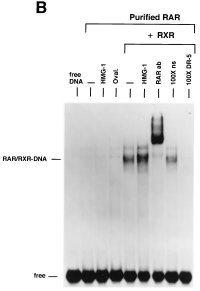
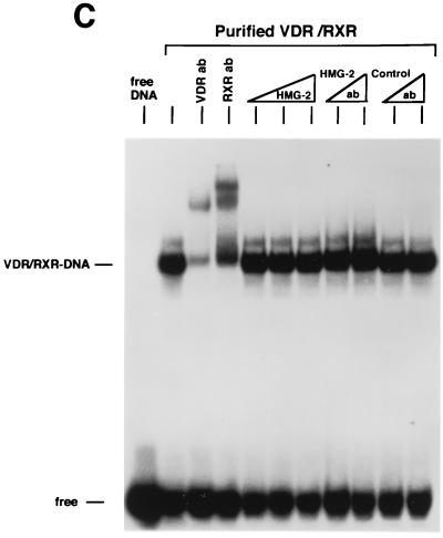
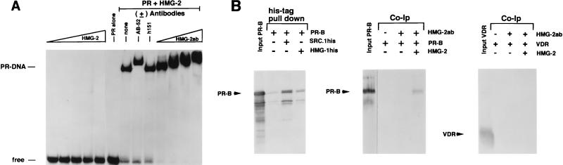
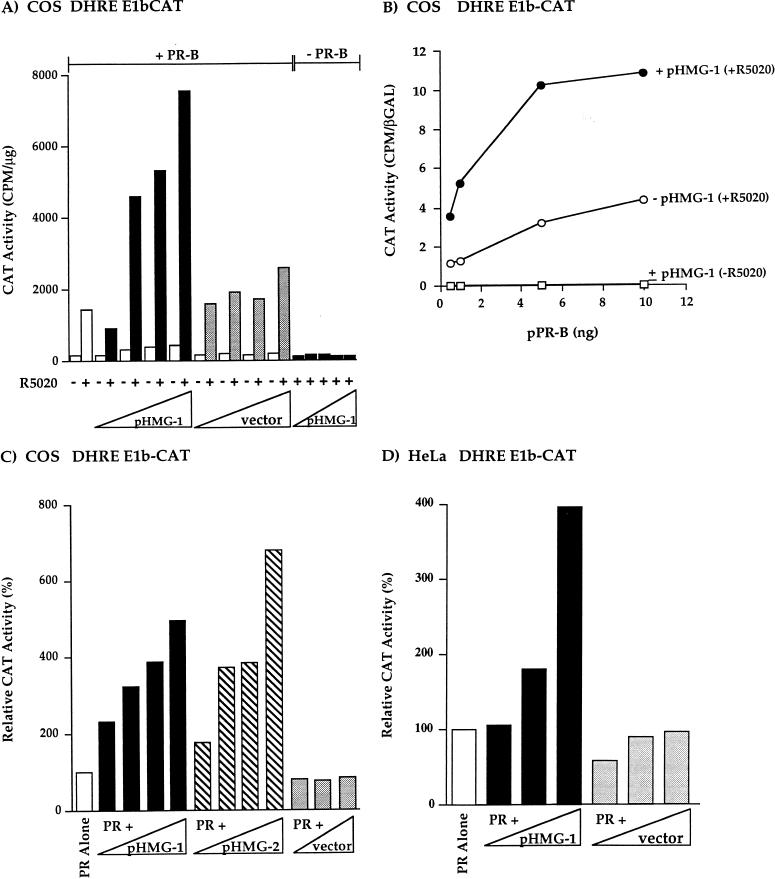
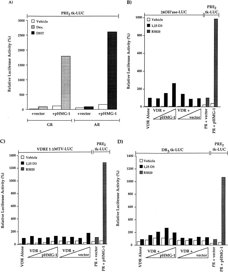
We previously reported that the chromatin high-mobility group protein 1 (HMG-1) enhances the sequence-specific DNA binding activity of progesterone receptor (PR) in vitro, thus providing the first evidence that HMG-1 may have a coregulatory role in steroid receptor-mediated gene transcription. Here we show that HMG-1 and the highly related HMG-2 stimulate DNA binding by other steroid receptors, including estrogen, androgen, and glucocorticoid receptors, but have no effect on DNA binding by several nonsteroid nuclear receptors, including retinoid acid receptor (RAR), retinoic X receptor (RXR), and vitamin D receptor (VDR). As highly purified recombinant full-length proteins, all steroid receptors tested exhibited weak binding affinity for their optimal palindromic hormone response elements (HREs), and the addition of purified HMG-1 or -2 substantially increased their affinity for HREs. Purified RAR, RXR, and VDR also exhibited little to no detectable binding to their cognate direct repeat HREs but, in contrast to results with steroid receptors, the addition of HMG-1 or HMG-2 had no stimulatory effect. Instead, the addition of purified RXR enhanced RAR and VDR DNA binding through a heterodimerization mechanism and HMG-1 or HMG-2 had no further effect on DNA binding by RXR-RAR or RXR-VDR heterodimers. HMG-1 and HMG-2 (HMG-1/-2) themselves do not bind to progesterone response elements, but in the presence of PR they were detected as part of an HMG-PR-DNA ternary complex. HMG-1/-2 can also interact transiently in vitro with PR in the absence of DNA; however, no direct protein interaction was detected with VDR. These results, taken together with the fact that PR can bend its target DNA and that HMG-1/-2 are non-sequence-specific DNA binding proteins that recognize DNA structure, suggest that HMG-1/-2 are recruited to the PR-DNA complex by the combined effect of transient protein interaction and DNA bending. In transient-transfection assays, coexpression of HMG-1 or HMG-2 increased PR-mediated transcription in mammalian cells by as much as 7- to 10-fold without altering the basal promoter activity of target reporter genes. This increase in PR-mediated gene activation by coexpression of HMG-1/-2 was observed in different cell types and with different target promoters, suggesting a generality to the functional interaction between HMG-1/-2 and PR in vivo. Cotransfection of HMG-1 also increased reporter gene activation mediated by other steroid receptors, including glucocorticoid and androgen receptors, but it had a minimal influence on VDR-dependent transcription in vivo. These results support the conclusion that HMG-1/-2 are coregulatory proteins that increase the DNA binding and transcriptional activity of the steroid hormone class of receptors but that do not functionally interact with certain nonsteroid classes of nuclear receptors.


Figures

FIG. 1
FIG. 1
Purification of baculovirus-expressed steroid receptors. Human PR-A, AR, and GR were expressed as polyhistidine-tagged proteins in baculovirus and were purified by metal ion affinity resins (Talon). Human ER was expressed as a nonfusion protein in baculovirus and purified by using a DNA affinity column. (A) (Left panel) Silver-stained SDS–7.5% PAGE of PR-A purification fractions. Lanes: 1, Sf9 whole-cell extract (WCE; 5 μl); 2, Talon resin flowthrough (F/T; 5 μl); 3, column wash (Wash; 50 μl); and 4, 100 mM imidazole (Idz) eluate (10 μl). (Right panel) Western blot of the same PR-A purification fractions (5 μl) with the AB-52 MAb. (B) (Left panel) Silver-stained SDS-PAGE of purified AR and GR eluted from Talon resins by 100 mM imidazole. ER is an NaCl eluate of a DNA affinity resin. (Right panel) Western blots of purified AR, GR, and ER were done with the following receptor specific antibodies: N441, an MAb to AR; a polyclonal antibody to GR; and h151, an MAb to human ER.
FIG. 2
FIG. 2
Purification of nonsteroid nuclear receptors. Human RXRα, RARγ, and VDR were expressed in yeast cells as polyhistidine-tagged proteins and were purified by using metal ion affinity resins. Purified products were analyzed by SDS-PAGE and silver staining (left panel) and by Western blotting with receptor-specific antibodies (right panel).
FIG. 3
FIG. 3
HMG-1 and HMG-2 purification and effects on PR-DNA binding in vitro. (A) HMG-1 and HMG-2 from calf thymus were purified and separated on a PBE94 chromatofocusing column as described in Materials and Methods. Coomassie blue-stained SDS-polyacrylamide gels (12%) and Western blot analysis of purified HMG-1/-2 fractions were prepared as follows: lane 1 (pool 1), HMG-2; lane 2 (pool 2), HMG-1 and HMG-2; lane 3 (pool 3), HMG-1; lane 4, purified recombinant polyhistidine-tagged HMG-1. Western blots were done with an IgM MAb (854/E10) that recognizes both HMG-1 and HMG-2. (B and C) The influence of purified calf thymus HMG-1 (pool 3) and HMG-2 (pool 1) on the binding of purified PR-A to a 32-bp PRE oligonucleotide probe as assessed by EMSA. A constant amount (30 nM) of purified PR-A was incubated with increasing amounts of purified HMG-1 (B) or HMG-2 (C). The amounts of HMG-1 and HMG-2 used were 30, 50, 70, 100, 200, 300, 400, 500, 700, and 1,000 μg/assay in lanes 3 to 12, respectively.
FIG. 3
FIG. 3
HMG-1 and HMG-2 purification and effects on PR-DNA binding in vitro. (A) HMG-1 and HMG-2 from calf thymus were purified and separated on a PBE94 chromatofocusing column as described in Materials and Methods. Coomassie blue-stained SDS-polyacrylamide gels (12%) and Western blot analysis of purified HMG-1/-2 fractions were prepared as follows: lane 1 (pool 1), HMG-2; lane 2 (pool 2), HMG-1 and HMG-2; lane 3 (pool 3), HMG-1; lane 4, purified recombinant polyhistidine-tagged HMG-1. Western blots were done with an IgM MAb (854/E10) that recognizes both HMG-1 and HMG-2. (B and C) The influence of purified calf thymus HMG-1 (pool 3) and HMG-2 (pool 1) on the binding of purified PR-A to a 32-bp PRE oligonucleotide probe as assessed by EMSA. A constant amount (30 nM) of purified PR-A was incubated with increasing amounts of purified HMG-1 (B) or HMG-2 (C). The amounts of HMG-1 and HMG-2 used were 30, 50, 70, 100, 200, 300, 400, 500, 700, and 1,000 μg/assay in lanes 3 to 12, respectively.
FIG. 4
FIG. 4
HMG-1/-2 stimulate the sequence-specific DNA binding activity of all steroid receptors tested. (A) Equal amounts (30 nM) of polyhistidine-tagged PR-A in Sf9 whole-cell extract (WCE) or affinity-purified PR-A were analyzed for binding to a 32P-labeled palindromic GRE-PRE oligonucleotide by EMSA. Lanes contain Sf9 whole-cell extract (WCE), purified PR alone (−), or purified PR plus 1 μg of ovalbumin (+Oval), 1 μg of nuclear extract (+NE) from calf thymus, 1 μg of insulin (+INS), or 300 ng of purified calf thymus HMG-1 (+HMG-1). The HMG-1-stimulated PR-DNA complexes were supershifted by the PR-specific MAb, AB-52, and were competed by a 100-fold molar excess of the PRE oligonucleotide (100×sp) but not by an ERE oligonucleotide (100×ns). (B) Equal amounts (70 nM) of baculovirus-expressed AR in WCE and affinity-purified AR were analyzed for binding to the GRE-PRE oligonucleotide by EMSA as described for panel A. (C) Equal amounts (10 nM) of baculovirus-expressed ER in WCE and affinity-purified ER were analyzed for binding to an ERE oligonucleotide by EMSA as described for panel A with or without the addition of 300 ng of purified calf thymus HMG-1. (D) Equal amounts (200 nM) of baculovirus-expressed GR in WCE and affinity-purified GR were analyzed for binding to the GRE-PRE by EMSA as described for panel A. All receptors were bound to their cognate (see Materials and Methods) hormonal ligands in Sf9 cells during expression.
FIG. 5
FIG. 5
Saturation DNA binding analysis of purified nuclear receptors in the presence or absence of HMG-1/-2. The concentrations of purified receptors indicated in the figure were varied in DNA binding reactions against a constant amount (0.3 ng) of 32P-labeled oligonucleotide probe, and DNA binding was analyzed by EMSA. Each receptor was assayed alone (−HMG-1 or −HMG-2) or with the addition of 150 to 300 ng of HMG-1, HMG-2, or ovalbumin (+OVAL). The oligonucleotide probe for PR-A (panel A), GR (panel B), and AR (panel C) was a palindromic GRE-PRE. A palindromic ERE oligonucleotide was used as the probe for ER (panel D), a DR-3 oligonucleotide was used for the VDR-RXR heterodimers (panel E), and a DR-1 probe was used for the RXR homodimers (panel F). Purified receptors were quantitated by protein Bradford assay and by comparing silver-stained receptor band intensities with those of known amounts of purified molecular weight protein standards. The specific DNA complexes at each concentration of receptor were quantitated by phosphorimage analysis and plotted as the percentage of total DNA.
FIG. 6
FIG. 6
HMG-1/-2 has no influence on sequence-specific DNA binding activity of purified human VDR and RARγ. (A) Human VDR expressed and purified from yeast cells was incubated for 1 h at 4°C with 0.3 ng of a [32P]DR-3 probe, and samples were analyzed by EMSA. A single concentration of purified VDR (60 nM) was incubated alone (−) or with the addition of 150 ng of purified HMG-1, 1 μg of ovalbumin (Oval), 50 ng of purified RXRα, or 50 ng of RXRα plus 150 ng of purified HMG-1. A supershift of RXR-induced DNA complexes with an antibody to RXRα (RXRab) shows that RXR is a component of the stimulated DNA complex. The specificity of the DNA complexes is shown by the lack of competition with a 100-fold molar excess of an unlabeled nonspecific DNA (100×ns) compared to an unlabeled DR-3 (100×DR-3). (B) Purified human RARγ (60 nM) expressed and purified from yeast cells was incubated with 0.3 ng of a [32P]DR-5 probe, and samples were analyzed by EMSA as for panel A. (C) Purified VDR and RXR were incubated with the DR-3 probe as for panel A. Supershifts of the RXR-VDR heterodimer-DNA complex are shown with antibodies to VDR (VDR ab) and RXR (RXR ab). Increasing amounts of purified HMG-2 (300, 600, and 900 ng) were added to the VDR-RXR heterodimer-DNA binding reaction. At the intermediate amount of HMG-2 (i.e., 600 ng), DNA binding reactions were incubated with two concentrations of HMG-2 antibody (HMG-2 ab) or a control unrelated antibody (Control ab).
FIG. 6
FIG. 6
HMG-1/-2 has no influence on sequence-specific DNA binding activity of purified human VDR and RARγ. (A) Human VDR expressed and purified from yeast cells was incubated for 1 h at 4°C with 0.3 ng of a [32P]DR-3 probe, and samples were analyzed by EMSA. A single concentration of purified VDR (60 nM) was incubated alone (−) or with the addition of 150 ng of purified HMG-1, 1 μg of ovalbumin (Oval), 50 ng of purified RXRα, or 50 ng of RXRα plus 150 ng of purified HMG-1. A supershift of RXR-induced DNA complexes with an antibody to RXRα (RXRab) shows that RXR is a component of the stimulated DNA complex. The specificity of the DNA complexes is shown by the lack of competition with a 100-fold molar excess of an unlabeled nonspecific DNA (100×ns) compared to an unlabeled DR-3 (100×DR-3). (B) Purified human RARγ (60 nM) expressed and purified from yeast cells was incubated with 0.3 ng of a [32P]DR-5 probe, and samples were analyzed by EMSA as for panel A. (C) Purified VDR and RXR were incubated with the DR-3 probe as for panel A. Supershifts of the RXR-VDR heterodimer-DNA complex are shown with antibodies to VDR (VDR ab) and RXR (RXR ab). Increasing amounts of purified HMG-2 (300, 600, and 900 ng) were added to the VDR-RXR heterodimer-DNA binding reaction. At the intermediate amount of HMG-2 (i.e., 600 ng), DNA binding reactions were incubated with two concentrations of HMG-2 antibody (HMG-2 ab) or a control unrelated antibody (Control ab).
FIG. 7
FIG. 7
Association of HMG-2 with the PR-DNA complex and direct binding of HMG-1/-2 to PR in the absence of DNA. (A) HMG-2 is recruited to the PR-DNA complex. A 43-bp oligonucleotide containing the distal-most GRE-PRE and flanking sequences of MMTV was used as the DNA probe for EMSA. The DNA probe (0.3 ng) was incubated with increasing amounts of purified HMG-2 alone (100 to 300 ng), with purified PR-A alone (30 nM), or with purified PR-A (30 nM) plus 300 ng of HMG-2, either without (none) or with antibody to PR (AB-52), ER (h151), or HMG-2 (HMG-2ab; rabbit antisera added in increasing amounts). (B) (Left panel) Baculovirus-expressed polyhistidine-tagged HMG-1 was incubated with nonfusion recombinant PR-B, and the samples were bound to Talon resins. Resins were washed six times in a buffer containing 100 mM NaCl and 15 mM imidazole, and bound proteins were eluted with SDS-sample buffer and analyzed by Western blotting with the 1294 MAb to PR. As a control for nonspecific binding, PR-B was incubated with Talon resins in the absence of polyhistidine-tagged HMG-1. As a positive control for a known interacting protein, PR-B was incubated with polyhistidine-tagged SRC-1. Purified PR-B (middle panel) or VDR (right panel) was incubated with calf thymus HMG-2, and samples were immunoprecipitated with a rabbit antibody to HMG-2 by using protein A-Sepharose as an absorbent. The protein A-Sepharose beads were washed six times in a buffer containing 100 mM NaCl, and bound proteins were eluted with SDS-sample buffer and analyzed by Western blotting with the 1294 MAb to PR (middle panel) or the 9A7 MAb to human VDR (right panel). The leftmost lane in each of these two panels is the assay input of PR-B or VDR.
FIG. 8
FIG. 8
Ectopically expressed HMG-1 or HMG-2 stimulates the transcriptional activity of human PR in mammalian cells. (A) COS1 cells were transfected with a PR-B expression plasmid (phPR-B; 0.5 ng/well), increasing amounts of an HMG-1 expression plasmid (pHMG-1; 1 to 20 ng/well) or an empty vector control (1 to 20 ng/well), and the DHRE-E1b-CAT reporter plasmid (500 ng/well). Cells were treated with or without R5020 (100 nM) for the last 24 h of transfection. In some wells, cells were cotransfected with increasing amounts of pHMG-1 (1 to 20 ng/well) and no PR-B (−PR-B). CAT activity was calculated as counts per minute of 3H-labeled acetyl coenzyme A converted per microgram of total protein, and the values shown are averages from three independent experiments. (B) COS1 cells were transfected with various amounts of phPR-B expression plasmid in the presence or absence of pHMG-1 (20-fold molar excess over phPR-B). At 24 h after transfection, cells were treated for the next 24 h with or without R5020 (100 nM), and CAT activity was measured and calculated as the ratio of CAT activity to β-Gal activity of the internal control plasmid. The results are average values from duplicate transfection wells from a single experiment and are representative of two independent experiments. (C) COS1 cells were transfected with phPR-B (1 ng/well), DHRE-E1b-CAT reporter (500 ng/well), various amounts of pHMG-1 and pHMG-2, or an empty control vector (0.5- to 20-fold molar excess over the PR expression plasmid). The CAT activity in each treatment group was calculated as the fold R5020 induction over that with no hormone. The data were expressed as relative CAT activity, setting the hormone-induced level obtained in cells transfected with PR alone as the 100% value. The values are averages from four independent experiments. (D) HeLa cells were transfected with phPR-B (10 ng/well), increasing amounts of either pHMG-1 or the empty control vector (1.0- to 20-fold molar excess over phPR-B), and the DHRE-E1b-CAT (500 ng/well). The relative CAT activity was calculated as for panel C. The values are averages from duplicate transfections from a single experiment that is representative of three independent experiments.
FIG. 9
FIG. 9
HMG-1 stimulates transcriptional activity of other steroid receptors in vivo but has minimal effects on VDR-mediated transcription. COS1 cells were cotransfected with human AR (p5HBL-AR-A) or GR (pSVGR) expression plasmids (10 ng/well), pHMG-1 (400 ng/well) or an empty vector control, and the PRE2-tk-LUC (200 ng/well) reporter. Cells were incubated for 24 h prior to harvest with vehicle, dexamethasone (Dex.; 10−6 M) or dihydrotestosterone (DHT) (10−7 M). LUC was measured and calculated as relative activity, setting the hormone-induced levels in cells transfected with receptor alone (plus empty vector control) at 100%. The values are averages from three independent experiments for AR and from four independent experiments for GR. (B) COS1 cells were transfected with a human VDR expression plasmid (1 ng/well), increasing amounts of pHMG-1 or an empty vector control (4- to 40-fold excess over VDR), and the VDRE-24(OH)ase-LUC reporter (200 ng/well). At 24 h after transfection, cells were treated for another 24 h without and with 1,25-(OH)2 D3 (10 nM). Within the same experiments, COS1 cells were cotransfected with phPR-B (1 ng/well), pHMG-1 or empty vector (40 ng/well), and PRE2-tk-LUC (200 ng/well). At 24 h after transfection, the cells were treated for another 24 h without and with R5020 (10 nM). LUC activity was measured and calculated as the relative activity as for panel A. Values are averages from seven independent experiments. (C and D) COS1 cells were cotransfected with VDR expression plasmid (1 ng/well), increasing amounts of pHMG-1, or empty control vector (4- to 400-fold excess over VDR) and VDRE-1ΔMTV-LUC (200 ng/well) or DR3-tk-LUC (200 ng/well) reporter plasmids. Cells were treated for the last 24 h prior to harvest with 10 mM 1,25-(OH)2 D3, and luciferase was assayed and calculated as the relative activity as for panels A and B. Within the same transfections, the effect of pHMG-1 on the PR-mediated activation of PRE2-tk-LUC was assessed as for panel B. The values are averages from four independent experiments for VDRE-1ΔMTV-LUC and from two independent experiments for DR3-tk-LUC.

Similar articles

Cited by

References

    1. Adachi Y, Mizuno S, Yoshida M. Efficient large scale purification of non-histone chromosomal proteins HMG-1 and HMG-2 by using polybuffer exchanger PBE94. J Chromatogr. 1990;530:39–46. - PubMed
    1. Aizawa S, Nishino H, Saito K, Kimura K, Shirakawa H, Yoshida M. Stimulation of transcription in cultured cells by high mobility group protein 1: essential role of the acidic carboxyl-terminal region. Biochemistry. 1994;33:14690–14695. - PubMed
    1. Allegretto E A, McClurg M R, Lazarchik S B, Clemm D L, Kerner S A, Elgort M G, Boehm M F, White S K, Pike J W, Heyman R A. Transactivation properties of retinoic acid and retinoid X receptors in mammalian cells and yeast: correlation with hormone binding and effects of metabolism. J Biol Chem. 1993;268:26625–26633. - PubMed
    1. Allgood V E, Zhang Y, O’Malley B W, Weigel N L. Analysis of chicken progesterone receptor function and phosphorylation using an adenovirus mediated procedure for high efficiency DNA transfer. Biochemistry. 1997;36:224–232. - PubMed
    1. Burns L, Duggan B, Atkinson E A, Famulski K S, Nemer M, Bleackley R C, Michalak M. Modulation of gene expression by calreticulin binding to the glucocorticoid receptor. Nature. 1994;376:476–480. - PubMed

Publication types

MeSH terms

LinkOut - more resources