. 1998 Aug;18(8):4639-50.
doi: 10.1128/MCB.18.8.4639.

Transcription factor Pip can enhance DNA binding by E47, leading to transcriptional synergy involving multiple protein domains

Affiliations

Transcription factor Pip can enhance DNA binding by E47, leading to transcriptional synergy involving multiple protein domains

S Nagulapalli et al. Mol Cell Biol. 1998 Aug.

Abstract

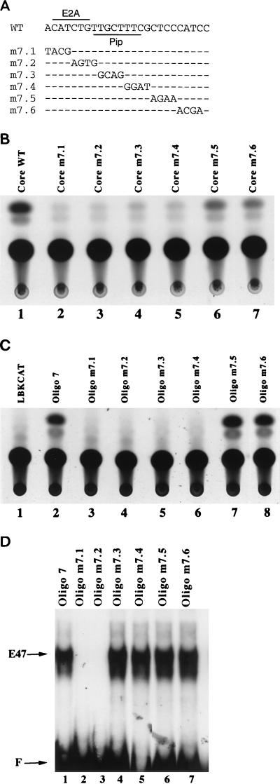
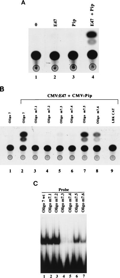
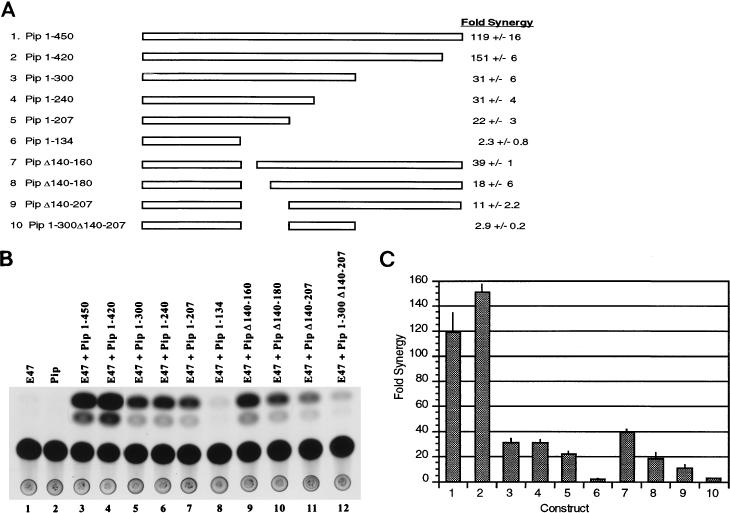
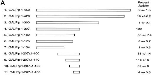
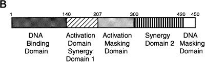
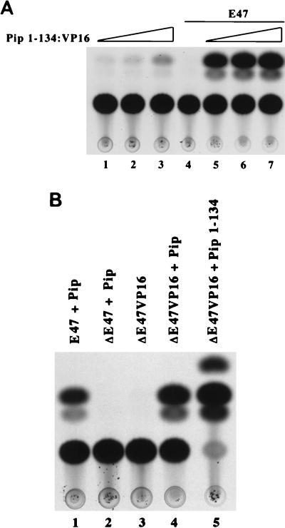
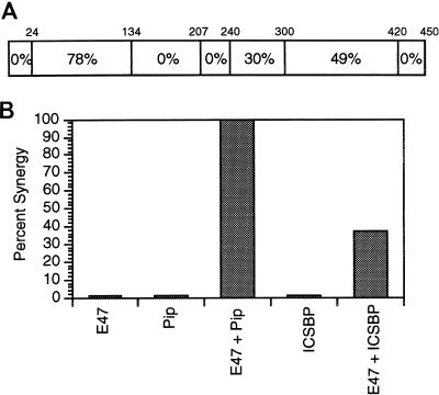
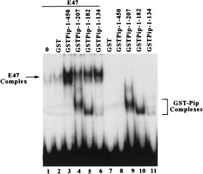
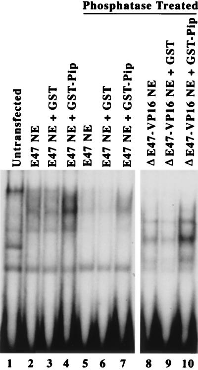
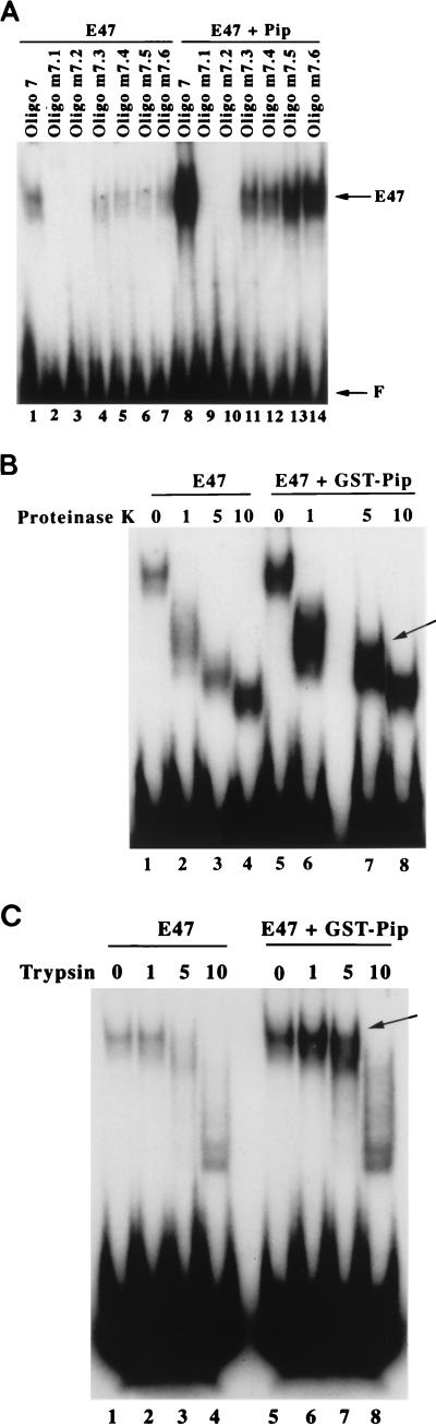
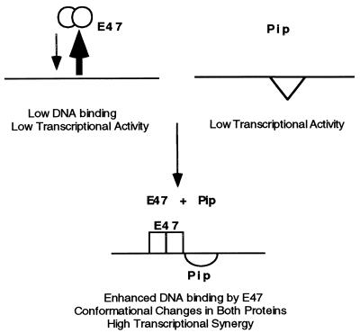
The transcription factors E2A (E12/E47) and Pip are both required for normal B-cell development. Each protein binds to regulatory sequences within various immunoglobulin enhancer elements. Activity of E2A proteins can be regulated by interactions with other proteins which influence their DNA binding or activation potential. Similarly, Pip function can be influenced by interaction with the protein PU.1, which can recruit Pip to bind to DNA. We show here that a previously unidentified Pip binding site resides adjacent to the E2A binding site within the immunoglobulin kappa 3' enhancer. Both of these binding sites are crucial for high-level enhancer activity. We found that E47 and Pip can functionally interact to generate a very potent 100-fold transcriptional synergy. Through a series of mutagenesis experiments, we identified the Pip sequences necessary for transcriptional activation and for synergy with E47. Two synergy domains (residues 140 to 207 and 300 to 420) in addition to the Pip DNA binding domain (residues 1 to 134) are required for maximal synergy with E47. We also identified a Pip domain (residues 207 to 300) that appears to mask Pip transactivation potential. Part of the synergy mechanism between E47 and Pip appears to involve the ability of Pip to increase DNA binding by E47, perhaps by inducing a conformational change in the E47 protein. E47 may also induce a conformational change in Pip which unmasks sequences important for transcriptional activity. Based upon our results, we propose a model for E47-Pip transcriptional synergy.


Figures

FIG. 1
FIG. 1
Identification of a functional enhancer sequence adjacent to the E-box motif. (A) The Ig(κ) 3′ enhancer sequence that includes the E-box and adjacent sequences is shown at the top (WT). The sequences of mutants m7.1 to m7.6 are shown below it. Positions of E2A and Pip binding sites are indicated. (B) S194 plasmacytoma cells were transfected with the wild-type enhancer core reporter plasmid or mutant 7.1 to 7.6 in the context of the enhancer core. CAT assays of extracts from the transfected cells show that enhancer activity is greatly reduced in mutants m7.1 to m7.4. (C) S194 plasmacytoma cells were transfected with reporter plasmids containing 25-bp oligonucleotide multimers (four copies) of the wild-type enhancer sequence (oligonucleotide 7) or multimers of mutants m7.1 to m7.6. CAT assays of the transfected cell extracts show that enhancer activity is absent in mutants m7.1 to m7.4. (D) E47-DNA binding was assayed by EMSA with the multimerized wild-type 25-bp oligonucleotide enhancer probe (oligonucleotide 7) or with each mutant oligonucleotide probe (oligonucleotides m7.1 to m7.6). E47 binding to probes oligonucleotides m7.1 and m7.2 was abolished. The probes used are indicated above each lane, and the positions of free probe (F) and E47 bound to DNA are indicated by arrows.
FIG. 2
FIG. 2
Pip can synergize with E47 to activate transcription. (A) NIH 3T3 cells were transfected with the Oligo7LBKCAT reporter plasmid alone (○) or with CMV:E47 (E47), CMV:Pip (Pip), or both (E47 + Pip). CAT assays of transfected cell extracts indicate that cotransfection of E47 and Pip results in a potent (100-fold) transcriptional synergy. The expression plasmids used in each transfection are indicated above each lane. (B) Transfections were performed with CMV:E47 plus CMV:Pip and either the wild-type reporter plasmid (oligonucleotide 7) or various mutant reporter plasmids (nucleotides m7.1 to m7.6). CAT assays of extracts isolated from transfected cells indicate that mutants m7.1 to m7.4 abolish synergy between E47 and Pip. The expression and reporter plasmids in each transfection are indicated above the lanes. (C) Pip can bind to a site adjacent to the E-box motif. Pip deletion protein GST–Pip 1-182 was used in EMSA with either the wild-type enhancer probe (oligo 7 wt) or with each enhancer mutant (oligonucleotides m7.1 to m7.6). Mutants m7.3 and m7.4 greatly reduce Pip DNA binding.
FIG. 3
FIG. 3
Identification of Pip sequences involved in synergy with E47. (A) Various Pip deletion mutant proteins are represented as rectangles. The Pip residues present in each construct are shown on the left. The fold synergy in NIH 3T3 cells for each protein in the presence of E47 is shown at the right. Error numbers represent standard deviations from three to five transfections. (B) Representative CAT assay. NIH 3T3 cells were cotransfected with plasmids expressing either wild-type Pip or various Pip mutants and wild-type E47. The plasmids used in each transfection are shown above the lanes. (C) Fold synergy for each construct (numbered as shown in panel A) is plotted. Synergy was defined as the percent acetylation observed with each Pip protein in the presence of E47 divided by the sum of the activities for each protein separately. Error bars represent standard deviations.
FIG. 4
FIG. 4
Identification of Pip activation domain sequences. (A) Various Pip sequences, represented as rectangles, were linked to the GAL4 DNA binding domain (residues 1 to 147). Pip residues present in each construct are listed on the left. Constructs were transfected into NIH 3T3 cells with the GALTKCAT reporter, and CAT activity was determined. The percent acetylation of the GAL–Pip 1-207 construct was defined as 100%, and all other levels are expressed relative to this value. Percent activity ± standard deviation is shown on the right. (B) Summary of the Pip functional domains. Pip residue positions are indicated. The DNA binding domain and the DNA masking domain were localized by Brass et al. (7).
FIG. 4
FIG. 4
Identification of Pip activation domain sequences. (A) Various Pip sequences, represented as rectangles, were linked to the GAL4 DNA binding domain (residues 1 to 147). Pip residues present in each construct are listed on the left. Constructs were transfected into NIH 3T3 cells with the GALTKCAT reporter, and CAT activity was determined. The percent acetylation of the GAL–Pip 1-207 construct was defined as 100%, and all other levels are expressed relative to this value. Percent activity ± standard deviation is shown on the right. (B) Summary of the Pip functional domains. Pip residue positions are indicated. The DNA binding domain and the DNA masking domain were localized by Brass et al. (7).
FIG. 5
FIG. 5
The Pip and E47 activation sequences can be replaced with a heterologous activation domain. (A) NIH 3T3 cells were transfected with GAL–Pip 1-134–VP16 either alone (lanes 1 to 3) or in the presence of 3 μg of E47 expression plasmid (lanes 5 to 7). Pip expression plasmid was included in quantities of 50 ng (lanes 1 and 5), 100 ng (lanes 2 and 6), or 250 ng (lanes 3 and 7). E47 alone (3 μg) is shown in lane 4. (B) NIH 3T3 cells were transfected with the various plasmids (3 μg) as indicated above the lanes. A representative CAT assay is shown.
FIG. 6
FIG. 6
ICSBP can synergize with E47. (A) Pip and ICSBP show a modular arrangement of identities. The rectangle represents the Pip protein sequence. The percent identity between Pip and ICSBP in the regions bounded by the Pip residues shown above the rectangle are indicated. (B) NIH 3T3 cells were transfected with E47, Pip, or ICSBP alone or with combinations of E47 plus Pip or E47 plus ICSBP. The level of synergy in the cotransfections was determined as described in the legend to Fig. 3. The level of synergy between E47 and Pip was defined as 100%, and the synergy between E47 and ICSBP is expressed relative to this value. The range in percent synergy for E47 plus ICSBP in two separate experiments was 1%.
FIG. 7
FIG. 7
Pip residues 1 to 134 can enhance E47 binding to DNA. EMSA was performed with the wild-type oligonucleotide 7 probe and E47 protein either alone (lane 1) or in the presence of various GST-Pip fusion proteins (lanes 3 to 6) or with GST protein alone (lane 2). Results for assays without E47 are also shown (lanes 7 to 11). The positions of the GST-Pip DNA and the E47-DNA complexes are indicated.
FIG. 8
FIG. 8
GST-Pip can increase DNA binding by transfected E47. NIH 3T3 cells were transfected with plasmids expressing either E47 (lanes 2 to 7) or ΔE47-VP16 (lanes 8 to 10). Mininuclear extracts (NE) were prepared and subjected to EMSA with the wild-type oligonucleotide 7 probe. The proteins included in each sample are shown above the lanes. Samples in lanes 5 to 10 were treated with alkaline phosphatase prior to EMSA.
FIG. 9
FIG. 9
(A) Maximal DNA binding by E47 requires E2A and Pip binding sites. EMSA was performed with wild-type or mutant oligonucleotide 7 probes and either E47 alone (lanes 1 to 7) or E47 plus Pip (lanes 8 to 14). The identity of each probe used in the EMSA is indicated above each lane. (B and C) The wild-type oligonucleotide 7 probe was incubated in an EMSA reaction mixture with either E47 alone or with E47 plus GST-Pip. After 30 min, various amounts of either proteinase K (B) or trypsin (C) were added to the reaction mixtures. Digestion was allowed to proceed for 5 min at room temperature. Samples were placed on ice and then subjected to electrophoresis. The amount, in nanograms, of each protease included in the reaction mixtures is shown above each lane. The position of complexes induced by GST-Pip are indicated with arrows.
FIG. 10
FIG. 10
Model of synergy between E47 and Pip. E47 dimers (empty circles) bind poorly to DNA. Pip (empty triangle) binds to DNA but in a transcriptionally inactive context. Pip can induce a conformational change in E47 (empty rectangles) which enhances E47 DNA binding. E47 induces a conformational change in Pip (empty semicircle) which exposes sequences necessary for transcription. These events result in high levels of synergy between E47 and Pip.

References

    1. Atchison M L, Perry R P. The role of the κ enhancer and its binding factor NF-κB in the developmental regulation of κ gene transcription. Cell. 1987;48:121–127. - PubMed
    1. Bain G, Gruenwald S, Murre C. E2A and E2-2 are subunits of B-cell-specific E2-box DNA-binding proteins. Mol Cell Biol. 1993;13:3522–3529. - PMC - PubMed
    1. Bain G, Maandag E C R, Izon D J, Amsen D, Kruisbeek A M, Weintraub B C, Krop I, Schlissel M S, Feeney A J, van Roon M, van der Valk M, te Riele H P J, Berns A, Murre C. E2A proteins are required for proper B cell development and initiation of immunoglobulin gene rearrangements. Cell. 1994;79:885–892. - PubMed
    1. Benezra R. An intermolecular disulfide bond stabilizes E2A homodimers and is required for DNA binding at physiological temperatures. Cell. 1994;79:1057–1067. - PubMed
    1. Benezra R, Davis R L, Lockshon D, Turner D L, Weintraub H. The protein Id: a negative regulator of helix-loop-helix DNA binding proteins. Cell. 1990;61:49–59. - PubMed

Publication types

MeSH terms

LinkOut - more resources