. 1998 Aug;18(8):4670-8.
doi: 10.1128/MCB.18.8.4670.

Distinct roles of RAG1 and RAG2 in binding the V(D)J recombination signal sequences

Affiliations

Distinct roles of RAG1 and RAG2 in binding the V(D)J recombination signal sequences

Y Akamatsu et al. Mol Cell Biol. 1998 Aug.

Abstract

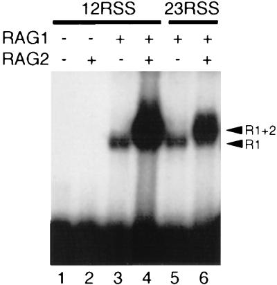
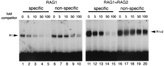
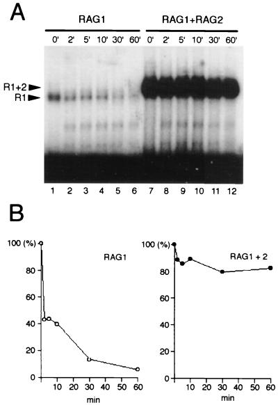
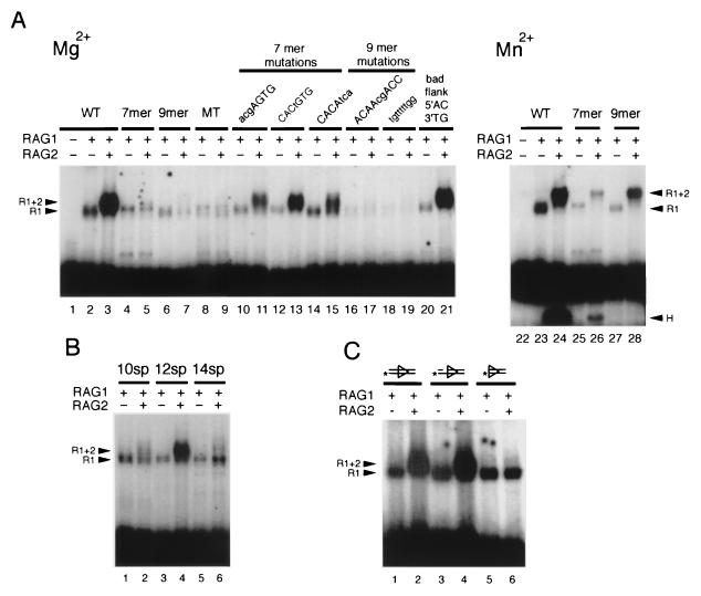
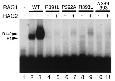
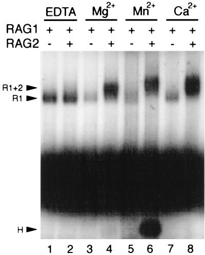
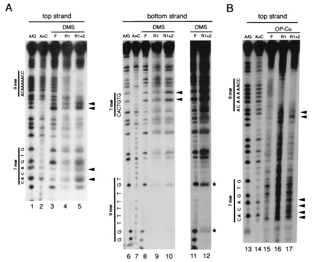
The RAG1 and RAG2 proteins initiate V(D)J recombination by introducing double-strand breaks at the border between a recombination signal sequence (RSS) and a coding segment. To understand the distinct functions of RAG1 and RAG2 in signal recognition, we have compared the DNA binding activities of RAG1 alone and RAG1 plus RAG2 by gel retardation and footprinting analyses. RAG1 exhibits only a three- to fivefold preference for binding DNA containing an RSS over random sequence DNA. Although direct binding of RAG2 by itself was not detected, the presence of both RAG1 and RAG2 results in the formation of a RAG1-RAG2-DNA complex which is more stable and more specific than the RAG1-DNA complex and is active in V(D)J cleavage. These results suggest that biologically effective discrimination between an RSS and nonspecific sequences requires both RAG1 and RAG2. Unlike the binding of RAG1 plus RAG2, RAG1 can bind to DNA in the absence of a divalent metal ion and does not require the presence of coding flank sequence. Footprinting of the RAG1-RAG2 complex with 1, 10-phenanthroline-copper and dimethyl sulfate protection reveal that both the heptamer and the nonamer are involved. The nonamer is protected, with extensive protein contacts within the minor groove. Conversely, the heptamer is rendered more accessible to chemical attack, suggesting that binding of RAG1 plus RAG2 distorts the DNA near the coding/signal border.


Figures

FIG. 1
FIG. 1
Detection of RAG1-DNA and RAG1-RAG2-DNA complexes. The indicated RAG proteins were incubated in the presence of 1 mM Mg2+ with 5′-end-labeled oligonucleotide substrates containing a 12-RSS or a 23-RSS, as indicated. The samples were fixed with 0.1% glutaraldehyde and electrophoresed through a 4 to 20% native polyacrylamide gel. The positions of the RAG1 (R1) and RAG1-RAG2 (R1+2) gel shifts are indicated by arrowheads.
FIG. 2
FIG. 2
The sequence specificity of RAG1 plus RAG2 (R1+2) is much greater than that of RAG1 (R1) alone. A comparison of the binding competition with RAG1 alone (left) and RAG1 plus RAG2 (right) is shown. Binding reactions were carried out in the presence of 1 mM Mg2+ and the specified ratios of unlabeled competitor DNA to labeled substrate DNA. VDJ176 (see Materials and Methods) was included as unlabeled, nonspecific single-stranded DNA. Specific competitor (lanes 1 to 5 and 11 to 15) was formed by annealing VDJ100/101 (5), and nonspecific competitor (lanes 6 to 10 and 16 to 20) was constructed from VDJ156/157 (see Materials and Methods).
FIG. 3
FIG. 3
Binding of RAG1 plus RAG2 (R1+2) is more stable than that of RAG1 (R1). (A) Dissociation of RAG1 versus RAG1 lus RAG2. RAG1 or RAG1 plus RAG2 proteins were allowed to bind to a labeled 12-RSS for 2 h at 25°C and then were challenged with a 1,000-fold excess of nonradioactive 12-RSS competitor for the times indicated before the addition of glutaraldehyde and subsequent electrophoresis. (B) Quantitation of dissociation over time. The gel from panel A was analyzed on a PhosphorImager, and the percentage of bound complex remaining at each time point is indicated.
FIG. 4
FIG. 4
Binding of RAG1 (R1) or RAG1 plus RAG2 (R1+2) to mutant substrates. The binding of RAG1 and RAG1 plus RAG2 to substrates with the indicated mutations was examined. Reactions were performed in the presence of 1 mM Mg2+, except as noted. The presence of RAG1 or RAG2 is indicated at the top of each lane. (A) Effects of mutations in the heptamer or nonamer. WT, 12-RSS; 7mer, intact heptamer with nonamer replaced; 9mer, intact nonamer with heptamer replaced; MT, both heptamer and nonamer replaced. The sequences of other mutants are as noted. (B) RAG1-RAG2 binding is sensitive to changes in spacer length. 10sp, 12sp, and 14sp are RSS with spacers of 10, 12, and 14 bp, respectively. Reactions mixtures were incubated for 15 min at 25°C. (C) Effects of structural variations of the substrate. Lanes: 1 and 2, 12-RSS; 3 and 4, top-strand nick; 5 and 6, RSS with no coding flank. Substrates were labeled on the 5′ end of the bottom strand in panel C and on the 5′ end of the top strand for panels A and B.
FIG. 5
FIG. 5
Mutations in the Hin domain disrupt binding of RAG1 (R1) and RAG1 plus RAG2 (R1+2). Binding of core RAG1 (WT) and RAG1 mutant proteins was examined under standard reaction conditions (the concentrations of WT and mutant RAG1 protein were adjusted to 1 pmol per reaction). Point mutations and deletions, as indicated, are described in the text.
FIG. 6
FIG. 6
RAG1-RAG2 dissociates from DNA after cleavage. (A) The structure of the DNA from gel-purified RAG1-RAG2-DNA complexes was examined by denaturing gel electrophoresis. Binding reactions were allowed to proceed for the times indicated in the presence of 1 mM Mn2+ to permit cleavage. The positions of the substrate and nicked or hairpinned products are indicated. (B) Graphic representation of the percentage of each species of DNA present in the bound complex. Quantitation of the bands in the gel from panel A was performed on a PhosphorImager. In addition, the amount of hairpinned product migrating below the unbound substrate was determined and included in the graph as open circles (free hairpin). Quantitation of free hairpin is given as the relative amount of radioactivity in the hairpin form compared to the total amount of bound complex.
FIG. 7
FIG. 7
Effects of divalent metal ions on binding of RAG1 (R1) or RAG1 plus RAG2 (R1+2). Binding in the presence of different divalent metal cations is analyzed. Binding reactions were carried out at 25°C for 15 min in the presence of 10 mM EDTA (lanes 1 and 2), 1 mM Mg2+ (lanes 3 and 4), 1 mM Mn2+ (lanes 5 and 6), or 1 mM Ca2+ (lanes 7 and 8). The cleaved hairpin product (H) can be seen below the free substrate in lane 6.
FIG. 8
FIG. 8
RAG1-RAG2 contacts the heptamer and the nonamer of the RSS. The binding of RAG1 (R1) and RAG1-RAG2 (R1+2) was analyzed by DMS protection and OP-Cu footprinting, as described in Materials and Methods. The RAG1 protein MR1 was used (see Materials and Methods). The positions of the heptamer and nonamer sequences are marked and the chemical sequencing ladder for A/G and A>C is shown. Sites of enhanced chemical cleavage are marked by arrowheads. (A) DMS protection. Top and bottom strands of 12-RSS oligonucleotides were labeled at the 5′ ends. The positions of the unprotected G residues are indicated by asterisks. Lanes 11 and 12 are longer exposures of lanes 8 and 10. (B) OP-Cu footprint. F, free substrate purified from the gel and treated in parallel with the bound complexes. R1 and R1+2 are the footprints of RAG1 and RAG1 plus RAG2, respectively.

References

    1. Akamatsu Y, Tsurushita N, Nagawa F, Matsuoka M, Okazaki K, Imai M, Sakano H. Essential residues in V(D)J recombination signals. J Immunol. 1994;153:4520–4529. - PubMed
    1. Akamatsu, Y. Unpublished observations.
    1. Akira S, Okazaki K, Sakano H. Two pairs of recombination signals are sufficient to cause immunoglobulin V-(D)-J joining. Science. 1987;238:1134–1138. - PubMed
    1. Ausubel F M, Brent R, Kingston R E, Moore D D, Seidman J G, Smith J A, Struhl K. Current protocols in molecular biology. New York, N.Y: Wiley Interscience; 1989.
    1. Cheung S, Arndt K, Lu P. Correlation of lac operator DNA imino proton exchange kinetics with its function. Proc Natl Acad Sci USA. 1984;81:3665–3669. - PMC - PubMed

Publication types

MeSH terms

LinkOut - more resources