. 1998 Aug;18(8):4732-43.
doi: 10.1128/MCB.18.8.4732.

Regulation of differentiation by HBP1, a target of the retinoblastoma protein

Affiliations

Regulation of differentiation by HBP1, a target of the retinoblastoma protein

H H Shih et al. Mol Cell Biol. 1998 Aug.

Abstract

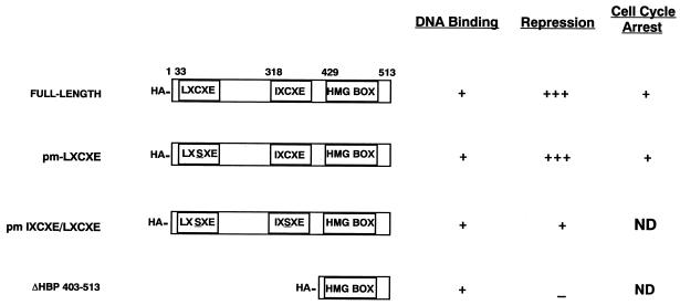
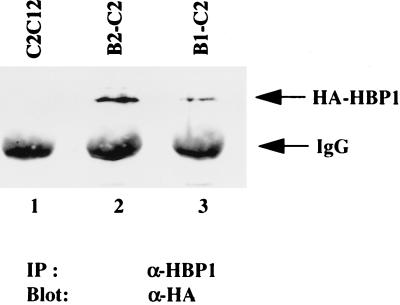
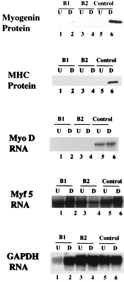
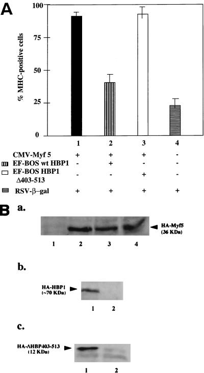
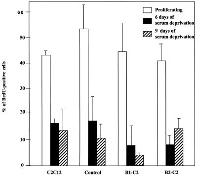
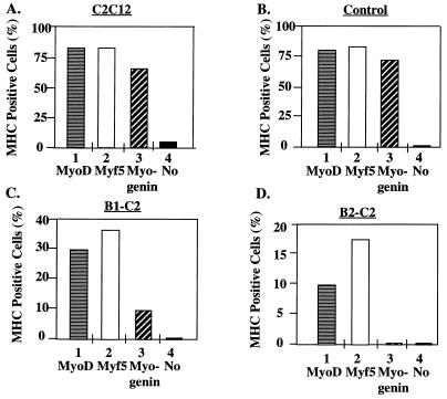
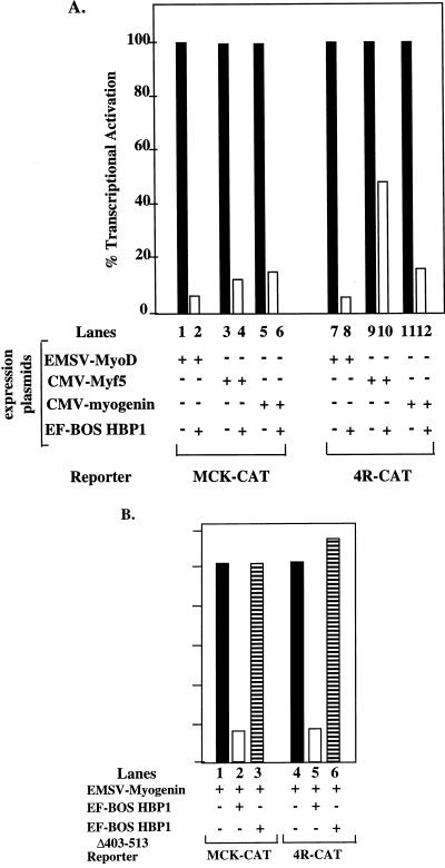
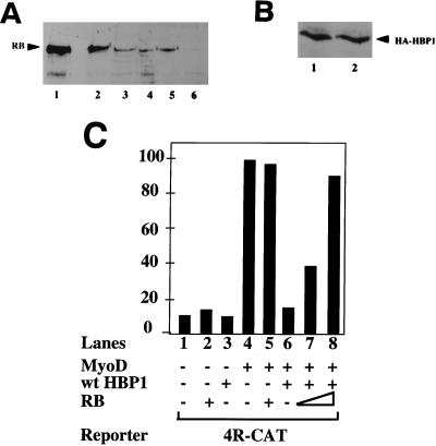
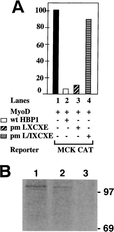
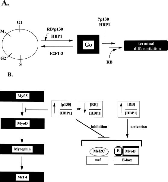
Differentiation is a coordinated process of irreversible cell cycle exit and tissue-specific gene expression. To probe the functions of the retinoblastoma protein (RB) family in cell differentiation, we isolated HBP1 as a specific target of RB and p130. Our previous work showed that HBP1 was a transcriptional repressor and a cell cycle inhibitor. The induction of HBP1, RB, and p130 upon differentiation in the muscle C2C12 cells suggested a coordinated role. Here we report that the expression of HBP1 unexpectedly blocked muscle cell differentiation without interfering with cell cycle exit. Moreover, the expression of MyoD and myogenin, but not Myf5, was inhibited in HBP1-expressing cells. HBP1 inhibited transcriptional activation by the MyoD family members. The inhibition of MyoD family function by HBP1 required binding to RB and/or p130. Since Myf5 might function upstream of MyoD, our data suggested that HBP1 probably blocked differentiation by disrupting Myf5 function, thus preventing expression of MyoD and myogenin. Consistent with this, the expression of each MyoD family member could reverse the inhibition of differentiation by HBP1. Further investigation implicated the relative ratio of RB to HBP1 as a determinant of whether cell cycle exit or full differentiation occurred. At a low RB/HBP1 ratio cell cycle exit occurred but there was no tissue-specific gene expression. At elevated RB/HBP1 ratios full differentiation occurred. Similar changes in the RB/HBP1 ratio have been observed in normal C2 differentiation. Thus, we postulate that the relative ratio of RB to HBP1 may be one signal for activation of the MyoD family. We propose a model in which a checkpoint of positive and negative regulation may coordinate cell cycle exit with MyoD family activation to give fidelity and progression in differentiation.


Figures

FIG. 1
FIG. 1
Schematic diagram of HBP1 mutants. The data presented here represent a summary of previous studies (49).
FIG. 2
FIG. 2
Detection of HBP1 transgene product in B1-C2 and B2-C2 cells. Cell lysates from each cell line were prepared, and the level of HBP1 transgene product in each cell line was detected by immunoprecipitation with anti-HBP1 antibodies followed by Western analysis with anti-HA (12CA5) antibody as described in Materials and Methods. The analyzed lysates are depicted in the figure as follows: lane 1, normal C2 cells; lane 2, B1-C2 HBP1 expressing line; lane 3, B2-C2 HBP1 expressing line.
FIG. 3
FIG. 3
Expression levels of terminal differentiation markers. The levels of MHC, myogenin, MyoD, and Myf5 were scored in B1-C2, B2-C2, and control cell lines. For myogenin and MHC, the protein levels were detected by Western blot analysis with monoclonal antimyogenin (F5D) and monoclonal anti-MHC (FS9) antibodies, respectively, in cell lysates prepared from each line. For MyoD and Myf5, the RNA levels were quantitated in a T2 RNase protection assay with total RNA that was isolated from each cell line. GAPDH (glyceraldehyde-3-phosphate dehydrogenase) was employed as an RNA loading control. The tested cell lines are indicated: lanes 1 and 2, B1-C2; and lanes 3 and 4, B2-C2; and lanes 5 and 6, hygromycin-resistant control line without HA-HBP1. The odd numbers represent undifferentiated conditions (15% serum) and are denoted “U.”. The even numbers represent differentiated conditions (2% serum) and are denoted “D.” A representative experiment is shown here, and identical results were obtained in two independent analyses.
FIG. 4
FIG. 4
Overexpression of HBP1 inhibited C2C12 differentiation in a transient-differentiation assay. A transient-differentiation assay was devised to score the effects of HBP1 on C2C12 differentiation. The basis of the assay was that expression of MyoD family members could accelerate C2 cell differentiation. (A) C2C12 cells grown on coverslips were transfected with plasmids encoding wild-type or mutant HBP1, Myf5, and β-Gal. After 24 h, the transfected cells were cultured in medium supplemented with 2% FCS for another 40 h. Cells were fixed and immunostained for β-Gal and MHC. The percentage of differentiated cells (MHC positive) among the transfected cells (β-Gal positive) was determined. Each transfection was repeated three times, and cells on two different coverslips from each experiment were counted. The total number of cells counted for each experiment was 200 to 300. All lanes contain β-Gal and the following additions: lane 1, Myf5 (filled column); lane 2, Myf5 and wild-type HBP1 (vertical stripes); lane 3, Myf5 and HA-ΔHBP403-513 (open column); and lane 4, no addition (horizontal stripes). (B) To measure protein expression levels, the lysates from a parallel experiment were analyzed for the expression of HA-Myf5, HA-ΔHBP403-513, and HA-HBP1 by using immunoprecipitation with anti-Myf5 or anti-HBP1 antibodies, respectively, followed by Western blot analyses of immune complexes with the anti-HA antibody. The constructs and antibodies are described in Materials and Methods. A representative experiment is shown. (Ba) Western blot showing Myf5 expression. The assay was performed as described for panel B. Cells were transfected with β-Gal and Myf5 (lane 4), Myf5 and HBP1 (lane 3), Myf5 and ΔHBP403-513 (lane 2), or no other expression vector (lane 1). (Bb) Western blot showing HBP1 expression. Cells were transfected with β-Gal, Myf5 and HBP1 (lane 1) or Myf5 (lane 2). (BC) Cells were transfected with β-Gal and Myf5 and ΔHBP403-513 (lane 1) or Myf5 (lane 2).
FIG. 5
FIG. 5
Suppression of S phase in HBP1-expressing cells upon serum deprivation. The HBP1-expressing cell lines (B1-C2 and B2-C2) and the control lines were assayed for the ability to exit the cell cycle upon serum deprivation. BrdU incorporation was used as a measure of the S phase, and quiescent cells should exhibit a reduction in BrdU-positive cells. All cell lines were grown on coverslips in differentiation medium for the indicated time period followed by a 1-h pulse of BrdU labeling. Cells were subsequently fixed and immunostained for BrdU. The percentages of BrdU-positive cells were determined by counting the cells from several random fields; approximately 200 to 300 cells were counted for each column. As controls, normal C2 and control hygromycin-resistant C2 cell lines were utilized. The open, filled, and diagonal striped bars represent the proliferating, 6-day-deprived, and 9-day-deprived cell populations for each cell line, respectively.
FIG. 6
FIG. 6
Expression of MyoD family members can overcome inhibition of differentiation in the B1-C2 and B2-C2 lines. The purpose of this experiment was to determine whether MyoD family members could rescue the differentiation defect imposed by HBP1. A modification of the transient-differentiation assay in Fig. 4 was used. Expression vectors encoding MyoD, Myf5, or myogenin were transiently transfected into either control or HBP1-expressing cell lines (B1-C2 or B2-C2). A β-Gal expression vector was cotransfected to identify the transfected cells in each experiment. At 24 h posttransfection, cells were grown in differentiation medium for an additional 40 h before they were stained with antibodies. Double immunostaining for differentiated and transfected cells was performed with anti-MHC monoclonal antibody and anti-β-Gal antisera, followed by staining with fluorescein-conjugated anti-mouse immunoglobulin G and rhodamine-conjugated anti-rabbit immunoglobulin G, respectively. The percentages of differentiated and transfected cells were quantitated from approximately 200 to 300 cells for each experimental point. The indicated cell lines are as described in the legend to Fig. 5. The percentage of MHC-positive cells was determined for MyoD (horizontal stripes), Myf5 (open column), myogenin (diagonal stripes), or β-Gal (filled; denoted “No”).
FIG. 7
FIG. 7
Inhibition by HBP1 of MyoD family transcriptional activation. (A) HBP1 can inhibit MyoD family activation of a natural differentiation-specific MCK-CAT or a simplified muscle-specific 4R-CAT reporter constructs. MCK-CAT denotes a CAT reporter construct driven by the ∼3 kb of the differentiation-specific muscle creatine kinase promoter (46). 4R-CAT denotes a simplified and muscle-specific reporter in which CAT expression is driven by four reiterated MyoD family binding sites (E-box elements) upstream of a minimal thymidine kinase promoter (51). The effect of HBP1 on transcriptional activation by either MyoD (lanes 1 and 2), Myf5 (lanes 3 and 4), or myogenin (lanes 5 and 6) was quantitated in C2 cells as described in Materials and Methods. In each set, transcriptional activities in the presence or absence of HBP1 were denoted by open or filled columns, respectively. (B) Inhibition of MyoD family transcriptional activation requires the N-terminal region of HBP1. Myogenin was used as a representative member of the MyoD family, and the relative inhibition by wild-type HBP1 (open column) and by ΔHBP403-513 (horizontal stripes) (see Fig. 1 for description) was compared by using assays similar to those described for panel A.
FIG. 8
FIG. 8
Expression of RB reverses the HBP1-mediated inhibition of differentiation and of MyoD transcriptional activation. Whereas Table 1 represents a direct quantitation of differentiation in the test cell lines, this figure depicts the relative expression levels of exogenous and endogenous RB protein. The presence of HA-RB transgene product was detected in lysates from each cell line by immunoprecipitation with anti-RB antibodies followed by Western analysis of immune complexes with a monoclonal anti-RB antibody (as described in Materials and Methods). This protocol allowed a direct comparison of “overexpressed” RB levels relative to endogenous RB. C2 cell line B1-B9 represents a line coexpressing HBP1 and RB; the C2 cell line B1-B2 represents a line expressing HBP1 only, but it was isolated with selection conditions identical to those for B1-B9. (A) Expression of RB and HA-RB in cell lines. Lanes: 1, C2C12 transiently transfected with HA-RB expression vector; 2, B1-B9 (HBP1+RB); 3, B1-B2 (control hygromycin- and puromycin-resistant line; HBP1 only); 4, B1-C2 (HBP1 only); 5, C2C12; 6, C2C12 cell extracts immunoprecipitated with anti-β-Gal antibodies as a negative control. The position of the RB protein is indicated and was determined in the positive control (lane 1). (B) Expression of HBP1 in cell lines. The levels were quantitated by Western blotting with an anti-HA antibody of an anti-HBP1 immunoprecipitation. This control experiment was performed to ensure that the reversal of the differentiation phenotype by RB was not due to the loss of HBP1 expression. Lanes: 1, B1-B9 (HBP1+RB); 2, B1-B2 (HBP1 only). (C) Effect of RB on HBP1-mediated inhibition of MyoD activation of 4R-CAT. The transcriptional activities were determined by transient-transfection assays in C2 cells by using specific promoter constructs together with wild-type or mutant HBP1 and RB expression vectors. Rous sarcoma virus–β-Gal was used as an internal transfection control to normalize transfection efficiency. The transfection output is expressed as a normalized ratio of CAT protein to β-Gal activity, and the combinations of expressed proteins are indicated. One representative experiment is shown in each graph, and each quantitation represents duplicate transfections that varied by <10%. Each experiment was repeated three to five times. Protein expression levels were equivalent in all transfections by Western blotting or immunoprecipitation followed by Western analyses (data not shown).
FIG. 9
FIG. 9
Regulation of MyoD family transcriptional activation by HBP1 and RB. (A) Effect of HBP1 mutants on MyoD activation of a MCK-CAT reporter construct. The role of RB binding in the inhibition of MyoD family activation was tested by using the indicated mutants of HBP1. As described in Fig. 1 and reference , the wild-type HBP1 and pm-LXCXE are functional in RB binding and in repression of the N-MYC promoter, but the pm-L/IXCXE is defective in both functions. The indicated proteins were expressed in conjunction with MyoD: lane 1, no HBP expression vector (filled bar); lane 2, wild-type HBP1 (open bar); lane 3, pm-LXCXE (diagonal stripes); and lane 4, pm-L/I XCXE (horizontal stripes). (B) In vivo association of RB and HBP1 in differentiated C2C12 myotubes. HBP1 was shown to interact with RB in differentiated C2C12 myotubes. C2C12 were completely differentiated for 4 days in DMEM supplemented with 2% FCS. Cells were metabolically labeled with 35S-methionine, and cell lysates were collected for double immunoprecipitations as described in Materials and Methods. The first immunoprecipitations were carried out with anti-RB antibodies (lane 1) or anti-HBP1 antibodies (lanes 2 and 3), and the second immunoprecipitations were carried out with anti-RB antibodies (lanes 1 and 2) and anti-β-Gal antibodies (lane 3). Note that the amount of extracts used in lane 1 was approximately one-sixth of that used in lane 2 or 3.
FIG. 10
FIG. 10
Summary model of HBP1 and RB functions in differentiation. We postulate that muscle differentiation can be divided into general cell cycle exit (G0) and tissue-specific gene expression (terminal differentiation) coordinated by the RB family (RB and p130) and their targets, such as HBP1. A high p130/HBP1 or low RB/HBP1 ratio may favor the cell cycle exit but act as a negative signal for terminal differentiation. This transient suspension of differentiation may eventually be relieved by activation of RB, resulting in a high RB/HBP1 ratio. This complex regulatory mechanism may be an effective means for ensuring fidelity in differentiation by ensuring that only viable and arrested cells proceed to the irreversible expression of genes that specify individual tissue phenotypes.

Similar articles

Cited by

References

    1. Barone M V, Crozat A, Tabaee A, Philipson L, Ron D. CHOP (GADD153) and its oncogenic variant, TLS-CHOP, have opposing effects on the induction of G1/S arrest. Genes Dev. 1994;8:453–464. - PubMed
    1. Batchvarova N, Wang X Z, Ron D. Inhibition of adipogenesis by the stress-induced protein CHOP (Gadd153) EMBO J. 1995;14:4654–4661. - PMC - PubMed
    1. Braun T, Rudnicki M, Arnold H-H, Jaenisch R. Targeted inactivation of the muscle regulatory gene Myf-5 results in abnormal rib development and perinatal death. Cell. 1992;71:369–382. - PubMed
    1. Chen P L, Riley D J, Chen Y, Lee W H. Retinoblastoma protein positively regulates terminal adipocyte differentiation through direct interaction with C/EBPs. Genes Dev. 1996;10:2794–2804. - PubMed
    1. Cheng T C, Tseng B S, Merlie J P, Klein W H, Olson E N. Activation of the myogenin promoter during mouse embryogenesis in the absence of positive autoregulation. Proc Natl Acad Sci USA. 1995;92:561–565. - PMC - PubMed

Publication types

LinkOut - more resources