. 1998 Aug;18(8):4752-60.
doi: 10.1128/MCB.18.8.4752.

A potential role for U2AF-SAP 155 interactions in recruiting U2 snRNP to the branch site

Affiliations

A potential role for U2AF-SAP 155 interactions in recruiting U2 snRNP to the branch site

O Gozani et al. Mol Cell Biol. 1998 Aug.

Abstract

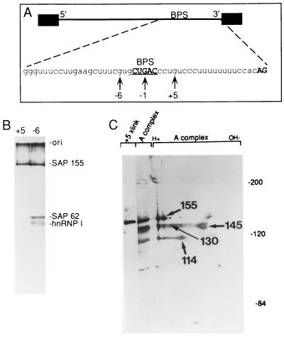
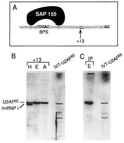
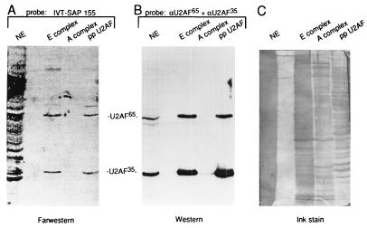
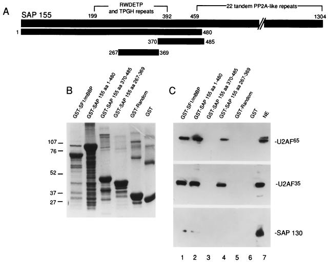
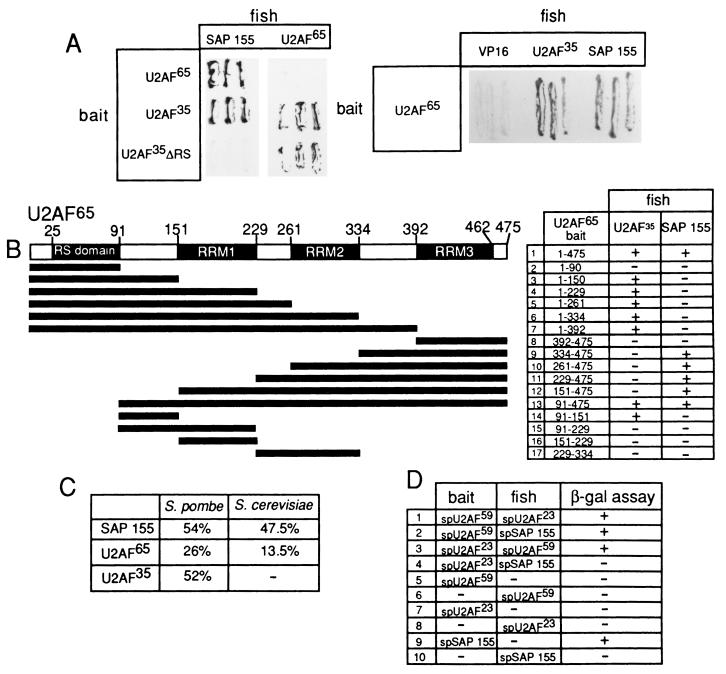
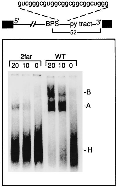
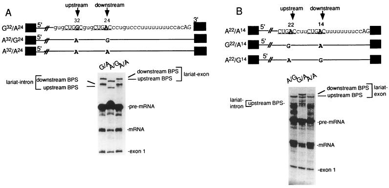
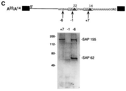
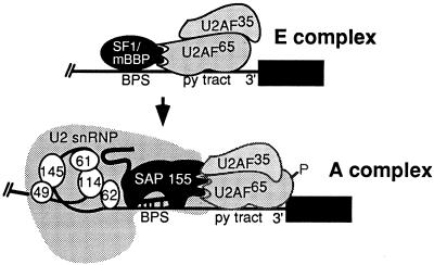
Base pairing between U2 snRNA and the branchpoint sequence (BPS) is essential for pre-mRNA splicing. Because the metazoan BPS is short and highly degenerate, this interaction alone is insufficient for specific binding of U2 snRNP. The splicing factor U2AF binds to the pyrimidine tract at the 3' splice site in the earliest spliceosomal complex, E, and is essential for U2 snRNP binding in the spliceosomal complex A. We show that the U2 snRNP protein SAP 155 UV cross-links to pre-mRNA on both sides of the BPS in the A complex. SAP 155's downstream cross-linking site is immediately adjacent to the U2AF binding site, and the two proteins interact directly in protein-protein interaction assays. Using UV cross-linking, together with functional analyses of pre-mRNAs containing duplicated BPSs, we show a direct correlation between BPS selection and UV cross-linking of SAP 155 on both sides of the BPS. Together, our data are consistent with a model in which U2AF binds to the pyrimidine tract in the E complex and then interacts with SAP 155 to recruit U2 snRNP to the BPS.


Figures

FIG. 1
FIG. 1
U2 snRNP protein SAP 155 UV cross-linking to pre-mRNA on both sides of the branch site. (A) Schematic of pre-mRNA. The nucleotide sequence of the 3′ portion of the AdML intron is shown. The BPS and AG dinucleotide at the 3′ splice site are in boldface, and the BPS is also underlined. The arrows indicate the G residues that were labeled, and the numbers are relative to the branch-site adenosine. (B) Pre-mRNAs labeled at −6 or +5 were assembled into the A complex, isolated by gel filtration, and UV cross-linked. After treatment with RNase A, proteins were fractionated on a 9% SDS gel (equal counts per minute of each pre-mRNA were loaded). The positions of SAPs 155 and 62 and hnRNP I are indicated. hnRNP I cross-linking is due to contamination of the A complex with the H complex in this experiment. (C) The +5 cross-linked sample was fractionated on a 6% SDS gel next to the A complex and next to the A complex separated in two dimensions. The A complex samples were detected by staining and the +5 cross-link (xlink) was detected by phosphorimager.
FIG. 2
FIG. 2
U2AF65 UV cross-linking next to SAP 155 in the A complex. (A) Nucleotide sequence of the 3′ portion of the AdML intron. The arrow indicates the G residue that was site-specifically labeled. The SAP 155 cross-linking sites in the A complex are indicated. At the +13 site, the UU was changed to GC in order to transcribe the 3′ RNA for site-specific labeling (see Materials and Methods). This alteration has no effect on A complex assembly or splicing. (B) The proteins that cross-link at the +13 site in the H, E, and A complexes are shown next to IVT U2AF65 (as a marker). (C) Pre-mRNA site-specifically labeled at +13 was assembled into the E complex and UV cross-linked, and then an immunoprecipitation (IP) with U2AF65 antibodies was carried out. IVT U2AF65 was run as a marker.
FIG. 3
FIG. 3
U2AF and SAP 155 interact directly. (A) IVT SAP 155 (aa 148 to 775) was used to probe a blot containing nuclear extract (NE), E complex, A complex, or partially purified U2AF (ppU2AF). U2AF65 and U2AF35 are indicated. (B) The blot in panel B was probed with U2AF65 and U2AF35 antibodies (α) (Western blotting). (C) An ink stain of the blot probed in panels A and B.
FIG. 4
FIG. 4
A specific amino-terminal region of SAP 155 is sufficient to mediate the interaction with U2AF. (A) Structure of SAP 155. The amino-terminal RWDETP and TPGH repeats and carboxy-terminal PP2A-like repeats are shown (29). Numbers indicate amino acids. The portions of SAP 155 that were used to make fusion proteins are indicated. (B) Coomassie stain of GST fusion proteins. Markers are indicated to the left in kilodaltons. (C) Lanes 1 to 6: GST fusion proteins bound to glutathione beads were incubated with a 25-μl aliquot of nuclear extract and washed, and then Western analysis of the bound proteins was carried out. Total nuclear extract (NE) (4 μl) is shown in lane 7. The blot was probed successively with U2AF65, U2AF35, and SAP 130 antibodies.
FIG. 5
FIG. 5
Analysis of the SAP 155-U2AF interaction by the yeast two-hybrid assay. (A) β-Galactosidase filter lift assays were done with cells transformed with the indicated bait and fish vectors. (B) Structure of U2AF65 and derivatives of U2AF65 that were used as baits for two-hybrid assays. U2AF35 and SAP 155 (aa 171 to 775) were used as fish. The interactions were assayed with the β-galactosidase filter assay. +, interactions between the fish and bait (blue color production on the filter). −, no interaction (relative to the VP16 negative control). (C) The percent identity between Homo sapiens, S. pombe, and S. cerevisiae SAP 155, U2AF35, U2AF65 homologs is shown. (D) Two-hybrid assays of S. pombe (sp) U2AF65, U2AF35, and SAP 155. The fish and baits are indicated. As with human SAP 155, S. pombe SAP 155 used as a bait transactivates the β-galactosidase (β-gal) promoter in the absence of a fish construct.
FIG. 6
FIG. 6
The distance between the BPS and pyrimidine (py) tract is critical for A complex assembly. A spacer of the sequence indicated was inserted immediately downstream of the BPS to generate the pre-mRNA 2far. The distance between the BPS and 3′ splice site is 52 nt. 2far or wild-type pre-mRNA was incubated under splicing conditions for the times indicated (in minutes) and then fractionated on a native gel. The H, A, and B complexes are indicated.
FIG. 7
FIG. 7
SAP 155 cross-linking to pre-mRNA correlates with BPS selection (A) The BPS closest to the 3′ splice site is selected. The structures of pre-mRNAs containing duplicated BPSs are shown. Pre-mRNAs were incubated under splicing conditions for 40 min, and total RNA was fractionated on a 15% polyacrylamide denaturing gel. The bands corresponding to spliced products and intermediates are indicated. (B) An optimal distance between the BPS and 3′ splice site is necessary for BPS selection. The structures of pre-mRNAs containing duplicated BPSs are indicated. The branch-site adenosine is in boldface, and the distance from this adenosine to the 3′ splice site is shown. The pre-mRNAs were incubated under splicing conditions for 45 min, and then total RNA was fractionated on a 13% polyacrylamide denaturing gel. The splicing intermediates and products are indicated. (C) SAP 155 UV cross-links on both sides of the functional BPS. A22/A14 pre-mRNA was 32P site-specifically labeled at the indicated guanosines, assembled into the A complex, isolated by gel filtration, and UV cross-linked. After RNase A treatment, cross-linked proteins were fractionated by SDS-polyacrylamide gel electrophoresis, and cross-linked proteins were detected by phosphorimager analysis. SAPs 155 and 62 are indicated.
FIG. 7
FIG. 7
SAP 155 cross-linking to pre-mRNA correlates with BPS selection (A) The BPS closest to the 3′ splice site is selected. The structures of pre-mRNAs containing duplicated BPSs are shown. Pre-mRNAs were incubated under splicing conditions for 40 min, and total RNA was fractionated on a 15% polyacrylamide denaturing gel. The bands corresponding to spliced products and intermediates are indicated. (B) An optimal distance between the BPS and 3′ splice site is necessary for BPS selection. The structures of pre-mRNAs containing duplicated BPSs are indicated. The branch-site adenosine is in boldface, and the distance from this adenosine to the 3′ splice site is shown. The pre-mRNAs were incubated under splicing conditions for 45 min, and then total RNA was fractionated on a 13% polyacrylamide denaturing gel. The splicing intermediates and products are indicated. (C) SAP 155 UV cross-links on both sides of the functional BPS. A22/A14 pre-mRNA was 32P site-specifically labeled at the indicated guanosines, assembled into the A complex, isolated by gel filtration, and UV cross-linked. After RNase A treatment, cross-linked proteins were fractionated by SDS-polyacrylamide gel electrophoresis, and cross-linked proteins were detected by phosphorimager analysis. SAPs 155 and 62 are indicated.
FIG. 8
FIG. 8
Model for recruitment of U2 snRNP. The 3′ portion of the intron containing the BPS and the pyrimidine (py) tract are shown. Only the factors discussed in this study are indicated. Proteins that interact directly are shown touching. P indicates the phosphorylation of U2AF65 that occurs in the A complex. Note that U2AF65 and U2AF35 are much less tightly bound in the A complex than in the E complex. SAPs 49, 61, 62, 114, 145, and 155 are shown.

Similar articles

Cited by

References

    1. Abovich N, Liao X C, Rosbash M. The yeast MUD2 protein: an interaction with PRP11 defines a bridge between commitment complexes and U2 snRNP addition. Genes Dev. 1994;8:843–854. - PubMed
    1. Abovich N, Rosbash M. Cross-intron bridging interactions in the yeast commitment complex are conserved in mammals. Cell. 1997;89:403–412. - PubMed
    1. Bennett M, Reed R. Correspondence between a mammalian spliceosome component and an essential yeast splicing factor. Science. 1993;262:105–108. - PubMed
    1. Bennett M, Michaud S, Kingston J, Reed R. Protein components specifically associated with prespliceosome and spliceosome complexes. Genes Dev. 1992;6:1986–2000. - PubMed
    1. Berglund J A, Chua K, Abovich N, Reed R, Rosbash M. The splicing factor BBP interacts specifically with the pre-mRNA branchpoint sequence UACUAAC. Cell. 1997;89:781–787. - PubMed

Publication types